. 2008 Dec 28;129(24):245101.
doi: 10.1063/1.3036928.

Proton assisted recoupling and protein structure determination

Affiliations

Proton assisted recoupling and protein structure determination

Gaël De Paëpe et al. J Chem Phys. .

Abstract

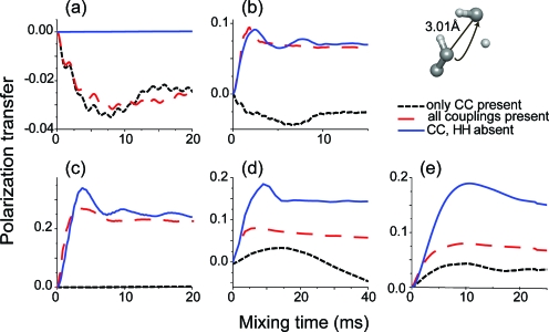
We introduce a homonuclear version of third spin assisted recoupling, a second-order mechanism that can be used for polarization transfer between (13)C or (15)N spins in magic angle spinning (MAS) NMR experiments, particularly at high spinning frequencies employed in contemporary high field MAS experiments. The resulting sequence, which we refer to as proton assisted recoupling (PAR), relies on a cross-term between (1)H-(13)C (or (1)H-(15)N) couplings to mediate zero quantum (13)C-(13)C (or (15)N-(15)N recoupling). In particular, using average Hamiltonian theory we derive an effective Hamiltonian for PAR and show that the transfer is mediated by trilinear terms of the form C(1) (+/-)C(2) (-/+)H(Z) for (13)C-(13)C recoupling experiments (or N(1) (+/-)N(2) (-/+)H(Z) for (15)N-(15)N). We use analytical and numerical simulations to explain the structure of the PAR optimization maps and to delineate the PAR matching conditions. We also detail the PAR polarization transfer dependence with respect to the local molecular geometry and explain the observed reduction in dipolar truncation. Finally, we demonstrate the utility of PAR in structural studies of proteins with (13)C-(13)C spectra of uniformly (13)C, (15)N labeled microcrystalline Crh, a 85 amino acid model protein that forms a domain swapped dimer (MW=2 x 10.4 kDa). The spectra, which were acquired at high MAS frequencies (omega(r)2pi>20 kHz) and magnetic fields (750-900 MHz (1)H frequencies) using moderate rf fields, exhibit numerous cross peaks corresponding to long (up to 6-7 A) (13)C-(13)C distances which are particularly useful in protein structure determination. Using results from PAR spectra we calculate the structure of the Crh protein.


Figures

Figure 1
Figure 1
(a) PAR pulse sequence for obtaining 2D homonuclear correlation spectra. The PAR mixing consist of continuous wave (cw) irradiation on the 1H and 13C channels that induces a second-order cross term between 1H–13C dipolar couplings [terms 2 and 3 of Eq. 6] in order to transfer polarization from 13C1 to 13C2. (b) The PAR subspace can be seen as a coupled basis between a fictitious ZQ operator involving two 13C’s and a 1H spin. The red arrows indicate PAR recoupling axis and longitudinal tilting field resulting from autocross terms (see Sec. 4).
Figure 2
Figure 2
PAR optimization map simulated using SPINEVOLUTION (Ref. 50). The initial magnetization is on the C1 spin (C2,X operator) and is detected after 3 ms PAR mixing on the three spins: (a) C1 spin, (b) C2 spin, and (c) H spin (C1,X, C2,X, HX operators, respectively). The spin system is composed of two directly bonded 13C’s and one 1H bonded to the 13C1 spin (see the inset of the figure). The distance between the 13C2 spin and the 1H is 2.15 Å. The angle between the two CH dipolar vectors is 42° and arises from the usual tetrahedral geometry. Simulations include typical chemical shift tensor values (see SI for details) (Ref. 69). The black dashed lines indicate the locations of the n=0 and ±1 and ±2 HH matching conditions.
Figure 3
Figure 3
PAR optimization maps where the polarization transfer between the 13C is monitored as a function of 13C and 1H irradiation strengths (in units of ωr) and a 1.5 ms mixing period. The spin system is identical to that in Fig. 2. No chemical shift interactions were included in these simulations. The three panels represent (a) analytical simulations of the 13C polarization transfer from 13C1 to 13C2 arising from only the TSAR term, (b) 13C signal intensity showing the analytical simulation obtained with the TSAR term and the longitudinal autocross-term contributions, and (c) 13C signal intensity depicting the numerical simulations performed with SPINEVOLUTION. The two white lines displayed on the contour plots in panels (b) and (c) represent points where χ(1,pC,pH)=0 and χ(2,pC,pH)=0 (i.e., autocross terms for spatial components m=1 and m=2 are equal to 0), described by equations pH=pC21 and pH=pC24, respectively.
Figure 4
Figure 4
Simulations of polarization transfer in the PAR experiment for a HC1C2 spin system. The chemical shift is not included in the simulations. The PAR 13C and 1H cw rf field strengths correspond to pC=2.6 and pH=2.35, respectively [see Fig. 3b]. The panels illustrate simulations that include the following couplings: (a) all dipolar couplings; (b) couplings C1H and C2H but not C1C2; (c) couplings C2H and C1C2, but not C1H (note that the polarization remains on C1); (d) couplings C1H and C1C2 but not C2H; (e) coupling C1H but not C2H or C1C2; (f) C1C2 but not C1H and C2H.
Figure 5
Figure 5
Dependence of PAR polarization transfer on the local geometry of the spin system. (a) The three-spin system geometry used in the simulations: the first 13C and the 1H are fixed in space, whereas the position of the second 13C is defined by θ and ϕ, the spherical coordinates with the origin at the 1H. The distances between the 13C’s and the 1H are constant and equal to 1.1 and 2.6 Å, respectively. The spherical map represents the 13C–13C polarization transfer efficiency for a PAR mixing time of 4.2 ms using pC=2.75 and pH=2.5. Polarization transfer for θ=0 orientation (aligned geometry) as a function of time is presented in panels (b) and (c). The buildup curves labeled (1)–(5) represent analytical simulations performed with the following contributions: (1) both m=1 and m=2 components without autocross terms, (2) both m=1 and m=2 with autocross terms, [(3) and (4)] only m=1 without and with autocross terms, [(5) and (6)] only m=2 without and with autocross terms.
Figure 6
Figure 6
Analytical contour plots of the PAR polarization transfer arising from the m=1 and m=2 components as a function of θ angle for a three spin system described in Fig. 5a with the mixing time and irradiation settings used in Fig. 5 with ϕ=0. (a) m=1 PAR component, (b) m=1 PAR term plus m=1 autocross-term components, (c) m=2 PAR component included, (d) m=2 PAR term and m=2 autocross-term components, (e) m=1 and m=2 PAR components, and (f) m=1 and m=2 PAR plus m=1 and m=2 autocross-term components.
Figure 7
Figure 7
Illustration of dipolar truncation in the CM5RR and PAR homonuclear recoupling schemes. The spin system 1 is composed of a directly bonded CαHα pair and Cremote spins of 4.5 and 3.56 Å distant from the Cα and Hα spins. In the spin system 2, a Cβ spin, directly bonded Cα, is also present. (a) The black dashed line depicts the polarization transfer (∼30%) from Cα to Cremote (rC−C=4.5 Å) using the broadband DQ CM5RR in the three spin system 1. When a directly bonded Cβ spin is added to the spin system (rC−C=1.5 Å), the polarization transfer to the Cremote (red dash-dot line) is quenched for CM5RR with most of the polarization being transferred to the directly bonded Cβ (blue solid line), thus demonstrating the phenomenon of dipolar truncation. (b) In the PAR simulation the presence of a third strongly coupled spin leads to a partial decrease in polarization transfer to Cremote (red dash-dot line) showing that dipolar truncation is attenuated in the TSAR transfer mechanism. Simulations were performed with SPINEVOLUTION (Ref. 50) ωr∕2π=20 kHz, ω0H∕2π=750 MHz 1H frequency and do not include chemical shifts.
Figure 8
Figure 8
(a) 2D 13C–13C correlation spectrum of [U–13C,15N]N-f-MLF-OH diluted to 10% in a natural abundance lattice. The spectrum was recorded at ω0H∕2π=750 MHz and ωr∕2π=20 kHz with τmix=7.5 ms, ω1C∕2π≈50 kHz, ω1H∕2π≈47 kHz, and the 13C offset at 101 ppm. The circled cross peaks in (a) correspond to ≥4 Å 13C–13C distances. (b) Numerical simulations of PAR polarization transfer between MCβ and LCα (highlighted 4.3 Å distance) using rf power levels specified in (a). The spin system includes nearby protons (2xMHβ, MCα, LHα, LH) (back solid line) and nearby protons plus MCα and MC’ (red dash line). The dotted blue line represents simulation on a spin system including nearby protons plus MCα and MC with the 1H–1H couplings removed from the calculation. Simulations include typical chemical shift tensor values (see SI for details) (Ref. 69). The plot illustrates that the contribution of the polarization relayed through MCα and MC to the polarization transfer between MCβ and LCα is negligible compared to the direct polarization transfer.
Figure 9
Figure 9
2D 13C–13C correlation spectra of [U–13C,15N]-Crh protein comparing two advanced recoupling pulse sequences at ω0H∕2π=750 MHz and ωr∕2π=20 kHz: (a) Broadband CM5RR spectrum recorded with τm=0.8 ms ω1C∕2π∼100 kHz and displaying only one-bond dipolar 13C–13C cross peaks (see the gray monomer of the Crh dimer structure representation in the inset). Note that the spectrum was acquired in ∼15 h without 1H irradiation during the CMRR mixing time. (b) PAR spectrum corresponding to τm=14 ms with ω1C∕2π∼53 kHz and ω1H∕2π∼50 kHz cw fields displaying 13C–13C cross peaks corresponding to medium (4.3 Å) and long (∼5.4 Å) distances (acquired in ∼40 h). Several illustrative examples are shown on the green monomer of the Crh dimer structure representation. A detailed description of the PAR optimization protocol can be found in the SI (Ref. 69).
Figure 10
Figure 10
PAR polarization transfer as a function of the 13C–13C distance. [(a)–(d)] A sampling of experimental 13C–13C PAR polarization transfer curves obtained on [U–13C,15N]-Crh protein with ωr∕2π=20 kHz, ω0H∕2π=900 MHz and a carrier frequency set to 38.9 ppm: (a) one-bond distance class; (b) 2.5–3.5 Å distance class; (c) 3.5–5 Å distance class; (d) >5 Å distance class. (e) Spin system used in the PAR polarization transfer simulations [(f) and (g)]. Atom coordinates and chemical shift tensors used in the simulations can be found in the SI (Table SI3).
Figure 11
Figure 11
Modification of the long distance polarization transfer in uniformly labeled systems when both first-order 13C–13C recoupling and second-order TSAR mechanism are simultaneously present. Note the different behavior for the simulations with only 13C–13C couplings included and the simulations also including 1H’s. The chosen 1H irradiations yield substantial TSAR mechanism contribution to overall polarization transfer (except CM5RR where no 1H irradiation was used in order to illustrate a case of pure second-order 13C–13C spin dynamics without TSAR contribution). (a) DQ CM5RR with 100 kHz 13C rf and no 1H rf. (b) DQ HORROR with 15 kHz 13C rf and 80 kHz 1H rf. (c) ZQ PAR with 56 kHz 13C rf and 54 kHz 1H rf. (d) ZQ SR62 (Ref. 6) with 20 kHz 13C rf and 82 kHz 1H rf. (e) ZQ RFDR with 12.5 kHz pulses and 69 kHz 1H rf. The simulations were performed at [(a), (c), (d), and (e)] ωr∕2π=20 kHz or (b) ωr∕2π=30 kHz and ω0∕2π=700 MHz and include isotropic chemical shift and CSA typical for the aliphatic sites (see SI) (Ref. 69). The spin system is based on the leucine sidechain in the structure of N-f-MLF-OH (Ref. 64). Note that all the pulse sequences except PAR [in (d)] are designed to reintroduce the 13C–13C dipolar coupling to the first order.
Figure 12
Figure 12
[(a) and (b)] Examples of 13C–13C correlation spectra of [U–13C,15N]-Crh protein at ω0H∕2π=900 MHz and ωr∕2π=20 kHz. Expansion of the aliphatic region for (a) CM5RR (0.8 ms) and (b) PAR (15 ms). The PAR spectrum contains numerous cross peaks corresponding to medium to long distances that involve methyl groups. As a comparison, the CM5RR spectrum displays only one-bond cross peaks. A detailed description of the PAR optimization protocol can be found in the SI (Ref. 69). (c) Ensemble of structures of a Crh monomer (residues 12–85) calculated using a unique 2.5–6 Å distance class for all the unambiguous 13C–13C cross peaks identified using the x-ray structure (Ref. 56) as a homology model. (e) Numerical simulations of the polarization transfer between CH3 and CH illustrating the influence of the threefold methyl group hopping on the overall polarization transfer. The coordinates for spin system (d) used in the simulations were taken for A20Cα and I47Cδ1 from the x-ray structure (Ref. 56) of the Crh protein. Simulations do not include chemical shift.

References

    1. Andrew E. R., Bradbury A., and Eades R. G., Nature (London) NATUAS10.1038/1821659a0 182, 1659 (1958) - DOI
    2. Andrew E. R., Bradbury A., and Eades R. G., Nature (London) NATUAS10.1038/1831802a0 183, 1802 (1959). - DOI
    1. Munowitz M. G., Griffin R. G., Bodenhausen G., and Huang T. H., J. Am. Chem. Soc. JACSAT10.1021/ja00400a007 103, 2529 (1981) - DOI
    2. Munowitz M. G. and Griffin R. G., J. Chem. Phys. JCPSA610.1063/1.443386 76, 2848 (1982) - DOI
    3. Raleigh D. P., Levitt M. H., and Griffin R. G., Chem. Phys. Lett. CHPLBC10.1016/0009-2614(88)85051-6 146, 71 (1988) - DOI
    4. Raleigh D. P., Creuzet F., Gupta S. K. D., Levitt M. H., and Griffin R. G., J. Am. Chem. Soc. JACSAT10.1021/ja00194a057 111, 4502 (1989) - DOI
    5. Tycko R. and Dabbagh G., Chem. Phys. Lett. CHPLBC10.1016/0009-2614(90)87235-J 173, 461 (1990) - DOI
    6. Tycko R. and Dabbagh G., J. Am. Chem. Soc. JACSAT10.1021/ja00025a003 113, 9444 (1991) - DOI
    7. Bayro M. J., Ramachandran R., Caporini M. A., Eddy M. T., and Griffin R. G., J. Chem. Phys. 128, (2008) - PubMed
    8. Brinkmann A., Eden M., and Levitt M. H., J. Chem. Phys. JCPSA610.1063/1.481458 112, 8539 (2000) - DOI
    9. Brinkmann A., Eden M., and Levitt M. H., J. Chem. Phys. JCPSA610.1063/1.1377031 115, 357 (2001). - DOI
    1. Gullion T. and Schaefer J., J. Magn. Reson. 81, 196 (1989).
    1. Bennett A. E., Ok J. H., Griffin R. G., and Vega S., J. Chem. Phys. JCPSA610.1063/1.462267 96, 8624 (1992). - DOI
    1. Bennett A. E., Rienstra C. M., Griffiths J. M., Zhen W. G., Lansbury P. T., and Griffin R. G., J. Chem. Phys. JCPSA610.1063/1.476420 108, 9463 (1998). - DOI

Publication types