Retinal blood flow measurement by circumpapillary Fourier domain Doppler optical coherence tomography
- PMID: 19123650
- PMCID: PMC2840042
- DOI: 10.1117/1.2998480
Retinal blood flow measurement by circumpapillary Fourier domain Doppler optical coherence tomography
Abstract
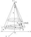
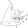
We present in vivo human total retinal blood flow measurements using Doppler Fourier domain optical coherence tomography (OCT). The scan pattern consisted of two concentric circles around the optic nerve head, transecting all retinal branch arteries and veins. The relative positions of each blood vessel in the two OCT conic cross sections were measured and used to determine the angle between the OCT beam and the vessel. The measured angle and the Doppler shift profile were used to compute blood flow in the blood vessel. The flows in the branch veins was summed to give the total retinal blood flow at one time point. Each measurement of total retinal blood flow was completed within 2 s and averaged. The total retinal venous flow was measured in one eye each of two volunteers. The results were 52.90+/-2.75 and 45.23+/-3.18 microlmin, respectively. Volumetric flow rate positively correlated with vessel diameter. This new technique may be useful in the diagnosis and treatment of optic nerve and retinal diseases that are associated with poor blood flow, such as glaucoma and diabetic retinopathy.
Figures


References
-
- Bouma B, Tearney GJ, Boppart SA, Hee MR, Brezinski ME, Fujimoto JG. High-resolution optical coherence tomographic imaging using a mode-locked Ti: Al2O3 laser source. Opt Lett. 1995;20:1486–1488. - PubMed
-
- Tearney GJ, Bouma BE, Fujimoto JG. High-speed phase- and group-delay scanning with a grating-based phase control delay line. Opt Lett. 1997;22:1811–1813. - PubMed
-
- Boppart SA, Bouma BE, Pitris C, Tearney GJ, Fujimoto JG. Forward-imaging instruments for optical coherence tomography. Opt Lett. 1997;22:1618–1620. - PubMed
-
- Tearney GJ, Boppart SA, Bouma BE, Brezinski ME, Weissman NJ, Southern JF, Fujimoto JG. Scanning single-mode fiber optic catheter-endoscope for optical coherence tomography. Opt Lett. 1996;21:543–545. - PubMed
Publication types
MeSH terms
Grants and funding
LinkOut - more resources
Full Text Sources
Other Literature Sources

