. 2009 Jan 5:9:2.
doi: 10.1186/1471-2180-9-2.

Cytokinesis is blocked in mammalian cells transfected with Chlamydia trachomatis gene CT223

Affiliations

Cytokinesis is blocked in mammalian cells transfected with Chlamydia trachomatis gene CT223

Damir T Alzhanov et al. BMC Microbiol. .

Abstract

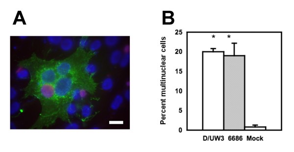
Background: The chlamydiae alter many aspects of host cell biology, including the division process, but the molecular biology of these alterations remains poorly characterized. Chlamydial inclusion membrane proteins (Incs) are likely candidates for direct interactions with host cell cytosolic proteins, as they are secreted to the inclusion membrane and exposed to the cytosol. The inc gene CT223 is one of a sequential set of orfs that encode or are predicted to encode Inc proteins. CT223p is localized to the inclusion membrane in all tested C. trachomatis serovars.

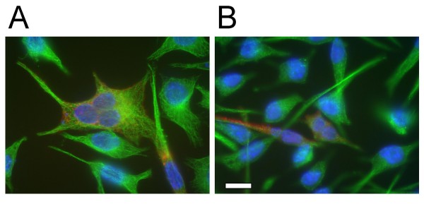
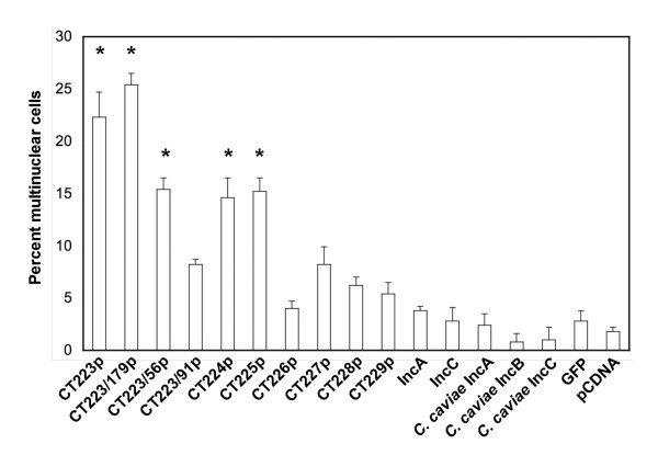
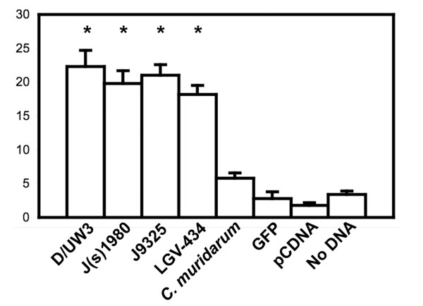
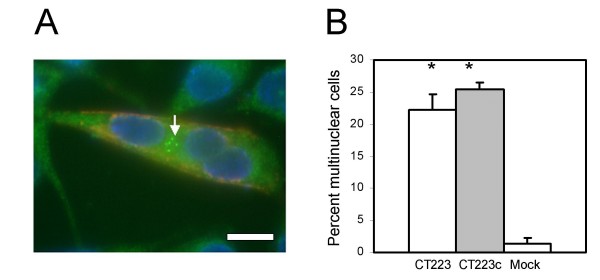
Results: A plasmid transfection approach was used to examine the function of the product of CT223 and other Inc proteins within uninfected mammalian cells. Fluorescence microscopy was used to demonstrate that CT223, and, to a lesser extent, adjacent inc genes, are capable of blocking host cell cytokinesis and facilitating centromere supranumeracy defects seen by others in chlamydiae-infected cells. Both phenotypes were associated with transfection of plasmids encoding the carboxy-terminal tail of CT223p, a region of the protein that is likely exposed to the cytosol in infected cells.

Conclusion: These studies suggest that certain Inc proteins block cytokinesis in C. trachomatis-infected cells. These results are consistent with the work of others showing chlamydial inhibition of host cell cytokinesis.


Figures

Figure 1
Figure 1
Confirmation of the polynuclear phenotype in cells infected with different C. trachomatis strains. Panel A: Fluorescence micrograph of C. trachomatis strain LGV-434 inclusion (anti-LPS, red) within a GFP-positive cell (green), showing three nuclei (blue). The scale bar indicates 10 microns. Panel B: The percentage of polynuclear cells 30 h after infection of HeLa cells with different C. trachomatis at an MOI of 3. Strains D/UW3 and J(s)6686 are shown, along with mock-infected cells. Statistical significance is indicated with the asterisk above the individual treatment groups, as compared to mock-transfected cells (Student's t-test, p < 0.001). Similar levels of significance were observed in a Kruskall-Wallis test (not shown).
Figure 2
Figure 2
Expression of CT223 at different times post infection and differential reactivity with specific antibodies. DNA in all panels is labeled with DAPI (blue) and the bar in panel F represents 10 microns in each image. Cells were infected at an MOI of approximately 0.2 and fixed with 100% methanol prior to antibody labeling. Panels A-D: Fluorescent microscopy of McCoy cells infected with either strain LGV-434 (A, B) or J/UW36 (C, D). Cells were fixed at different times p.i. (A: 12 h, C: 18 h, B, D: 38 h). In panels A-D, cells were labeled with monoclonal anti-CT223p antibody (green) and anti-HSP60 (red). Note that labeling of CT223p is patchy in each strain at the early times points p.i. (A, C) but the labeling is distinct between strains at 38 h p.i. (B, D). Panels E-H: Cells were infected with strain J/UW36 (E, F) or J(s)1980 (G, H) and fixed 30 hours p.i. Cells were then labeled with either polyclonal anti-CT223p antisera (E, G) or monoclonal anti-CT223p antibody (F, H), both of which are labeled red. Note that CT223p is labeled by the polyclonal antisera in each strain, while the monoclonal anti-CT223p does not label the protein in strain J(s)1980.
Figure 3
Figure 3
Cytosolic production of CT223p and CT223/179p from C. trachomatis serovar D/UW3 leads to a multinuclear phenotype within mammalian cells. The vector pcDNA4/HisMaxC was used in each construct. Full length CT223p (panel A) and CT223/179p (panel B) were produced within cells following transfection of pcDNA4-based plasmids. Each was detected with anti-6 × His monoclonal antibodies (red). Microtubules were detected by labeling with specific anti-tubulin antibodies (green). The nuclei are labeled with DAPI (blue). Panel A; McCoy cell transfected with pcDNA4/HisMaxC encoding CT223p. Three nuclei are localized inside of a single cell expressing CT223. Panel B; McCoy cells transfected with pcDNA4/HisMaxC encoding carboxy-terminal CT223/179p. The scale bar in B indicates 10 microns for each panel.
Figure 4
Figure 4
Quantification of multinuclear cells following expression of different inc genes in McCoy cells. This graph represents percentage of polynuclear cells among McCoy cells following transfection of pcDNA4/HisMaxC-based plasmids encoding different Inc proteins. Unless indicated, the sequences were derived from the published C. trachomatis D/UW3 genome sequence. Statistical significance is indicated with the asterisk above the individual treatment groups, as compared to pCDNA-transfected cells (Student's t-test, p < 0.01).
Figure 5
Figure 5
Quantification of multinuclear cells following expression in McCoy cells of CT223 alleles from different C. trachomatis strains. Statistical significance is indicated with the asterisk above the individual treatment groups, as compared to pcDNA-transfected cells (Student's t-test, p < 0.01).
Figure 6
Figure 6
Centrosome supranumeracy in cells transfected with plasmids encoding C. trachomatis serovar D CT223p and CT223/179p. The vector pcDNA4/HisMaxC was used in each construct. The proteins CT223p and CT223/179p were detected with anti-6 × His monoclonal antibody and are labeled in red. Structures of γ-tubulin were detected by labeling with anti γ-tubulin antibodies and are stained in green. The nuclei are labeled with DAPI (blue). Panel A; McCoy cell transfected with pcDNA4/HisMaxC encoding CT223p. Three nuclei are localized inside of a single cell expressing CT223. Multiple centrosomes are shown with an arrow. The scale bar indicates 10 microns. Panel B; The percentage of cells with multiple centrosomes among cells transfected with plasmids encoding CT223p or CT223/179p (CT223c), or cells transfected with the pcDNA4/HisMaxC vector only (Mock). The vertical axis indicates the percent of cells that had two or more centrosomes. At least 500 cells were tested for each construct. The proportions of cells containing 2 or more centrosomes were significantly different than the mock-transfected cells for both the full length and truncated CT223 sequences. Statistical significance is indicated with the asterisk above the individual treatment groups, as compared to mock-transfected cells (Student's t-test, p < 0.001).

References

    1. Valdivia RH. Chlamydia effector proteins and new insights into chlamydial cellular microbiology. Curr Opin Microbiol. 2008;11(1):53–59. - PubMed
    1. Fields KA, Hackstadt T. The chlamydial inclusion: escape from the endocytic pathway. Annu Rev Cell Dev Biol. 2002;18:221–245. - PubMed
    1. Mabey D. Trachoma: recent developments. Adv Exp Med Biol. 2008;609:98–107. - PubMed
    1. Stamm WE. Chlamydia trachomatis infections: progress and problems. J Infect Dis. 1999;179(Suppl 2):S380–383. - PubMed
    1. Alzhanov D, Barnes J, Hruby DE, Rockey DD. Chlamydial development is blocked in host cells transfected with Chlamydophila caviae incA. BMC Microbiol. 2004;4:24. - PMC - PubMed

Publication types

Substances