. 2009 Jan 5:6:1.
doi: 10.1186/1742-2094-6-1.

Beta amyloid oligomers and fibrils stimulate differential activation of primary microglia

Affiliations

Beta amyloid oligomers and fibrils stimulate differential activation of primary microglia

Cindy M Sondag et al. J Neuroinflammation. .

Abstract

Background: Beta amyloid (Abeta) peptides are the major constituents of the senile plaques present in Alzheimer's diseased brain. Pathogenesis has been associated with the aggregated form of the peptide as these fibrils are the conformation readily found in the plaques. However, recent studies have shown that the nonaggregated, soluble assemblies of Abeta have the potential to stimulate neuronal dysfunction and may play a prominent role in the pathogenesis of Alzheimer's disease.

Methods: Soluble, synthetic Abeta1-42 oligomers were prepared producing mainly dimer-trimer conformations as assessed by SDS-PAGE. Similar analysis demonstrated fibril preparations to produce large insoluble aggregates unable to migrate out of the stacking portion of the gels. These peptide preparations were used to stimulate primary murine microglia and cortical neuron cultures. Microglia were analyzed for changes in signaling response and secretory phenotype via Western analysis and ELISA. Viability was examined by quantifying lactate dehydrogenase release from the cultures.

Results: Abeta oligomers and fibrils were used to stimulate microglia for comparison. Both the oligomers and fibrils stimulated proinflammatory activation of primary microglia but the specific conformation of the peptide determined the activation profile. Oligomers stimulated increased levels of active, phosphorylated Lyn and Syk kinase as well as p38 MAP kinase compared to fibrils. Moreover, oligomers stimulated a differential secretory profile for interleukin 6, monocyte chemoattractant protein-1 and keratinocyte chemoattractant when compared to fibrils. Finally, soluble oligomers stimulated death of cultured cortical neurons that was exacerbated by the presence of microglia.

Conclusion: These data suggest that fibrils and oligomers stimulate unique signaling responses in microglia leading to discrete secretory changes and effects on neuron survival. This suggests that inflammation changes during disease may be the consequence of unique peptide-stimulated events and each conformation may represent an individual anti-inflammatory therapeutic target.


Figures

Figure 1
Figure 1
Oligomeric and fibrillar Aβ stimulated a qualitatively similar and dose-dependent increase in tyrosine phosphorylated protein levels. Aβ1–42 oligomers (Aβo) and Aβ1–42 fibrils (Aβf) were prepared to a 100 μM concentration via 4 degree incubation overnight (Aβo) or 37 degree incubation 7 days (Aβf) then diluted to 20 μM in DMEM/F12 media and incubated an additional 48 hours at 37 degree C to determine peptide states under bioassay conditions. 1 μg (A, C) or 0.2–2 μg (B) of peptides were separated by (A, B) 15% SDS-PAGE or (C) 15% non-denaturing gel electrophoresis and Western blotted with anti-Aβ antibody, 6E10. (D) Alternatively, prepared Aβo and Aβf were dot blotted (5 μg) each onto PVDF and blotted with anti-Aβ antibody, 6E10, or anti-oligomer antibody, A11. (E) Primary mouse microglia were unstimulated (control) or stimulated with increasing concentrations of Aβo, Aβf, or vehicle. Cells were lysed after 5 min with RIPA buffer. Cell lysates were separated by 7% SDS-PAGE and Western blotted with anti-phosphotyrosine antibody (4G10) and anti-ERK2 antibody (loading control). Antibody binding was visualized by chemiluminescence. Blots are representative of at least three independent experiments.
Figure 2
Figure 2
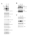
Aβ1–42 stimulated a conformation-specific MAP and tyrosine kinase signaling response along with increased COX-2 and CD68 protein levels. Primary mouse microglia were unstimulated (control) or stimulated with 20 μM Aβo or 20 μM Aβf. (A) Cells were lysed after 5 min with RIPA buffer and lysates were separated by 10% SDS-PAGE and Western blotted with anti-phosphotyrosine antibody (4G10), anti-phospho-JNK antibody, anti-JNK antibody (loading control), anti-phospho-ERK antibody, anti-ERK2 antibody (loading control), anti-phospho-p38 antibody, anti-p38 antibody (loading control), anti-phospho-Lyn antibody, or anti-Lyn antibody (loading control). (B) Cells were also lysed after 5 min with RIPA buffer and Syk was immunoprecipitated. Immunoprecipitates were separated by 7% SDS-PAGE and Western blotted with anti-phosphotyrosine antibody (4G10) and anti-Syk antibody. Arrowheads differentiate specific immunoreactivity from IgG heavy chain. (C) Cells were lysed after 24 hrs with RIPA buffer. Cell lysates were separated by 10% SDS-PAGE and Western blotted with anti-COX-2 antibody, anti-CD68 antibody, anti-CD45 antibody and anti-ERK2 antibody (loading control). Antibody binding was visualized by chemiluminescence. Blots are representative of at least three independent experiments.
Figure 3
Figure 3
Aβ1–42 stimulated a conformation-specific increase in proinflammatory cytokine and chemokine secretion. Primary microglia were unstimulated (control) or stimulated for 24 hours with 20 μM Aβo, 20 μM Aβf, or 25 ng/mL lipopolysaccharide (LPS, positive control). (A) Media were collected and used to perform a preliminary screen of a mouse inflammation antibody array to assess whether relative differences in secretion occurred. In order to quantitate select changes, media was collected and analyzed by (A) mouse IL-6 ELISA, (B) mouse KC ELISA, or (C) mouse MCP-1 ELISA. Data were analyzed by unpaired ANOVA with Tukey's post-test comparison and are expressed as mean +/- SD. Values are representative of three independent experiments (* = p < 0.05 over control, *** = p < 0.001 from control, ** = p < 0.001 from oligomer, # = p < 0.001 from fibril).
Figure 4
Figure 4
Aβ1–42 oligomers were only neurotoxic in neuron-microglia co-cultures. (A) Primary microglia, (B) 14 day in vitro primary cortical neuron, or (C, D) primary neuron-microglia co-cultures were unstimulated (control) or stimulated for 24 (A-C) or 48 (D) hours with increasing doses of Aβo, Aβf, or vehicle. Media were collected and analyzed by (A-C) LDH release assay to assess viability. Data were analyzed by unpaired ANOVA with Tukey's post-test comparison and are expressed as mean +/- SD. Values are representative of three independent experiments (* = p < 0.05 from control, ** = p < 0.001 from control, *** = p < 0.01 from Aβf+neurons). (D) Alternatively, neurons were fixed after stimulation and immunostained for MAP2 expression and counted to assess viability. Data were analyzed by unpaired ANOVA with Tukey's post-test comparison, expressed as mean +/- SEM, and representative of three independent experiments. (* = p < 0.01 from microglia+neurons, ** = p < 0.001 from microglia+neurons).

References

    1. Selkoe DJ. Amyloid beta-protein and the genetics of Alzheimer's disease. J Biol Chem. 1996;271:18295–18298. - PubMed
    1. Pike CJ, Walencewicz AJ, Glabe CG, Cotman CW. Aggregation-related toxicity of synthetic beta-amyloid protein in hippocampal cultures. Eur J Pharmacol. 1991;207:367–368. doi: 10.1016/0922-4106(91)90014-9. - DOI - PubMed
    1. Loo DT, Copani A, Pike CJ, Whittemore ER, Walencewicz AJ, Cotman CW. Apoptosis is induced by beta-amyloid in cultured central nervous system neurons. Proc Natl Acad Sci USA. 1993;90:7951–7955. doi: 10.1073/pnas.90.17.7951. - DOI - PMC - PubMed
    1. Lorenzo A, Yankner BA. Beta-amyloid neurotoxicity requires fibril formation and is inhibited by congo red. Proc Natl Acad Sci USA. 1994;91:12243–12247. doi: 10.1073/pnas.91.25.12243. - DOI - PMC - PubMed
    1. Terry RD, Peck A, DeTeresa R, Schechter R, Horoupian DS. Some morphometric aspects of the brain in senile dementia of the Alzheimer type. Ann Neurol. 1981;10:184–192. doi: 10.1002/ana.410100209. - DOI - PubMed

Publication types

MeSH terms