. 2009 Mar 15;46(6):791-8.
doi: 10.1016/j.freeradbiomed.2008.12.003. Epub 2008 Dec 24.

Genetic deficiency of NADPH oxidase does not diminish, but rather enhances, LPS-induced acute inflammatory responses in vivo

Affiliations

Genetic deficiency of NADPH oxidase does not diminish, but rather enhances, LPS-induced acute inflammatory responses in vivo

Wei-Jian Zhang et al. Free Radic Biol Med. .

Abstract

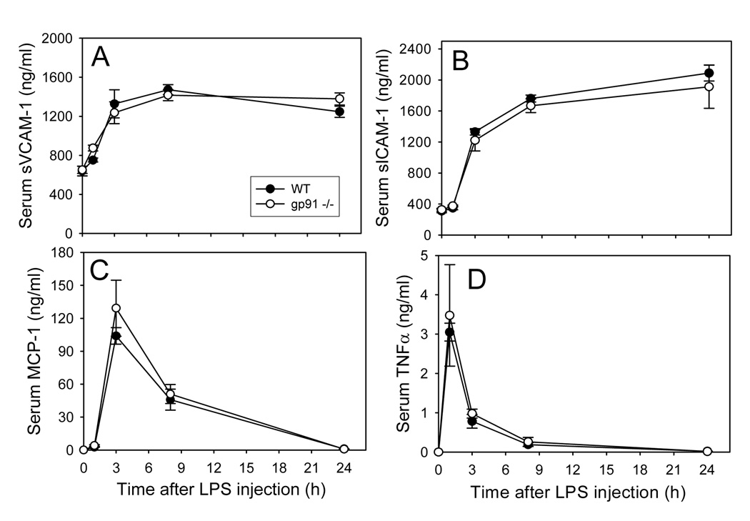
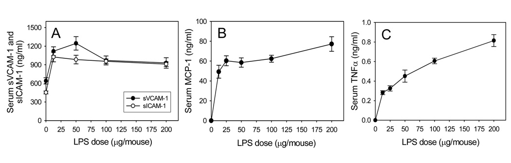
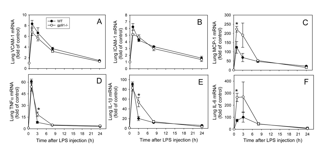
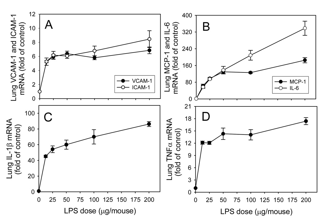
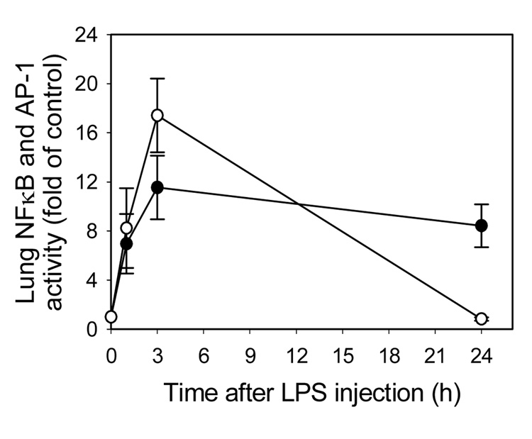
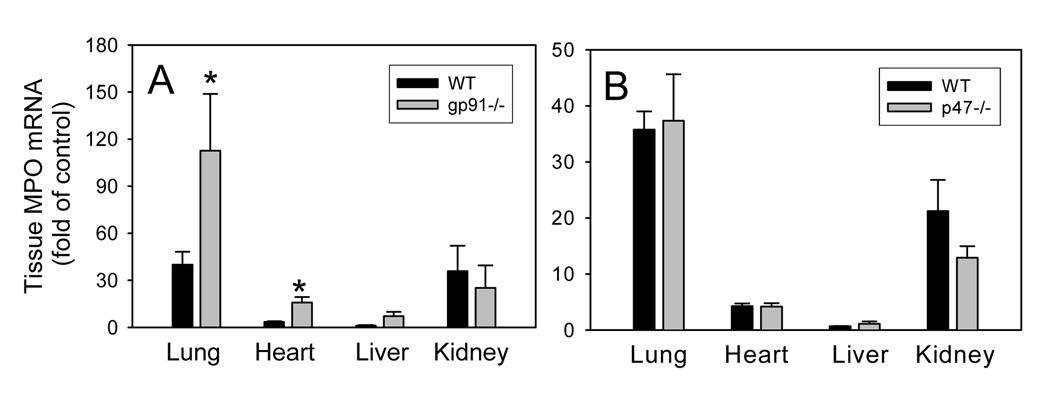
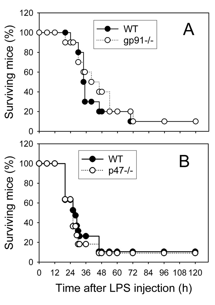
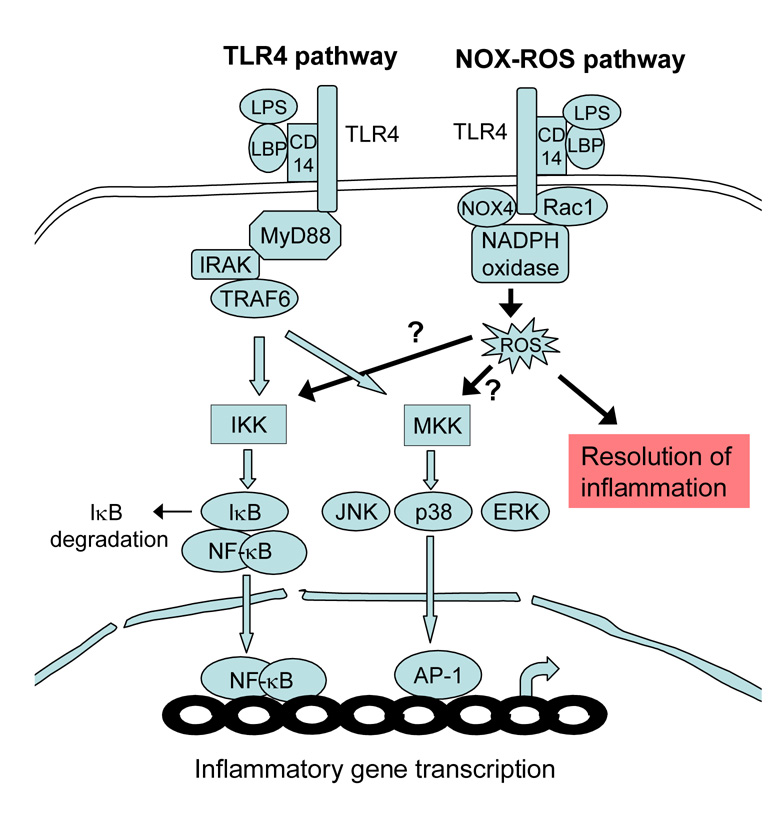
Reactive oxygen species (ROS) and oxidative stress are thought to play a central role in the etiology of cell dysfunction and tissue damage in sepsis. However, there is limited and controversial evidence from in vivo studies that ROS mediate cell signaling processes that elicit acute inflammatory responses during sepsis. Because NADPH oxidase is one of the main cellular sources of ROS, we investigated the role of this enzyme in lipopolysaccharide (LPS)-induced acute inflammation in vivo, utilizing mice deficient in the gp91(phox) or p47(phox) subunits of NADPH oxidase. Age-and body weight-matched C57BL/6J wild-type (WT) and gp91(phox-/-) and p47(phox-/-) mice were injected ip with 50 microg LPS or saline vehicle and sacrificed at various time points up to 24 h. We found that LPS-induced acute inflammatory responses in serum and tissues were not significantly diminished in gp91(phox-/-) and p47(phox-/-) mice compared to WT mice. Rather, genetic deficiency of NADPH oxidase was associated with enhanced gene expression of inflammatory mediators and increased neutrophil recruitment to lung and heart. Furthermore, no protection from LPS-induced septic death was observed in either knockout strain. Our findings suggest that NADPH oxidase-mediated ROS production and cellular redox signaling do not promote, but instead limit, LPS-induced acute inflammatory responses in vivo.


Figures

Fig. 1
Fig. 1. Time-course of LPS-induced increases in serum levels of sVCAM-1 (A), sICAM-1 (B), MCP-1 (C), and TNF (D) in wild-type and gp91phox−/− mice
Wild-type (solid circles) and gp91phox−/− (open circles) mice were injected i.p. with HBSS (0 time point control) or 50µ g LPS as described in Methods. At the indicated time points after LPS injection, the animals were sacrificed and blood was collected. Serum sVCAM-1, sICAM-1, MCP-1, and TNFα were measured by ELISA. Data shown are mean values ± SEM of 4–8 animals.
Fig. 2
Fig. 2. Dose-response of LPS-induced increases in serum levels of sVCAM-1 and sICAM-1 (A), MCP-1 (B), and TNFα (C) in wild-type mice
Wild-type mice were injected i.p. with HBSS (control) or different doses of LPS as described in Methods. Three hours after LPS injection, the animals were sacrificed and blood was collected. Serum sVCAM-1, sICAM-1, MCP-1, and TNFα were measured by ELISA. Data shown are mean values ± SEM of 4–8 animals.
Fig. 3
Fig. 3. Time-course of LPS-induced increases in lung VCAM-1 (A), ICAM-1 (B), MCP-1 (C), TNFα (D), IL-1β (E), and IL-6 (F) mRNA levels in wild-type and gp91phox−/− mice
Wild-type (solid circles) and gp91phox−/− (open circles) mice were injected i.p. with HBSS (0 time point control) or 50µ g LPS as described in Methods. At the indicated time points after LPS injection, the animals were sacrificed and total RNA was isolated from lung. Real-time quantitative PCR analysis was performed for VCAM-1, ICAM-1, MCP-1, TNFα, IL-1β, IL-6, and GAPDH mRNA. Data were expressed as fold of HBSS-treated control after normalization to the internal control gene, GAPDH, and are presented as mean values ± SEM of 4–8 animals. *P<0.05 compared to WT mice.
Fig. 4
Fig. 4. Dose-response of LPS-induced increases in lung VCAM-1 and ICAM-1 (A), MCP-1 and IL-6 (B), IL-1β (C), and TNFα (D) mRNA levels in wild-type mice
Wild-type mice were injected i.p. with HBSS (control) or different doses of LPS as described in Methods. Three hours after LPS injection, the animals were sacrificed and total RNA was isolated from lung. Real-time quantitative PCR analysis was performed for VCAM-1, ICAM-1, MCP-1, TNFα, IL-1β, IL-6, and GAPDH mRNA. Data were expressed as fold of HBSS-treated control after normalization to the internal control gene, GAPDH, and are presented as mean values ± SEM of 4–8 animals.
Fig. 5
Fig. 5. Time-course of LPS-induced activation of lung NFκB and AP-1 in wild-type mice
Wild-type mice were injected i.p. with HBSS (0 time point control) or 50µ g LPS as described in Methods. One, 3, and 24 hours after LPS injection, the animals were sacrificed and nuclear extracts were isolated from lung. NFκB (p65)/DNA (closed circles) and AP-1 (c-fos)/DNA (open circles) binding activities were quantified by ELISA. Data were expressed as fold of HBSS-treated control and are presented as mean values ± SEM of 4–8 animals.
Fig. 6
Fig. 6. LPS-induced increases in myeloperoxidase mRNA levels in various organs of wild-type, gp91phox−/− (A), and p47phox−/− (B) mice
Wild-type, gp91phox−/−, and p47phox−/− mice were injected i.p. with HBSS (control) or 50µ g LPS as described in Methods. Three hours after LPS injection, the animals were sacrificed and total RNA was isolated from various organs. Real-time quantitative PCR analysis was performed for MPO and GAPDH mRNA. After normalization to the internal control gene, GAPDH, the results for the MPO gene were expressed as fold of control. Data shown are mean values ± SEM of 4–8 animals. *P<0.05 compared to WT mice
Fig. 7
Fig. 7. LPS-induced endotoxemic death in wild-type, gp91phox−/− (A), and p47phox−/− mice
Wide-type (closed circles), gp91phox−/− (open circles, panel A) and p47phox−/− mice (open circles, panel B) were injected i.p. with 1.5 mg LPS as described in Methods. The animals were monitored for survival three times daily for up to five days. The numbers of animals used were: 10 and 19 wild-type mice, respectively, in Panels A and B; 10 gp91phox−/− mice; and 11 p47phox−/− mice.
Scheme 1
Scheme 1. Diagram depicting the TLR4 and NOX-ROS pathways
LPS binds to LPS-binding protein (LBP) and CD14, thus activating TLR4. TLR4 activates two major kinases: IκB kinase (IKK), which phosphorylates IκB, leading to its ubiquitylation and subsequent degradation and NFκB translocation to the nucleus; and mitogen-activated protein kinase kinases (MKK), which phosphorylate and activate c-Jun kinase (JNK), extracellular receptor-activated kinase (ERK), and p38 kinase, leading to AP-1 activation. Activated NFκB and AP-1 in the nucleus bind to DNA promoter regions to induce inflammatory gene transcription. In the NOX-ROS pathway, LPS activates TLR4, which in turn activates NADPH oxidase through Rac1 and Nox4. While it has been hypothesized that ROS generation by NADPH oxidase enhances IKK and MKK activity (arrows marked “?”), the results presented in this paper indicate that the NOX-ROS pathway contributes to the resolution of LPS-induced inflammation.

References

    1. Ulevitch RJ, Tobias PS. Recognition of gram-negative bacteria and endotoxin by the innate immune system. Curr Opin Immunol. 1999;11:19–22. - PubMed
    1. Guha M, Mackman N. LPS induction of gene expression in human monocytes. Cell Signal. 2001;13:85–94. - PubMed
    1. Aird WC. The role of the endothelium in severe sepsis and multiple organ dysfunction syndrome. Blood. 2003;101:3765–3777. - PubMed
    1. Salvemini D, Cuzzocrea S. Oxidative stress in septic shock and disseminated intravascular coagulation. Free Radic Biol Med. 2002;33:1173–1185. - PubMed
    1. Victor VM, Rocha M, Esplugues JV, De la Fuente M. Role of free radicals in sepsis: antioxidant therapy. Curr. Pharm. Des. 2005;11:3141–3158. - PubMed

Publication types

MeSH terms