

Computer-aided detection of endobronchial valves using volumetric CT
- PMID: 19124102
- PMCID: PMC2659554
- DOI: 10.1016/j.acra.2008.07.009
Computer-aided detection of endobronchial valves using volumetric CT
Abstract
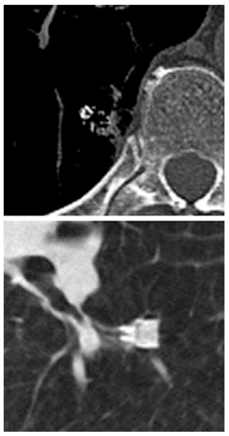
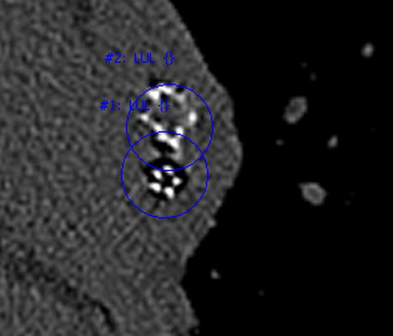
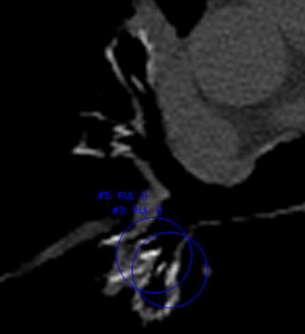
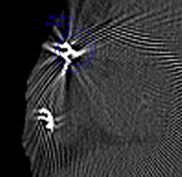
Rationale and objectives: The ability to automatically detect and monitor implanted devices may serve an important role in patient care by aiding the evaluation of device and treatment efficacy. The purpose of this research was to develop a system for the automated detection of one-way endobronchial valves that were implanted for less invasive lung volume reduction.
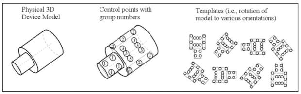
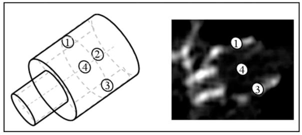
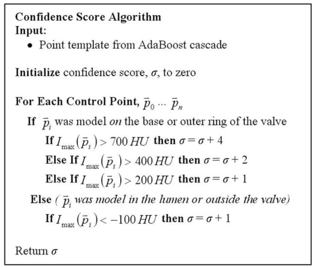
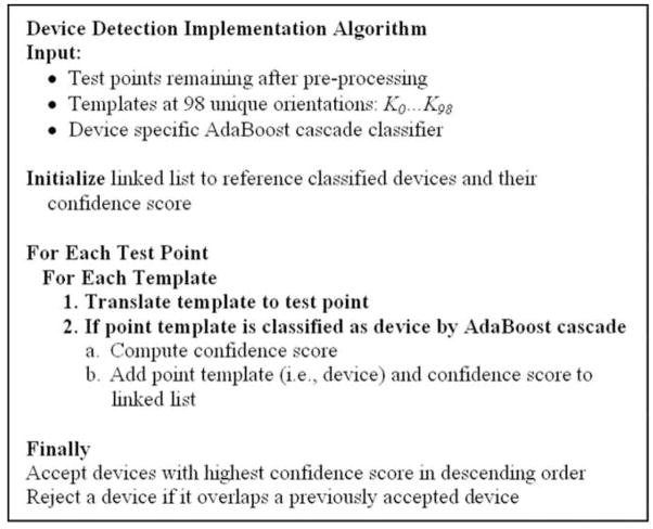
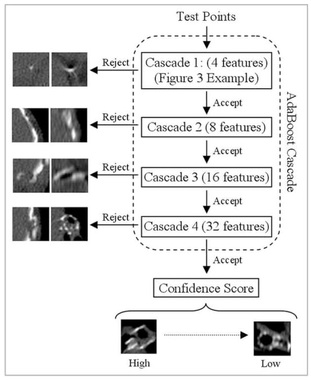
Materials and methods: Volumetric thin-section computed tomographic data was obtained for 194 subjects; 95 subjects implanted with 246 devices were used for system development and 99 subjects implanted with 354 devices were reserved for testing. The detection process consisted of preprocessing, pattern recognition based detection, and a final device selection. Following the preprocessing, a set of classifiers was trained using AdaBoost to discriminate true devices from false positives. The classifiers in the cascade used two simple features (either the mean or maximum attenuation) of a local region computed at multiple fixed landmarks relative to a template model of the valve.
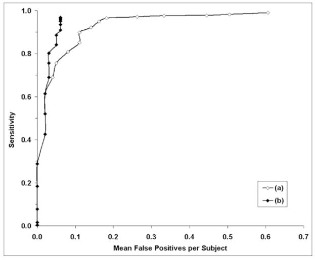
Results: Free-response receiver-operating characteristic analysis was performed for the evaluation; the system could be set so the mean sensitivity was 96.5% with a mean of 0.18 false positives per subject. If knowledge of the number of implanted devices were incorporated, the sensitivity would be 96.9% with a mean of 0.061 false positives per subject; this corresponds to a total of 12 false negatives and six false positives for the 99 subjects in the test dataset.
Conclusion: Software was developed for automated detection of endobronchial valves on volumetric computed tomography. The proposed device modeling and detection techniques may be applicable to other devices as well as useful for evaluation of treatment response.
Figures
Similar articles
-
Computer aided detection of epidural masses on computed tomography scans.Comput Med Imaging Graph. 2014 Oct;38(7):606-12. doi: 10.1016/j.compmedimag.2014.04.007. Epub 2014 May 9. Comput Med Imaging Graph. 2014. PMID: 24908192 Free PMC article.
-
Computer-aided detection of lung nodules: false positive reduction using a 3D gradient field method and 3D ellipsoid fitting.Med Phys. 2005 Aug;32(8):2443-54. doi: 10.1118/1.1944667. Med Phys. 2005. PMID: 16193773 Free PMC article. Clinical Trial.
-
Automatic coregistration of volumetric images based on implanted fiducial markers.Med Phys. 2008 Oct;35(10):4513-23. doi: 10.1118/1.2975153. Med Phys. 2008. PMID: 18975698
-
Automated detection of mass lesions in dedicated breast CT: a preliminary study.Med Phys. 2012 Feb;39(2):866-73. doi: 10.1118/1.3678991. Med Phys. 2012. PMID: 22320796 Free PMC article.
-
Intrathoracic airway trees: segmentation and airway morphology analysis from low-dose CT scans.IEEE Trans Med Imaging. 2005 Dec;24(12):1529-39. doi: 10.1109/TMI.2005.857654. IEEE Trans Med Imaging. 2005. PMID: 16353370 Free PMC article.
References
-
- Toma T, Hopkinson N, Hillier J, Hansell D, Morgan C, Goldstraw P, Polkey M, Geddes D. Bronchoscopic volume reduction with valve implants in patients with severe emphysema. Lancet. 2003;361:931–933. - PubMed
-
- Wan IYP, Toma TP, Geddes DM, et al. Chest. 2006;129:518–526. - PubMed
-
- Yim A, Hwong T, Lee T, Li W, Lam S, Yeung T, Hui D, Ko F, Sihoe A, Thung K, Arifi A. Early results of endoscopic lung volume reduction for emphysema. J Thorac Cardiovasc Surg. 2004;127:1564–1573. - PubMed
-
- Maxfield RA. New and Emerging Minimally Invasive Techniques for Lung Volume Reduction. Chest. 2004;125:777–783. - PubMed
-
- Harris E, McNair H, Evans P. Feasibility of fully automated detection of fiducial markers implanted into the prostate using electronic portal imaging: A comparison of methods. Int J Radiation Oncology Biol Phys. 2006;66:1263–1270. - PubMed
