. 2009 May;23(5):1358-65.
doi: 10.1096/fj.08-110296. Epub 2009 Jan 5.

Premature senescence of highly proliferative endothelial progenitor cells is induced by tumor necrosis factor-alpha via the p38 mitogen-activated protein kinase pathway

Affiliations

Premature senescence of highly proliferative endothelial progenitor cells is induced by tumor necrosis factor-alpha via the p38 mitogen-activated protein kinase pathway

Yanmin Zhang et al. FASEB J. 2009 May.

Abstract

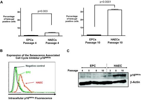
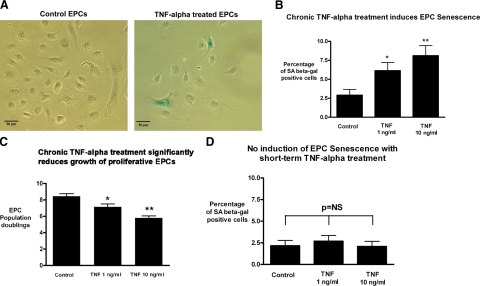
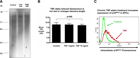
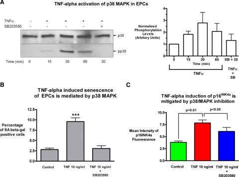
Senescence of endothelial cells increases with systemic aging and is thought to contribute to the development of atherosclerosis. Cell therapy with highly proliferative endothelial progenitor cells (EPCs) is an emerging therapeutic option to promote endothelial regeneration, but little is known about their senescence and their vulnerability to inflammatory stressors. We therefore studied the senescence of proliferative human EPCs and investigated the effects of the proinflammatory cytokine tumor necrosis factor-alpha (TNF-alpha) on their senescence. Human EPCs had a significantly lower rate of senescence at baseline, compared with that of mature endothelial cells. However, EPCs up-regulated the expression of the senescence-associated cell cycle arrest protein p16(INK4a) and markedly increased measured senescence levels when exposed to chronic TNF-alpha treatment. Analysis of telomere length showed that the increases in senescence were not related to changes in telomere length. Inhibition of the p38 mitogen-activated protein kinase pathway blocked the induction of p16(INK4a) and cellular senescence. In conclusion, highly proliferative EPCs have a low rate of intrinsic senescence but are vulnerable to premature senescence induction by chronic proinflammatory stimulation. These findings will lead to a better understanding of physiological endothelial regeneration as well as to targeted therapies with the aim of promoting endothelial regeneration through endothelial progenitor cells.


Figures

Figure 1.
Figure 1.
Differences in cellular senescence between cord blood-derived EPCs and HAECs. A) Percentages of cells positive for SA-β-Gal in human cord blood-derived EPCs and mature HAECs. HAECs and EPCs are compared at a low passage (passage 4, P=0.003 by t test, n=4) as well as at a high passage (passage 10, P<0.0001 by t test, n=4). Quantification demonstrates a marked increase in the percentage of senescent cells among HAECs, and relatively minor increases in senescence among EPCs with passaging. B) Intracellular flow cytometric staining for the senescence-associated cell cycle inhibitor p16INK4a was performed. Cell numbers are depicted on the y axis and fluorescence intensity on a logarithmic scale is used as an indicator of expression on the x axis. At baseline, p16INK4a was expressed in HAECs (orange) and not in EPCs. The level of p16INK4a staining in EPCs (green) is the same as that of the negative isotype control (gray). C) Representative Western blot staining for the senescence-associated cell cycle inhibitor p16INK4a in HAEC and EPC samples demonstrates that p16INK4a expression markedly increases in HAECs as cells reach passages 8–10.
Figure 2.
Figure 2.
Chronic exposure to TNF-α increases the cellular senescence of cord blood-derived EPCs independent of changes in telomere length. EPCs were treated with varying doses of TNF-α for 1 week, and SA-β-Gal staining was measured. A) Representative phase-contrast images of control EPCs and cells treated with 10 ng/ml TNF-α. Scale bars = 50 μm. B) Degree of cell senescence was quantified as percentage of SA-β-Gal-positive cells. Continuous treatment with TNF-α for 1 wk resulted in a dose-dependent increase in EPC senescence, which was nearly double the baseline senescence rate at the low dose of 1 ng/ml (n=5, P<0.05) and was nearly triple the baseline EPC senescence rate at the higher dose of 10 ng/ml (n=5, P<0.01). To investigate whether TNF-α treatment also reduced growth of highly proliferative EPCs, cell numbers were assessed. C) Cell numbers were converted to cumulative population doublings. Treatment with TNF-α resulted in a marked, dose-dependent drop in EPC proliferation (P<0.05 at 1 ng/ml and P<0.01 at 10 ng/ml). D) Short-term treatment with TNF-α for only 3 days did not increase EPC senescence.
Figure 3.
Figure 3.
Chronic exposure to TNF-α increases the cellular senescence of cord blood-derived EPCs independent of changes in telomere length. EPCs were chronically treated with TNF-α for 1 wk at the indicated concentrations, and cells were collected for analyses of TRF lengths. Genomic DNA was digested with restriction enzymes that do not cut telomeric sequences and analyzed via a modified Southern method. Results yield a smear that corresponds to the distribution of all telomere lengths within the population of cells collected. A) Representative gel. B) Statistical analysis of TRFs (n=3) showed no significant difference between control and TNF-α treatments. C) To assess whether TNF-α increased the expression of the senescence-associated cell cycle inhibitor p16INK4a intracellular flow cytometry was performed; representative histogram with a control-treated cell sample (green) and TNF-α-treated cell sample (red) is shown. Cell numbers are on the y axis; fluorescence intensity is used as a marker of expression on the x axis.
Figure 4.
Figure 4.
Chronic TNF-α-induced senescence in EPCs is mediated by activation of the p38 MAPK pathway. A) To investigate whether the p38/MAPK pathway was involved in mediating TNF-α-induced EPC senescence, an immunoblot analysis with an anti-phospho-p38 MAPK antibody and a total p38 MAPK antibody was performed in EPCs treated with 10 ng/ml TNF-α at the indicated times after stimulation in the presence or absence of the specific p38 MAPK inhibitor SB203580 at a concentration of 10 μM. B) Quantification of fold change in activated MAPK was analyzed by densitometry and normalized to total protein levels. A representative Western blot and average densitometry with sem are shown (n=5). To assess whether p38 MAPK inhibition could prevent the induction of cellular senescence, cells were treated with TNF-α in the presence or absence of the specific inhibitor SB203580 (10 μM) for 1 wk, and senescence was quantified by SA-β Gal staining. Inhibition of the p38 MAPK pathway reduced the highly significant induction of cellular senescence by TNF-α in EPCs (P<0.001, n=4) down to the low baseline level. C) EPCs were also stained for the senescence-associated cell cycle inhibitor p16INK4a using intracellular flow cytometry. Measurement of mean fluorescence intensity showed the expected significant increase p16INK4a expression with chronic TNF-α treatment (P<0.01, control vs. treatment, n=3), while also showing a significant decrease of p16INK4a expression with p38 MAPK inhibition (TNF vs. TNF+10 μM SB203580, P<0.05, n=3).

References

    1. Minamino T, Komuro I. Vascular cell senescence: contribution to atherosclerosis. Circ Res. 2007;100:15–26. - PubMed
    1. Gulati R, Jevremovic D, Peterson T E, Chatterjee S, Shah V, Vile R G, Simari R D. Diverse origin and function of cells with endothelial phenotype obtained from adult human blood. Circ Res. 2003;93:1023–1025. - PubMed
    1. Yoder M C, Mead L E, Prater D, Krier T R, Mroueh K N, Li F, Krasich R, Temm C J, Prchal J T, Ingram D A. Redefining endothelial progenitor cells via clonal analysis and hematopoietic stem/progenitor cell principals. Blood. 2007;109:1801–1809. - PMC - PubMed
    1. Yoon C H, Hur J, Park K W, Kim J H, Lee C S, Oh I Y, Kim T Y, Cho H J, Kang H J, Chae I H, Yang H K, Oh B H, Park Y B, Kim H S. Synergistic neovascularization by mixed transplantation of early endothelial progenitor cells and late outgrowth endothelial cells: the role of angiogenic cytokines and matrix metalloproteinases. Circulation. 2005;112:1618–1627. - PubMed
    1. Murasawa S, Llevadot J, Silver M, Isner J M, Losordo D W, Asahara T. Constitutive human telomerase reverse transcriptase expression enhances regenerative properties of endothelial progenitor cells. Circulation. 2002;106:1133–1139. - PubMed

Publication types

MeSH terms