. 2009 Mar;191(6):1777-86.
doi: 10.1128/JB.01437-08. Epub 2009 Jan 5.

Contact-dependent growth inhibition causes reversible metabolic downregulation in Escherichia coli

Affiliations

Contact-dependent growth inhibition causes reversible metabolic downregulation in Escherichia coli

S K Aoki et al. J Bacteriol. 2009 Mar.

Abstract

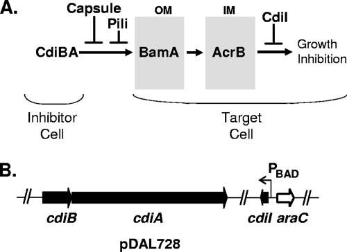
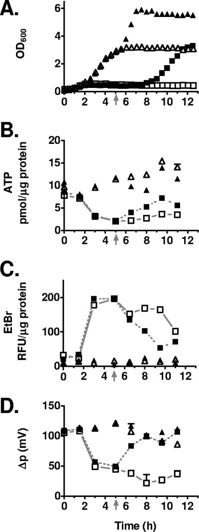
Contact-dependent growth inhibition (CDI) is a mechanism identified in Escherichia coli by which bacteria expressing two-partner secretion proteins encoded by cdiA and cdiB bind to BamA in the outer membranes of target cells and inhibit their growth. A third gene in the cluster, cdiI, encodes a small protein that is necessary and sufficient to confer immunity to CDI, thereby preventing cells expressing the cdiBA genes from inhibiting their own growth. In this study, the cdiI gene was placed under araBAD promoter control to modulate levels of the immunity protein and thereby induce CDI by removal of arabinose. This CDI autoinhibition system was used for metabolic analyses of a single population of E. coli cells undergoing CDI. Contact-inhibited cells showed altered cell morphology, including the presence of filaments. Notably, CDI was reversible, as evidenced by resumption of cell growth and normal cellular morphology following induction of the CdiI immunity protein. Recovery of cells from CDI also required an energy source. Cells undergoing CDI showed a significant, reversible downregulation of metabolic parameters, including aerobic respiration, proton motive force (Deltap), and steady-state ATP levels. It is unclear whether the decrease in respiration and/or Deltap is directly involved in growth inhibition, but a role for ATP in the CDI mechanism was ruled out using an atp mutant. Consistent with the observed decrease in Deltap, the phage shock response was induced in cells undergoing CDI but not in recovering cells, based on analysis of levels of pspA mRNA.


Figures

FIG. 1.
FIG. 1.
Construction of a contact-dependent autoinhibition system in Escherichia coli. (A) CDI model. CdiB and CdiA (CdiBA) expressed by one cell (inhibitor cell) binds to BamA in the outer membrane (OM) of an adjacent cell (target cell) (3). A signal, such as a CdiA peptide, is transferred through BamA to AcrB located in the inner membrane (IM), causing inhibition of cell growth by an unknown mechanism. CDI is modulated at the cell surface by certain pili, including pyelonephritis-associated pili (4), and by colanic acid capsule (3) and inside cells by CdiI immunity protein (4). (B) Map of the CDI autoinhibition plasmid (pDAL728).
FIG. 2.
FIG. 2.
Characterization of the CDI autoinhibition system. Reversibility and carbon source requirement. (A) CDI+ E. coli LMG194 Δara714 with pDAL728 (DL5263) were prepared and incubated at 37°C with shaking at 225 rpm as described in Materials and Methods for the CDI autoinhibition assay. Cultures were incubated in LBM (open squares), LBM plus arabinose (closed circles), LBM plus maltose (open circles), or LBM plus maltose plus arabinose (closed squares). Maltose (0.2%) was present at time zero, and arabinose (0.2%) was added at 5 h, where indicated. (B) CDI+ DL5263 (squares) and CDI control E. coli DL5400 cells (triangles) were prepared as described for panel A for the CDI autoinhibition assay. Maltose and arabinose (0.2% each) were added to the indicated cultures at 5 h (closed squares and triangles; shown by arrow on x axis). (C) Extended time course. CDI+ DL5263 was grown and treated as described for panel A in LBM. After 20 h of incubation, cultures were supplemented with 0.2% arabinose (closed circles) or with 0.2% arabinose and maltose (closed squares). A no-supplement control was included (open squares). (D) Inhibition of CDI by Pap pili. CDI+ E. coli cells expressing Pap21 pili (half-shaded squares) or not expressing Pap21 pili (open squares) were prepared as described for panel A in LBM without supplements.
FIG. 3.
FIG. 3.
E. coli cell morphology during CDI-mediated autoinhibition and recovery. CDI+ E. coli (DL5263) and CDI control E. coli (DL5400) cells were grown in arabinose-free LBM, as shown in Fig. 2B, performed in parallel with the morphological analysis shown here. After 5 h, the cultures were supplemented with 0.2% arabinose and 0.2% maltose, as indicated at the left of each row of panels (+ara +mal). At the times indicated, cells were stained with DAPI and imaged at the same magnification (×1,000), using phase optics and fluorescence overlay by using MicroFire imaging software (Optronix, Goleta, CA). Multiple nucleoids are indicated with arrows, and ghosts are indicated with blunt arrows.
FIG. 4.
FIG. 4.
Measurement of membrane potential (Δp), EtBr export, and ATP level in cells entering and exiting CDI. CDI+ (DL5417; squares) and CDI (DL5418; triangles) E. coli cells were prepared and incubated in LBM as described for Fig. 2. Arabinose and maltose (closed symbols) were added to two cultures at 5 h (as indicated by an arrow on the x axis). For clarity, data points for CDI+ cells receiving arabinose are connected by dotted lines and CDI+ cells without arabinose are connected by solid lines. Error bars show the standard deviation for duplicate measurements. (A) Growth curve. (B) Intracellular ATP level (pmol/μg protein). (C) EtBr fluorescence (relative fluorescence units [RFU per μg protein]). (D) Estimated Δp.
FIG. 5.
FIG. 5.
Analysis of CDI in E. coli lacking ATP synthase. Results for CDI+ E. coli DL5283 (with ATPase [squares]) and CDI+ DL5285 (without ATPase [inverted triangles]) cells are shown in the left panels, and results for E. coli CDI control strains DL5414 (with ATPase [triangles]) and DL5415 (without ATPase [diamonds]) are shown in the right panels. Strains receiving arabinose and maltose at 5 h are indicated with closed symbols, and no-addition controls are indicated with open symbols. For clarity, data points for CDI+ atp+ cells with arabinose are connected by solid lines, and CDI+ atp401 cells with arabinose are connected by dotted lines. (A) OD600. (B) Intracellular ATP level. (C) EtBr fluorescence.
FIG. 6.
FIG. 6.
Reversible downregulation of respiration in CDI. CDI+ E. coli DL5417 strains (left panels [squares]), CDI E. coli DL5418 control strains (right panels [closed triangles]), and CDI+ acrB mutant E. coli DL5711 strains (open circles) were prepared and incubated as described for Fig. 2, with maltose (0.2%) added at time zero. Cells receiving arabinose at 5 h (arrows) are indicated by closed symbols. Error bars show the standard deviation of the mean calculated from two independent experiments.
FIG. 7.
FIG. 7.
Induction of pspA transcription during CDI. CDI+ E. coli DL5417 (squares; panels A and B) and CDI+ E. coli DL5735 ΔpspA740::kan (circles; panel A) were grown in LBM with 0.2% maltose. Cultures receiving arabinose (0.2%) at 5 h are shown as closed symbols. (A) OD600. (B) pspA mRNA expression level relative to expression at 1 h, as determined using qPCR.

Similar articles

Cited by

References

    1. Ahmed, S., and I. R. Booth. 1981. Quantitative measurements of the proton-motive force and its relation to steady state lactose accumulation in Escherichia coli. Biochem. J. 200573-581. - PMC - PubMed
    1. Alam, M., M. Sultana, G. B. Nair, A. K. Siddique, N. A. Hasan, R. B. Sack, D. A. Sack, K. U. Ahmed, A. Sadique, H. Watanabe, C. J. Grim, A. Huq, and R. R. Colwell. 2007. Viable but nonculturable Vibrio cholerae O1 in biofilms in the aquatic environment and their role in cholera transmission. Proc. Natl. Acad. Sci. USA 10417801-17806. - PMC - PubMed
    1. Aoki, S., J. Malinverni, K. Jacoby, B. Thomas, R. Pamma, B. Trinh, S. Remers, J. Webb, B. Braaten, T. Silhavy, and D. Low. 2008. Contact-dependent growth inhibition requires the essential outer membrane protein BamA (YaeT) and the inner membrane transport protein AcrB. Mol. Microbiol. 70323-340. - PMC - PubMed
    1. Aoki, S. K., R. Pamma, A. D. Hernday, J. E. Bickham, B. A. Braaten, and D. A. Low. 2005. Contact-dependent inhibition of growth in Escherichia coli. Science 3091245-1248. - PubMed
    1. Baba, T., T. Ara, M. Hasegawa, Y. Takai, Y. Okumura, M. Baba, K. A. Datsenko, M. Tomita, B. L. Wanner, and H. Mori. 2006. Construction of Escherichia coli K-12 in-frame, single-gene knockout mutants: the Keio collection. Mol. Syst. Biol. 22006.0008. - PMC - PubMed

Publication types

MeSH terms

LinkOut - more resources