. 2009 Mar;29(6):1506-14.
doi: 10.1128/MCB.00857-08. Epub 2009 Jan 5.

Tropomyosin isoform expression regulates the transition of adhesions to determine cell speed and direction

Affiliations

Tropomyosin isoform expression regulates the transition of adhesions to determine cell speed and direction

Cuc T T Bach et al. Mol Cell Biol. 2009 Mar.

Abstract

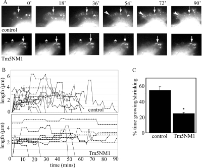
The balance of transition between distinct adhesion types contributes to the regulation of mesenchymal cell migration, and the characteristic association of adhesions with actin filaments led us to question the role of actin filament-associating proteins in the transition between adhesive states. Tropomyosin isoform association with actin filaments imparts distinct filament structures, and we have thus investigated the role for tropomyosins in determining the formation of distinct adhesion structures. Using combinations of overexpression, knockdown, and knockout approaches, we establish that Tm5NM1 preferentially stabilizes focal adhesions and drives the transition to fibrillar adhesions via stabilization of actin filaments. Moreover, our data suggest that the expression of Tm5NM1 is a critical determinant of paxillin phosphorylation, a signaling event that is necessary for focal adhesion disassembly. Thus, we propose that Tm5NM1 can regulate the feedback loop between focal adhesion disassembly and focal complex formation at the leading edge that is required for productive and directed cell movement.


Figures

FIG. 1.
FIG. 1.
Tm5NM1 overexpression promotes adhesions. (A) B35 neuroblastoma cells immunostained with paxillin antibodies. Focal adhesions are indicated by arrows. (B and C) Histograms show lengths and numbers of focal adhesions, expressed relative to control cells (mean of 20 cells). (D) Paxillin immunostaining of B35/Tm5NM1 cells treated with scrambled control (Scr cont.) or anti-Tm5NM1 siRNA. Arrows show centrally located focal adhesions. Insets show images of the same regions of the coverslip viewed with the green filter set; note the positive staining of the Tm5NM1 cells that have received the Alexa488-labeled Tm5NM1 siRNA (indicated with arrowheads). Scale bar, 20 μm. (E) Numbers of focal adhesions in cells treated with anti-Tm5NM1 siRNA expressed relative to the number in control (Scr cont.) cells. Graphs show the average from three independent repeats (n = 3, minimum of 17 cells per experiment). *, P < 0.05. NS, not significant.
FIG. 2.
FIG. 2.
Enhanced fibrillar adhesion formation in cells overexpressing Tm5NM1. (A) Cells plated onto fibronectin- and laminin-coated coverslips, fixed and coimmunostained for paxillin and tensin. The two right-hand panels show a merged image of the costained cells. The boxed region in the B35/Tm5NM1 merged cell image is shown at a higher magnification below. Arrows indicate paxillin (green) and tensin (red) positive fibrillar adhesions. (B) Cells plated in serum onto glass coverslips, fixed and costained for fibronectin and F-actin. The two right-hand panels show a merged image of the costained cells. The boxed region in the B35/Tm5NM1 merged cell image is shown at higher magnification below. Arrows indicate regions of coincident fibronectin (green) and F-actin (red) staining.
FIG. 3.
FIG. 3.
Actin filaments are stabilized by Tm5NM1. (A) Cells plated on fibronectin and laminin and coimmunostained with CY5-phalloidin to detect filamentous actin and paxillin antibodies. Panels on the right-hand side show merged images of the coimmunostained cells. Arrows on images point to the leading edge actin meshwork (M), transverse arcs of actin (TA), ventral stress fibers (V), focal complexes (FC), focal adhesions (FA), and fibrillar adhesions (FB). (B) Cells exposed to 1 μM Latrunculin A for the indicated times and immunostained with TRITC-phalloidin to detect filamentous actin, indicated by white arrows. Scale bar, 20 μm. (C) Percentage of stress fiber-positive cells in latrunculin-treated cells for the indicated times, expressed relative to the number at time zero (n = 23 to 72 cells).
FIG. 4.
FIG. 4.
Tm5NM1 reduces cell persistence and speed. (A) Migration traces for control B35, B35/Tm5NM1, and B35/Tm3 cells. (B and C) Histograms showing average (B35, n = 17; Tm5NM1, n = 34; Tm3, n = 24) speed and persistence ratios, respectively, and the SEM. *, P < 0.05. NS, not significant. (D) Migration traces illustrating the movement patterns in the first 3 h as cells migrate into the wound in a scratch wound healing assay. (E) Cumulative persistence ratios (upper panel) and velocities (lower panel) (n = 20).
FIG. 5.
FIG. 5.
Tm5NM1 expression inhibits focal adhesion turnover. (A) Cropped regions from time-lapse series of cells transfected with YFP.p130Cas. Stable adhesions are indicated by arrows, a newly formed adhesion is indicated by an arrowhead, and adhesions that turn over are indicated by an asterisk. (B) Representative plots of the lengths of 10 YFP.p130Cas-positive focal adhesion lengths over time from each cell line. (C) Histogram showing the percentage of total time focal adhesions are observed to either form or turn over during the time-lapse series and the SEM values. (17 individual adhesions from 5 B35 cells and 33 individual adhesions from 10 B35/Tm5NM1 cells). *, P < 0.05.
FIG. 6.
FIG. 6.
Downregulation of adhesion signaling pathways. (A and B) Western blot analysis of p130Cas (A) and Src protein (B) expression. Relative levels of hyperphosphorylated (pp130Cas, upper form) and hypophosphorylated (p130Cas, lower form) p130Cas and total Src protein levels were determined by densitometry from triplicate repeats and are represented in histograms below the immunoblot data. *, P < 0.05. NS, not significant.
FIG. 7.
FIG. 7.
Reduced paxillin tyrosine phosphorylation in cells with elevated Tm5NM1 expression. (A) Western blot analysis of phosphorylated and total paxillin levels and tubulin to demonstrate equal loading. The histogram shows the ratio of phosphopaxillin divided by paxillin. Values were first normalized to tubulin. The data show the mean of four independent repeats. *, P < 0.05. (B) Paxillin and phosphopaxillin immunostaining of cells plated on fibronectin- and laminin-coated coverslips. Merged images show color overlays of paxillin (green) and phosphopaxillin (red) images. (C) Paxillin phosphorylation represented by ratio imaging. Adhesions that have high levels of phosphorylated paxillin are shown in red hues, and areas of low paxillin phosphorylation are shown in blue. Arrowed regions in each ratio image are magnified in the boxed insets. Scale bar, 5 μm. (D) Line scans showing fluorescence intensities of focal adhesions. Phosphopaxillin intensities are shown in red, and paxillin intensities are shown in green. (E) Distribution of the ratios of phosphorylated paxillin at individual focal adhesions (n = 100, from at least five different cells). Horizontal bars indicate the mean of each population. p-pax, phosphopaxillin; pax, total paxillin.
FIG. 8.
FIG. 8.
Focal complex stimulation and enhanced paxillin phosphorylation in cells lacking Tm5NM1 expression. (A) Paxillin staining of wild-type (MEF) and knockout (MEF/Tm5NM1−/−) cells. Focal complexes are indicated by arrowheads (inset i). (B) Histogram showing the average percentage of focal complex positive cells from three independent experiments. *, P < 0.05. (C) MEF/Tm5NM1−/− cells transfected with either GFP alone (vector) or dominant-negative Rac fused to GFP (GFP.RacN17). Panels on the left show paxillin staining of transfected cells (arrows indicate focal complexes). (D) Percentage of cells displaying focal complexes in GFP control and GFP.RacN17 transfected cells. The data show the average from three independent experiments. *, P < 0.05. (E) Western blot analysis of phosphopaxillin and total paxillin levels. Membranes were probed with anti-actin antibodies to demonstrate equivalent loading. The data shown are representative of two independent repeats.
FIG. 9.
FIG. 9.
Tm5NM1 expression regulates cell persistence. (A) Migration traces for wild-type MEF and MEF/Tm5NM1−/− cells. (B and C) Histograms showing average velocities and persistence ratios, respectively (MEF, n = 27; MEF/Tm5NM1, n = 28). (D) Migration traces illustrating the movement patterns in the first 6 h as cells migrate into the wound in a scratch wound healing assay. (E) Graph showing cumulative persistence ratios as wild-type MEF or MEF/Tm5NM1−/− migrate in an in vitro scratch wound assay. The data points are averages of 10 cells per experiment repeated in triplicate. (F) Average speeds of MEF/Tm5NM1−/− cells transfected with YFP control or YFP-fused Tm5NM1. Only cells expressing above a threshold of YFP.Tm5NM1 fluorescence intensity were selected for analysis (see Materials and Methods for details). Experiments were carried out on three separate occasions, and the averages of the triplicate data sets are reported. *, P < 0.05; NS, not significant.

References

    1. Bargon, S. D., P. W. Gunning, and G. M. O'Neill. 2005. The Cas family docking protein, HEF1, promotes the formation of neurite-like membrane extensions. Biochim. Biophys. Acta 1 746143-154. - PubMed
    1. Beningo, K. A., M. Dembo, I. Kaverina, J. V. Small, and Y. L. Wang. 2001. Nascent focal adhesions are responsible for the generation of strong propulsive forces in migrating fibroblasts. J. Cell Biol. 153881-888. - PMC - PubMed
    1. Bryce, N. S., G. Schevzov, V. Ferguson, J. M. Percival, J. J. Lin, F. Matsumura, J. R. Bamburg, P. L. Jeffrey, E. C. Hardeman, P. Gunning, and R. P. Weinberger. 2003. Specification of actin filament function and molecular composition by tropomyosin isoforms. Mol. Biol. Cell 141002-1016. - PMC - PubMed
    1. Carragher, N. O., and M. C. Frame. 2004. Focal adhesion and actin dynamics: a place where kinases and proteases meet to promote invasion. Trends Cell Biol. 14241-249. - PubMed
    1. Cau, J., and A. Hall. 2005. Cdc42 controls the polarity of the actin and microtubule cytoskeletons through two distinct signal transduction pathways. J. Cell Sci. 1182579-2587. - PubMed

Publication types

LinkOut - more resources