. 2009 Jan 12;184(1):101-12.
doi: 10.1083/jcb.200805155. Epub 2009 Jan 5.

Progenitor cell proliferation in the retina is dependent on Notch-independent Sonic hedgehog/Hes1 activity

Affiliations

Progenitor cell proliferation in the retina is dependent on Notch-independent Sonic hedgehog/Hes1 activity

Dana S Wall et al. J Cell Biol. .

Abstract

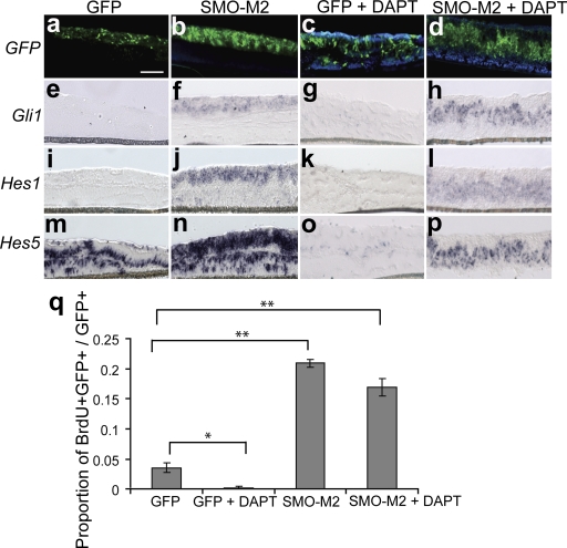
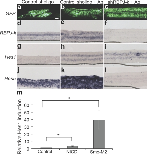
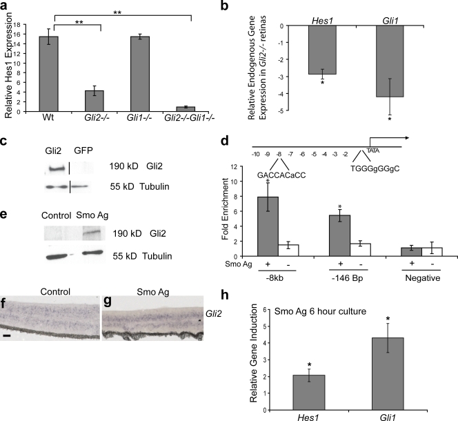
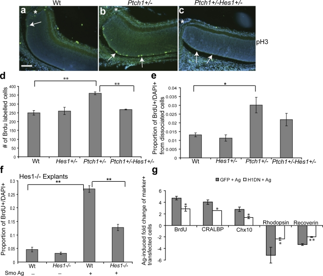
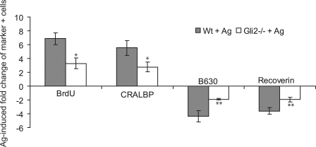
Sonic hedgehog (Shh) is an indispensable, extrinsic cue that regulates progenitor and stem cell behavior in the developing and adult mammalian central nervous system. Here, we investigate the link between the Shh signaling pathway and Hes1, a classical Notch target. We show that Shh-driven stabilization of Hes1 is independent of Notch signaling and requires the Shh effector Gli2. We identify Gli2 as a primary mediator of this response by showing that Gli2 is required for Hh (Hedgehog)-dependent up-regulation of Hes1. We also show using chromatin immunoprecipitation that Gli2 binds to the Hes1 promoter, which suggests that Hes1 is a Hh-dependent direct target of Gli2 signaling. Finally, we show that Shh stimulation of progenitor proliferation and cell diversification requires Gli2 and Hes1 activity. This paper is the first demonstration of the mechanistic and functional link between Shh, Gli, and Hes1 in the regulation of progenitor cell behavior.


Figures

Figure 1.
Figure 1.
Shh is required to maintain Hes1 protein and mRNA in postnatal retinal explants. (a) Diagram of the retinal explant culture method. Once the retina is surgically detached from the lens and surrounding ocular tissues, it is flattened by making four incisions and cultured on a membrane in the presence of a Smo agonist to activate the Hh signaling pathway. A cross section of a postnatal retinal explant is shown demonstrating Gli1 transcript expression in the Hh-responsive progenitor cells of the neuroblast region. The RGC layer is comprised of a population of postmitotic neurons that are not responsive to Hh signaling. Bar, 100 μm. (b) Retinal explants were treated with and without a Smo agonist at E14 (n = 3) and P0 (n = 3) for 3 d in culture and analyzed for Hes1, Hes5, and Gli1 mRNA by RT-qPCR. Values represent fold mRNA induction in Smo agonist–treated explants relative to untreated explants. Error bars represent SEM. *, P < 0.05. (c) Western blot for Hes1 from P0 retinal explants cultured for 3 d from untreated and Smo agonist–treated explants; β-tubulin protein level was used as a loading control.
Figure 2.
Figure 2.
Shh activation of Hes1 and Hes5 is independent of Notch signaling. Retinas at P0 were electroporated with SMO-M2 cotransfected with pUB-GFP or pUB-GFP alone and cultured for 3 d with DAPT or DMSO control. (a–d) GFP fluorescence localizes the transfected cells. ISH was performed for Gli1 (e–h), Hes1 (i–l), and Hes5 (m–p). Differences in the localization of transfected cells within the explants are caused by folding and twisting during tissue processing. Bars, 100 μm. (q) Retinal explants (P0 + 3 days in vitro [DIV]) were electroporated with Smo-M2/pUb-GFP, treated with DAPT, dissociated, and scored for the proportion of BrdU+GFP+/GFP+ cells. The magnitude of Smo-M2–induced proliferation is not changed with DAPT treatment. Error bars represent SEM. *, P < 0.05; **, P < 0.005.
Figure 3.
Figure 3.
RBPJ-κ signaling is not required for Shh induction of Hes1 but is necessary for Shh induction of Hes5. Retinal explants were electroporated with shRBPJ-κ or a control short hairpin plasmid at P0 and cultured for 4 d with or without a Smo agonist. (a–c) GFP fluorescence localizes the transfected cells. ISH was performed for RBPJ-κ (d–f), Hes1 (g–i), and Hes5 (j–l). Bar, 100 μm. (m) Retinal explants were coelectroporated with an empty vector control, NICD, or Smo-M2 and pUB-GFP, and cultured for 3 d, then Hes1 expression was analyzed by RT-qPCR. Values represent the relative induction of Hes1 expression normalized to GFP. Error bars represent SEM. *, P < 0.005.
Figure 4.
Figure 4.
Shh induction of Hes1 requires Gli2. (a) Retinal explants were cultured from wild-type (Wt; n = 5), Gli1−/− (n = 3), Gli2−/− (n = 6), and Gli2−/−Gli1−/− (n = 3) mice with or without a Smo agonist at E18 for 3 d, then analyzed for Hes1 expression by RT-qPCR. Values represent fold mRNA induction in Smo agonist–treated explants relative to untreated explants. (b) RT-qPCR on acutely dissected retinas from E18 wild-type (n = 5) and Gli2−/− (n = 5) animals. Values represent fold mRNA induction in Gli2−/− retinas compared with the wild type. The black lines in the Western blot indicate that intervening lanes have been spliced out. (c) Western blot analysis of protein lysates from Myc-Gli2–transfected or control COS cells blotted with an anti-Gli2 antibody. The β-tubulin protein level was used as a loading control. (d) Schematic of the 10-kb region of the Hes1 promoter. The Gli2-binding sites are indicated with the mismatched nucleotides relative to the ideal Gli consensus sequence in small letters. ChIP reveals enrichment of Gli2 at sites −7,808 bp and −146 bp upstream of the transcription start site in the Hes1 promoter in retinal explants treated with a Smo agonist. No enrichment of Gli2 was detected at these sites in untreated retinal explants. Association of Gli2 at a region of the Hes1 promoter that does not contain a Gli consensus sequence was used as a negative control. (e) Western blot analysis for Gli2 on retinal explants treated with or without a Smo agonist (P0 + 3 DIV). The β-tubulin protein level was used as a loading control. (f and g) Retinal explants cultured with or without a Smo agonist for 3 DIV and subjected to ISH for Gli2. Bars, 100 μm. (h) Retinal explants (P0) were cultured with (n = 5) or without a Smo agonist (n = 4) for 6 h and analyzed for Hes1 and Gli1 expression by RT-qPCR. Values represent fold mRNA induction in Smo agonist–treated explants relative to untreated explants. Error bars represent SEM. *, P < 0.05; **, P < 0.001.
Figure 5.
Figure 5.
Shh-mediated RPC proliferation and cell fate specification requires Hes1. (a–c) In vivo anti-pH3 staining of the central retina adjacent to the optic nerve (asterisks) in P5 wild-type (Wt), PtchlacZ+/−, and PtchlacZ+/−Hes1+/− retinas. Arrows indicate pH3-positive cells. Note that pH3+ cells in the vicinity of the optic nerve are rare in Wt and compound heterozygous mice. Bar, 100 μm. (d) Quantitative analysis of BrdU incorporation in vivo from P5 Wt (n = 3), Hes1+/− (n = 3), PtchlacZ+/− (n = 3), and PtchlacZ+/−Hes1+/− (n = 6) retinas. Values represent the mean number of BrdU-positive cells counted from three sections per animal. (e) Quantification of the proportion of BrdU+ cells in single-cell dissociates from the retinas of Wt (n = 5), Hes1+/− (n = 3), PtchlacZ+/− (n = 8), and PtchlacZ+/−Hes1+/− (n = 7) retinas at P5. (f) Retinal explants from Hes1−/− (n = 3) or Wt (n = 3) animals were treated with a Smo agonist for 3 d, dissociated, and scored for the proportion of BrdU+DAPI+ cells. (g) Quantitative analysis for BrdU, CRALBP, Chx10, rhodopsin, and recoverin-positive cells in Smo agonist–treated P0 retinal explants electroporated with GFP and Hes1DN. Values are based on scoring marker+ cells among the transfected cohort in dissociates from retinal explants and represent the fold induction of double-positive (marker+GFP+) cells in GFP + Ag and Hes1DN + Ag cultures compared with double-positive cells in GFP-transfected untreated explants. There is no difference in proliferation or cell type composition in GFP and Hes1DN-transfected cells in untreated explants. Error bars represent SEM. *, P < 0.05; **, P < 0.01.
Figure 6.
Figure 6.
Gli2 is required for the Shh effects on proliferation and cell fate. Retinal explants were cultured from wild-type (Wt; n = 3) and Gli2−/− (n = 3) mice at E18 for 3 d in culture with or without a Smo agonist. IHC was performed on dissociated cells using anti-BrdU, anti-CRALBP, anti-rhodopsin, and anti-recoverin antibodies. Values represent the fold induction of positive cells in Wt + Ag or Gli2−/− + Ag cultures compared with nontreated explants. Error bars represent SEM. *, P < 0.05; **, P < 0.01.

References

    1. Bai, C.B., W. Auerbach, J.S. Lee, D. Stephen, and A.L. Joyner. 2002. Gli2, but not Gli1, is required for initial Shh signaling and ectopic activation of the Shh pathway. Development. 129:4753–4761. - PubMed
    1. Barolo, S., R.G. Walker, A.D. Polyanovsky, G. Freschi, T. Keil, and J.W. Posakony. 2000. A notch-independent activity of suppressor of hairless is required for normal mechanoreceptor physiology. Cell. 103:957–969. - PubMed
    1. Belliveau, M.J., T.L. Young, and C.L. Cepko. 2000. Late retinal progenitor cells show intrinsic limitations in the production of cell types and the kinetics of opsin synthesis. J. Neurosci. 20:2247–2254. - PMC - PubMed
    1. Beres, T.M., T. Masui, G.H. Swift, L. Shi, R.M. Henke, and R.J. MacDonald. 2006. PTF1 is an organ-specific and Notch-independent basic helix-loop-helix complex containing the mammalian Suppressor of Hairless (RBP-J) or its paralogue, RBP-L. Mol. Cell. Biol. 26:117–130. - PMC - PubMed
    1. Black, G.C., C.J. Mazerolle, Y. Wang, K.D. Campsall, D. Petrin, B.C. Leonard, K.F. Damji, D.G. Evans, D. McLeod, and V.A. Wallace. 2003. Abnormalities of the vitreoretinal interface caused by dysregulated Hedgehog signaling during retinal development. Hum. Mol. Genet. 12:3269–3276. - PubMed

Publication types

MeSH terms