. 2009 Jan 16;206(1):221-32.
doi: 10.1084/jem.20082044. Epub 2009 Jan 5.

Down-regulation of CYLD expression by Snail promotes tumor progression in malignant melanoma

Affiliations

Down-regulation of CYLD expression by Snail promotes tumor progression in malignant melanoma

Ramin Massoumi et al. J Exp Med. .

Abstract

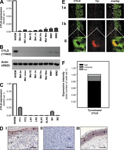
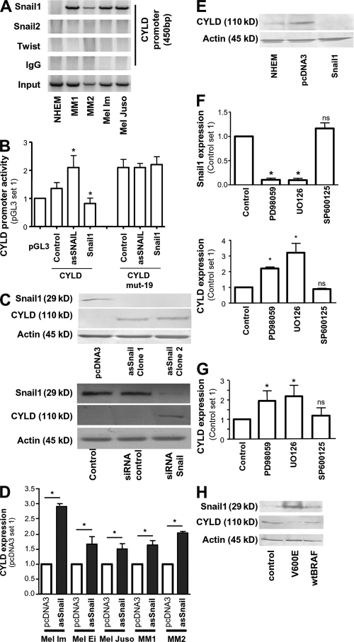
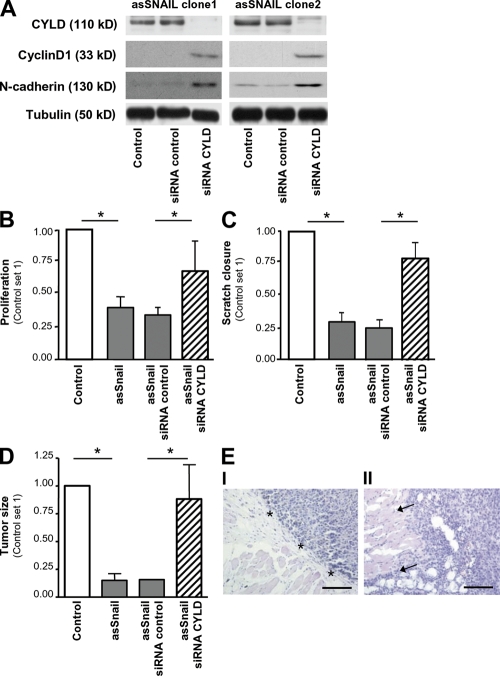
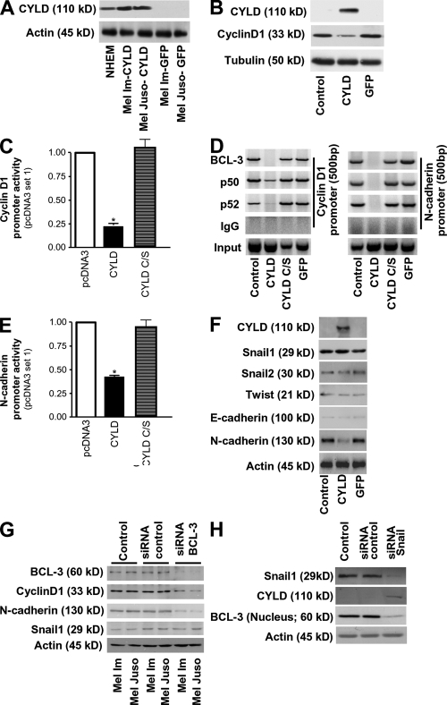
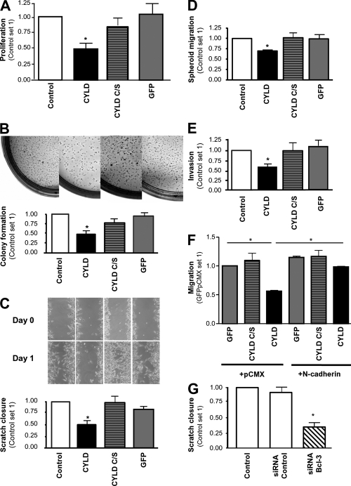
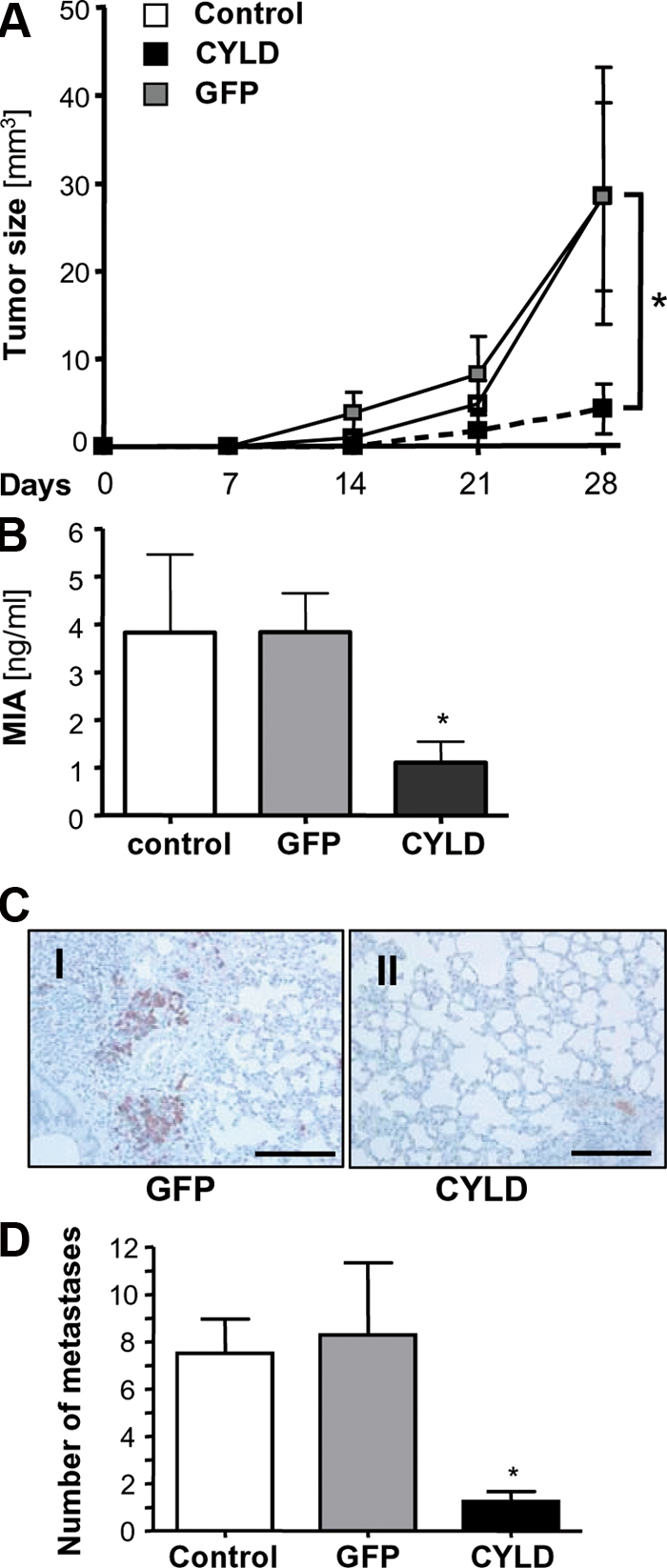
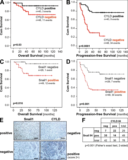
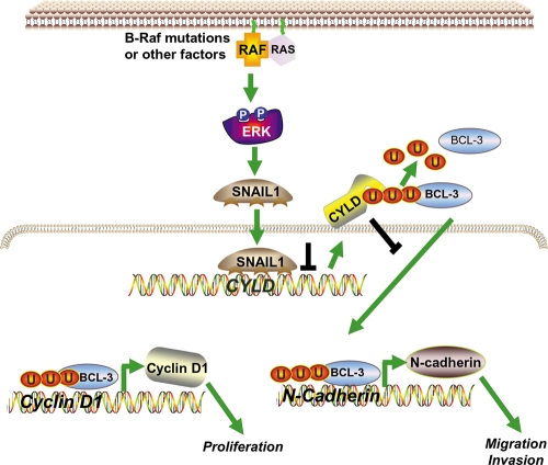
High malignancy and early metastasis are hallmarks of melanoma. Here, we report that the transcription factor Snail1 inhibits expression of the tumor suppressor CYLD in melanoma. As a direct consequence of CYLD repression, the protooncogene BCL-3 translocates into the nucleus and activates Cyclin D1 and N-cadherin promoters, resulting in proliferation and invasion of melanoma cells. Rescue of CYLD expression in melanoma cells reduced proliferation and invasion in vitro and tumor growth and metastasis in vivo. Analysis of a tissue microarray with primary melanomas from patients revealed an inverse correlation of Snail1 induction and loss of CYLD expression. Importantly, tumor thickness and progression-free and overall survival inversely correlated with CYLD expression. Our data suggest that Snail1-mediated suppression of CYLD plays a key role in melanoma malignancy.


Figures

Figure 1.
Figure 1.
Reduced CYLD expression in malignant melanoma. Quantitative RT-PCR (A) and immunoblot analysis (B) showing CYLD expression in six human melanoma cell lines (Mel Im, Mel Juso, Mel Ei, Mel Ju, Mel Ho, and Mel Wei) and freshly isolated primary melanoma cells from two donors (MM1 and MM2) compared with NHEMs. (C) CYLD mRNA expression in NHEMs, primary melanomas (PT), and metastases of lymph nodes (LN), sentinel lymph nodes (SLN), and two distant metastases of malignant melanomas (M). Experiments in A–C were repeated at least three times. Data are the mean ± SEM. (D) Immunohistochemistry showing strong down-regulation or loss of CYLD (red staining) in primary malignant melanoma (indicated by arrows; I) and in melanoma metastasis (II). In III, CYLD staining in normal skin is presented. Strong CYLD staining is shown in keratinocytes in normal epidermis (I, III), which is indicated by *. For counterstaining, hematoxylin was used. Experiments were performed four times independently. (E) CYLD immunoreactivity of normal skin showing CYLD staining (green) and tyrosinase-marked melanocytes (red; Ia and Ib). Higher magnification images (II) indicate a melanocyte displaying expression of both proteins. Experiments were performed three times independently. (F) Quantification of CYLD and tyrosinase costaining in epidermal melanocytes. Fraction of tyrosinase-expressing cells (melanocytes) that are expressing CYLD at low, moderate, or high levels (Tyrosinase/CYLD). Bars: (D) 100 μm; (E, Ia and Ib) 100 μm; (E, II) 5 μm.
Figure 2.
Figure 2.
Snail1 inhibits transcriptional expression of CYLD in melanocytic cells. (A) Lysates from NHEMs, primary melanoma cells (MM1 and MM2), and melanoma cell lines (Mel Im, Mel Juso) were examined by ChIP assay using an anti-Snail1, anti-Twist, and anti-Snail2–specific antibody, respectively, as well as a PCR primer pair corresponding to the promoter of the CYLD gene. Recruitment to the CYLD promoter was only shown for Snail1. Snail1, Twist, Snail2, immunoprecipitation (IP) using specific antibodies; IgG, IP using negative control rabbit immunoglobulin; Input, 10% of the cell lysate used for the IP is shown. (B) Reporter assays revealing inducible CYLD promoter (−550 to −1 bp) activity in Mel Im cells after transient transfection with asSnail (asSnail), whereas transfection with Snail1 expression plasmid (Snail1) completely abrogated CYLD promoter activity compared with the control (pGL3). Mutation of the consensus Snail1 binding site (CYLD mut-19) led to strong promoter activity that was not affected by transfection with asSnail or Snail1. Data are given as mean ± SEM. *, P < 0.05 compared with CYLD control. (C) Immunoblot analysis showing that CYLD protein is up-regulated in Mel Im cells after stable transfection with asSnail expression plasmids (clone 1 and clone 2; top) and after transient transduction with Snail1 siRNA nucleotides (siRNA Snail1) or scrambled siRNA control (siRNA control; bottom). Nontransduced Mel Im cells were used as control. (D) Quantitative RT-PCR analysis showing increased CYLD mRNA expression in three different melanoma cell lines (Mel Im, Mel Ei, and Mel Juso) and melanoma cells (MM1 and MM2) transiently transfected with asSnail expression plasmids compared with mock-transfected (pcDNA3) cells. Data are given as mean ± SEM. *, P < 0.05. (E) Western blot analysis showing decreased CYLD protein expression in NHEM cells transiently transfected with Snail1 expression plasmids compared with mock-transfected (pcDNA3) or nontransfected (NHEM) cells. (F) ERK inhibition by treatment with MEK inhibitors PD98059 and UO126 significantly affects Snail1 and CYLD mRNA expression in Mel Im cells. JNK inhibition (SP600125) exhibits no effect compared with vehicle (DMSO) control. Data are given as mean ± SEM. *, P < 0.05. ns, not significant, both compared with Control. (G) Quantitative RT-PCR revealing that inhibition of ERK by PD98059 or UO126 further induced CYLD expression in melanocytes. JNK inhibition (SP600125) exhibits no effect compared with vehicle (DMSO) control. Data are given as mean ± SEM. *, P < 0.05. Experiments in A–G were repeated at least three times. (H) Detection of CYLD expression by Western blot analysis. Transfection of NHEMs with mutated BRAF (V600E) induced Snail1 protein expression and, consequently, CYLD protein suppression, whereas no changes were found after transfection of wild-type BRAF (wtBRAF). Experiments were repeated three times with cells from two different donors.
Figure 3.
Figure 3.
Snail1 regulates tumorigenicity via CYLD repression. (A) Western blot analysis of two Mel Im asSnail clones (clone 1 and clone 2) applying CYLD, Cyclin D1, and N-cadherin antibodies. Stable transduction with viral vectors encoding siRNA against CYLD (siRNA CYLD) results in the complete depletion of CYLD, but induction of Cyclin D1 and N-cadherin in both clones. Transduction with control siRNA (siRNA control) exhibits no effect. Proliferation (B) and migration (C; in monolayer scratch assays) of Mel Im control cells (Control) and Mel Im asSnail clone 1 either stably transduced with viral vectors encoding siRNA against CYLD (siRNA CYLD), control siRNA (siRNA control), or without transduction (asSnail). Data represent proliferation after 72 h, and areas between scratch fronts (after 2 d in monolayer scratch assays) and are given as mean ± SEM. *, P < 0.05. Experiments in A–C were repeated at least three times. (D) Growth of Mel Im asSnail clone 1 (asSnail) transduced with siRNA against CYLD (siRNA CYLD) or control siRNA (siRNA control) and Mel Im control cells (Control) after s.c. implantation into nude mice (106 cells/mouse). Bars represent mean tumor size (± SEM) 3 wk after implantation. *, P < 0.05. (E) Histological analysis of the growths of Mel Im asSnail clone 1 without transduction (I) or stably transduced with siRNA against CYLD (II) 3 wk after s.c. implantation into nude mice. Pictures are representative and arrows indicate diffuse infiltration in II, whereas nodular growth (*) appears in I. Bar, 200 μm. Experiments and analysis in D and E, respectively, have been performed with 10 mice/group.
Figure 4.
Figure 4.
CYLD regulates N-cadherin and Cyclin D1 expression via BCL-3 in melanoma. (A) Western blot analysis revealing CYLD expression in NHEMs and Mel Im and Mel Juso cells stably transduced with CYLD, but not in melanoma cells stably transduced with GFP. Actin labeling is used as loading control. (B) Western blot detecting Cyclin D1 expression in Mel Im cells stably transduced with CYLD, but not in nontransduced control cells or cells stably transduced with GFP. Tubulin labeling is used as loading control. (C) Effect of transfection of a CYLD expression vector carrying wild-type (CYLD) or mutated CYLD (CYLD C/S) on Cyclin D1 promoter activity in Mel Im cells. Data are given as mean ± SEM. *, P < 0.05 versus pcDNA3 empty vector used as control. (D) Lysates from Mel Im cells (Control) and CYLD, mutant CYLD (CYLD C/S), or GFP stably expressing Mel Im cells were examined by ChIP assay using specific polyclonal antibodies against BCL-3, p50, or p52, and PCR primer pairs corresponding to the promoter of the CyclinD1 or N-cadherin gene to analyze recruitment of BCL-3 to the Cyclin D1 and N-cadherin promoter. BCL-3, p50, and p52 immunoprecipitation (IP) using polyclonal antibodies as indicated. IgG, negative control rabbit IgG (DAKO); Input, 10% of the cell lysate used for the IP is shown. (E) Effect of transduction of a CYLD expression vector carrying wild-type (CYLD) or a catalytic inactive mutant of CYLD (CYLD C/S) on N-cadherin promoter activity in Mel Im cells. Data are given as mean ± SEM. *, P < 0.05 versus pcDNA3 empty vector used as control. (F) Western blot analysis of CYLD, Snail1, Snail2, Twist, E-cadherin, N-cadherin, and Actin expression in nontransduced Mel Im cells (Control) or Mel Im cells transduced with GFP or CYLD. (G) Western blot analysis of BCL-3, Cyclin D1, N-cadherin, Snail1, and Actin expression in nontransduced Mel Im and Mel Juso cells (Control), cells transduced with the BCL-3 siRNA nucleotides (siRNA BCL-3), or scrambled siRNA control (siRNA control). (H) Western blot analysis of Snail1, CYLD, (nuclear) BCL-3, and Actin expression in nontransduced Mel Im cells (Control), and cells transduced with Snail1 siRNA nucleotides (siRNA Snail1) or scrambled siRNA control (siRNA control). Experiments in A–H were repeated at least three times.
Figure 5.
Figure 5.
CYLD regulates proliferation and N-cadherin–mediated migration and invasion. (A) Proliferation of Mel Im cells expressing CYLD, CYLD C/S, or GFP versus the parental Mel Im cell line (Control) after 72 h. Data are given as mean ± SEM. *, P < 0.05. (B) Colony formation in soft agar 3 wk after plating 104 Mel Im cells expressing CYLD, CYLD C/S, or GFP versus the parental (Control) cell line. (C) Migration of melanoma cells in monolayer scratch assays. Mel Im cells transduced with lentiviral vectors carrying CYLD, CYLD C/S, or GFP and noninfected control cells (Control) were used. Representative pictures (at day 0–2) and calculation (mean ± SEM; *, P < 0.05) of the areas between scratch fronts (after 2 d). (D) Migration of Mel Im cells (Control) compared with CYLD, CYLD C/S, or GFP stably transduced cells in spheroid migration assays. Data are given as mean ± SEM. *, P < 0.05. (E) Invasion of Mel Im (Control) cells compared with CYLD, CYLD C/S, or GFP stably transduced cells (after 24 h) in Boyden chamber assays. Data are given as mean ± SEM. *, P < 0.05 compared with Control and GFP. (F) Migration of melanoma cells in Boyden chamber assays. Comparison of Mel Im cells stably transduced with CYLD, CYLD C/S or GFP, and transiently cotransfected with an N-cadherin expression vector (+N-cadherin) or control vector (+pCMX). Data are given as mean ± SEM. *, P < 0.05. (G) Migration of melanoma cells in monolayer scratch assays. Comparison of nontransduced Mel Im cells (Control), cells transfected with BCL-3 siRNA nucleotides (siRNA BCL-3), or scrambled siRNA control (siRNA Control). Data are given as mean ± SEM. *, P < 0.05 compared with Control or siRNA control. Experiments in A–G were repeated at least three times.
Figure 6.
Figure 6.
CYLD inhibits proliferation and metastasis of melanoma cells in vivo. (A) Growth kinetic of tumors formed by Mel Im control cells (Control) or cells transduced with viral vectors carrying CYLD or GFP after subcutaneous implantation into nude mice (106 cells/mouse). Data represent mean tumor size (± SEM) at different time points. *, P < 0.05 versus both GFP and Control. (B) MIA serum levels in nude mice after i.v. injection of Mel Im control cells (Control) or cells transduced with viral vectors carrying CYLD or GFP (106 cells/mouse; 8–10 mice/group). Data represent mean MIA level (± SEM) 4 wk after injection. (*, P < 0.05 versus both GFP and Control). (C) Immunohistochemical MART1 staining of pulmonary tissue 4 wk after inoculation. Photomicrographs showing representative sections of the lung of mice receiving Mel Im cells transduced with CYLD or GFP. Bar, 200 μm. (D) Number of micrometastatic lesions per one representative cross section of the lungs from each mouse. Data are given as mean ± SEM. *, P < 0.05 compared with control and GFP. Experiments and analysis in A–D have been performed with 8–10 mice/group.
Figure 7.
Figure 7.
CYLD expression in melanoma has prognostic implication. Kaplan-Meier curves for overall survival (A) and progression-free survival (B) in melanoma patients with a positive immunosignal for CYLD (CYLD positive) or without detectable CYLD protein expression (CYLD negative). CYLD immunohistochemical staining of primary malignant melanoma tissue was performed using a tissue microarray consisting of 88 cases. Investigation of CYLD protein expression was informative in all specimens. Kaplan-Meier curves for overall survival (C) and progression-free survival (D) in melanoma patients with a positive immunosignal for Snail1 (Snail1 positive) or without detectable Snail1 protein expression (Snail1 negative). Snail1 immunohistochemical staining of primary malignant melanoma tissue was performed using a tissue microarray consisting of 88 cases. Immunohistochemical analysis of Snail1 was informative in 78 samples. (E) Examples of tissues with positive and negative CYLD and Snail1 staining, respectively. Bar, 100 μm. (F) Comparison of CYLD and Snail1 immunoreactivity. An inverse correlation of CYLD and Snail1 staining was observed.
Figure 8.
Figure 8.
Model of the regulation of Snail and how Snail affects proliferation, as well as migration and invasion, via suppression of CYLD. In melanoma cells, constitutively high ERK-activity (potentially caused by B-Raf mutations) leads to high Snail1 expression, which in turn causes strong suppression of CYLD. The repression of CYLD allows nuclear translocation of Bcl-3 and recruitment to the NF-κB binding sites of the Cyclin D1 and N-cadherin promoter. Activation of Cyclin D1 and N-cadherin leads to an increased proliferation rate of melanoma cells and contributes to progression and metastasis of tumors.

References

    1. Chin, L., L.A. Garraway, and D.E. Fisher. 2006. Malignant melanoma: genetics and therapeutics in the genomic era. Genes Dev. 20:2149–2182. - PubMed
    1. Haass, N.K., K.S. Smalley, L. Li, and M. Herlyn. 2005. Adhesion, migration and communication in melanocytes and melanoma. Pigment Cell Res. 18:150–159. - PubMed
    1. Attis, M.G., J.L. Burchette, M.A. Selim, T. Pham, and A.P. Soler. 2006. Differential expression of N-cadherin distinguishes a subset of metastasizing desmoplastic melanomas. Hum. Pathol. 37:899–905. - PubMed
    1. Qi, J., N. Chen, J. Wang, and C.H. Siu. 2005. Transendothelial migration of melanoma cells involves N-cadherin-mediated adhesion and activation of the beta-catenin signaling pathway. Mol. Biol. Cell. 16:4386–4397. - PMC - PubMed
    1. Smalley, K.S., P. Brafford, N.K. Haass, J.M. Brandner, E. Brown, and M. Herlyn. 2005. Up-regulated expression of zonula occludens protein-1 in human melanoma associates with N-cadherin and contributes to invasion and adhesion. Am. J. Pathol. 166:1541–1554. - PMC - PubMed

Publication types

MeSH terms