. 2009 Jan 16;206(1):61-8.
doi: 10.1084/jem.20082058. Epub 2009 Jan 5.

Efferocytosis impairs pulmonary macrophage and lung antibacterial function via PGE2/EP2 signaling

Affiliations

Efferocytosis impairs pulmonary macrophage and lung antibacterial function via PGE2/EP2 signaling

Alexandra I Medeiros et al. J Exp Med. .

Abstract

The ingestion of apoptotic cells (ACs; termed "efferocytosis") by phagocytes has been shown to trigger the release of molecules such as transforming growth factor beta, interleukin-10 (IL-10), nitric oxide, and prostaglandin E(2) (PGE(2)). Although the antiinflammatory actions of these mediators may contribute to the restoration of homeostasis after tissue injury, their potential impact on antibacterial defense is unknown. The lung is highly susceptible to diverse forms of injury, and secondary bacterial infections after injury are of enormous clinical importance. We show that ACs suppress in vitro phagocytosis and bacterial killing by alveolar macrophages and that this is mediated by a cyclooxygenase-PGE(2)-E prostanoid receptor 2 (EP2)-adenylyl cyclase-cyclic AMP pathway. Moreover, intrapulmonary administration of ACs demonstrated that PGE(2) generated during efferocytosis and acting via EP2 accounts for subsequent impairment of lung recruitment of polymorphonuclear leukocytes and clearance of Streptococcus pneumoniae, as well as enhanced generation of IL-10 in vivo. These results suggest that in addition to their beneficial homeostatic influence, antiinflammatory programs activated by efferocytosis in the lung have the undesirable potential to dampen innate antimicrobial responses. They also identify an opportunity to reduce the incidence and severity of pneumonia in the setting of lung injury by pharmacologically targeting synthesis of PGE(2) or ligation of EP2.


Figures

Figure 1.
Figure 1.
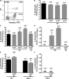
Efferocytosis inhibits FcR-mediated phagocytosis and bacterial killing by AMs. (A) Jurkat T cells were incubated with 8 μg/ml camptothecin for 5 h and apoptotic cells were detected by AnnexinV-FITC/PI and analyzed by flow cytometry. Early ACs represent 25.69% of cells. (B) Phagocytosis of IgG RBCs or IgG E. coli was determined after a 90-min pretreatment with ACs at the indicated AC/AM ratios. (C) Phagocytosis of IgG RBCs was determined after pretreatment for the indicated times with ACs added at a ratio of 3:1. (D) Phagocytosis of IgG RBCs was determined after a 90-min pretreatment with viable (VC) or necrotic (NC) Jurkat cells added at a ratio of 3:1. (E) AMs were preincubated with or without ACs (3:1) for 90 min and then infected with K. pneumoniae (50:1). Microbicidal activity was determined and expressed as the percentage survival of ingested bacteria. Results represent the mean ± SEM from three independent experiments, each performed in quintuplicate (B–D) or the mean ± SEM of quintuplicate values from a single experiment representative of three independent experiments (A and E). *, P < 0.05 versus control.
Figure 2.
Figure 2.
PGE2 mediates the suppressive effects of efferocytosis on AM antimicrobial functions via EP2. (A) AMs were pretreated with culture supernatant derived from parallel incubations of ACs/AMs (3:1), with 5 μM PGE2, or with 3:1 ACs in the absence or presence of 6 μg/ml of anti–TGF-β blocking antibody or 5 μM of the COX inhibitors indomethacin (Indo) and 200 μM of aspirin (Asp). They were subsequently challenged with IgG RBCs and phagocytosis was determined. (B) AMs were incubated with medium alone or with ACs in the presence or absence of aspirin. PGE2 in supernatant was quantitated by immunoassay after 30 min. (C) AM phagocytosis of IgG RBCs was determined after a 90-min pretreatment with medium alone or with ACs (3:1) in the absence or presence of 100 μM of the EP2 antagonist AH-6809. (D) AMs from EP2−/− or WT control mice were preincubated with or without apoptotic thymocytes (5:1) for 90 min before challenge with IgG RBCs and phagocytosis was determined. Results represent the mean ± SEM from three independent experiments, each performed in quintuplicate (A–C) or the mean ± SEM of quintuplicate values from one experiment representative of three independent experiments (D). *, P < 0.05 versus control; #, P < 0.05 versus AC.
Figure 3.
Figure 3.
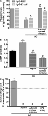
Adenylyl cyclase generation of cAMP mediates the suppressive effects of efferocytosis on AM antimicrobial functions. (A) Phagocytosis of IgG RBCs or IgG E. coli was determined after 90-min pretreatment with ACs (3:1) in the absence or presence of 10 μM of the adenylyl cyclase inhibitor SQ 22536. (B) AMs were pretreated or not with 200 μM of aspirin or 10 μM of the adenylyl cyclase inhibitor SQ 22536 before addition of ACs or vehicle for 30 min. Intracellular cAMP concentrations were determined by enzyme immunoassay. (C) AMs were preincubated with 100 μM of the EP2 antagonist AH-6809, 200 μM of aspirin, 10 μM of the adenylyl cyclase inhibitor SQ 22536, or vehicle before the addition of K. pneumoniae (50:1). Microbicidal activity was assessed and intracellular survival of bacteria is expressed as a percentage of the control, to which no AC were added. Results represent the mean ± SEM from three independent experiments, each performed in quintuplicate. *, P < 0.05 versus control; #, P < 0.05 versus AC.
Figure 4.
Figure 4.
Intrapulmonary administration of ACs impairs host defense in a mouse model of pneumococcal pneumonia. (A) Thymocytes were incubated with 1 μM dexamethasone for 6 h and ACs were detected by AnnexinV-FITC/PI and analyzed by flow cytometry. Early ACs comprise 40.3% of total cells. (B) 106 CFU of S. pneumoniae and varying numbers of apoptotic thymocytes were coadministered intratracheally in WT mice. Lung homogenates were assessed for bacterial CFUs 48 h later. (C) Indicated numbers of apoptotic or viable thymocytes were instilled intranasally in WT mice and, 16 h later, 106 CFU S. pneumoniae were administered intratracheally. Lung homogenates were assessed for bacterial CFUs 48 h after S. pneumoniae challenge. (D) Bacterial CFUs were determined in blood obtained 48 h after S. pneumoniae challenge from the same WT mice studied in C. (E) WT and EP2−/− mice were subjected to intranasal administration of apoptotic thymocytes 16 h before intratracheal challenge with S. pneumoniae as described in C. Lung homogenate CFUs 48 h after bacterial challenge are presented. (F) Bacterial CFUs were determined in blood obtained 48 h after S. pneumoniae challenge from the same EP2−/− mice studied in E. Results represent the mean ± SEM of one experiment representative of two. The number of animals analyzed in each group is indicated above each bar. ND, none detected. *, P < 0.05 versus control; #, P < 0.05 versus AC.
Figure 5.
Figure 5.
PGE2/EP2 signaling impairs PMN recruitment and promotes in vivo generation of IL-10 in a mouse model of pneumococcal pneumoniae. 106 apoptotic thymocytes were instilled intranasally in WT and EP2−/− mice and, 16 h later, 106 CFU S. pneumoniae were administered intratracheally. (A–C) PGE2 (A), total TGF-β (B), and IL-10 levels (C) were quantified in the supernatant of lung homogenates from animals studied in Fig. 4 (C and E). (D) PMNs in BALF from WT and EP2−/− mice were counted. Results represent the mean ± SEM of one experiment representative of two (A–C) or of one experiment (D). The number of animals analyzed in each group is indicated above each bar. *, P < 0.05 versus control; #, P < 0.05 versus AC.

Similar articles

Cited by

References

    1. Wheeler, A.P., and G.R. Bernard. 2007. Acute lung injury and the acute respiratory distress syndrome: a clinical review. Lancet. 369:1553–1564. - PubMed
    1. Matute-Bello, G., and T.R. Martin. 2003. Science review: apoptosis in acute lung injury. Crit. Care. 7:355–358. - PMC - PubMed
    1. Fadok, V.A., D.L. Bratton, A. Konowal, P.W. Freed, J.Y. Westcott, and P.M. Henson. 1998. Macrophages that have ingested apoptotic cells in vitro inhibit proinflammatory cytokine production through autocrine/paracrine mechanisms involving TGF-beta, PGE2, and PAF. J. Clin. Invest. 101:890–898. - PMC - PubMed
    1. Voll, R.E., M. Herrmann, E.A. Roth, C. Stach, J.R. Kalden, and I. Girkontaite. 1997. Immunosuppressive effects of apoptotic cells. Nature. 390:350–351. - PubMed
    1. McDonald, P.P., V.A. Fadok, D. Bratton, and P.M. Henson. 1999. Transcriptional and translational regulation of inflammatory mediator production by endogenous TGF-beta in macrophages that have ingested apoptotic cells. J. Immunol. 163:6164–6172. - PubMed

Publication types

MeSH terms