. 2009 Jan;66(1):51-63.
doi: 10.1001/archgenpsychiatry.2008.504.

Deficient activity in the neural systems that mediate self-regulatory control in bulimia nervosa

Affiliations

Deficient activity in the neural systems that mediate self-regulatory control in bulimia nervosa

Rachel Marsh et al. Arch Gen Psychiatry. 2009 Jan.

Abstract

Context: Disturbances in neural systems that mediate voluntary self-regulatory processes may contribute to bulimia nervosa (BN) by releasing feeding behaviors from regulatory control.

Objective: To study the functional activity in neural circuits that subserve self-regulatory control in women with BN.

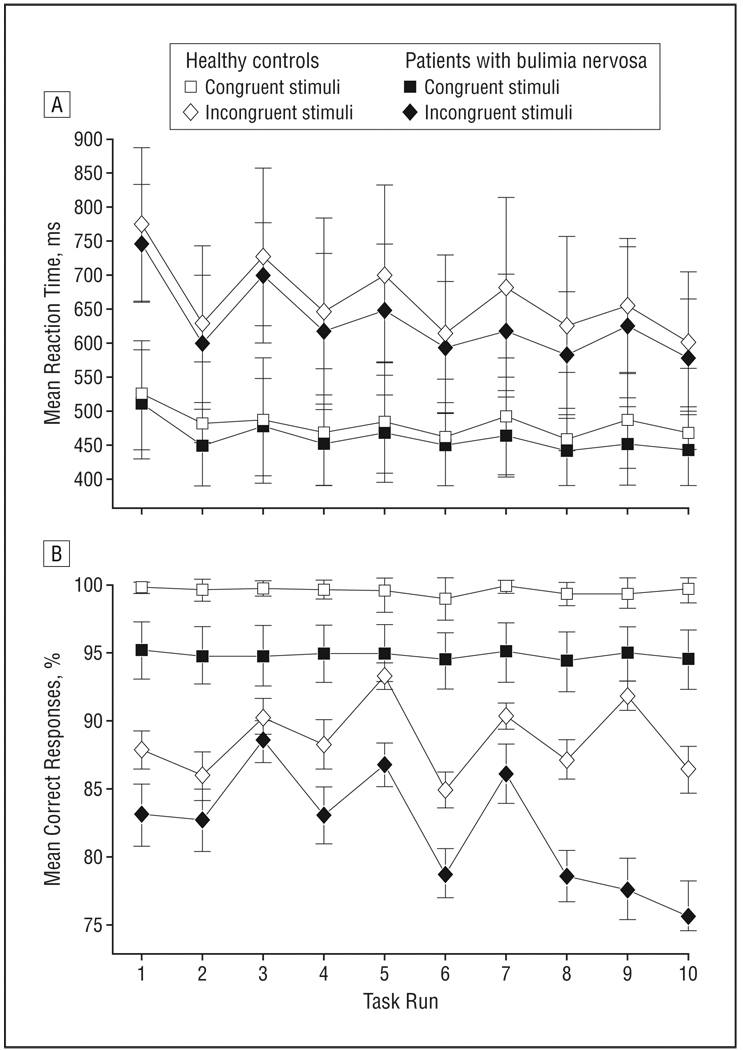
Design: We compared functional magnetic resonance imaging blood oxygenation level-dependent responses in patients with BN with healthy controls during performance of the Simon Spatial Incompatibility task.

Setting: University research institute.

Participants: Forty women: 20 patients with BN and 20 healthy control participants. Main Outcome Measure We used general linear modeling of Simon Spatial Incompatibility task-related activations to compare groups on their patterns of brain activation associated with the successful or unsuccessful engagement of self-regulatory control.

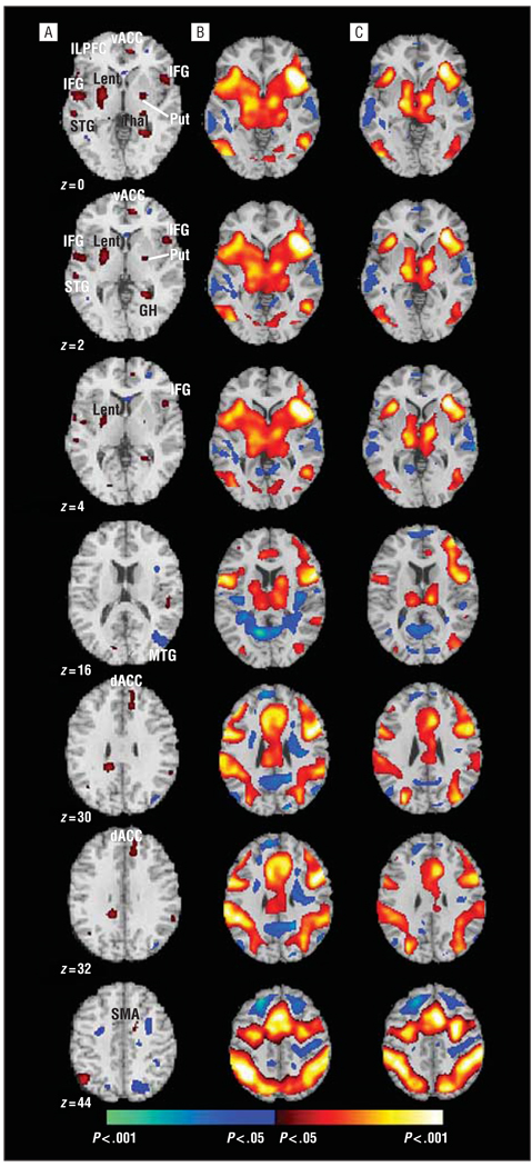
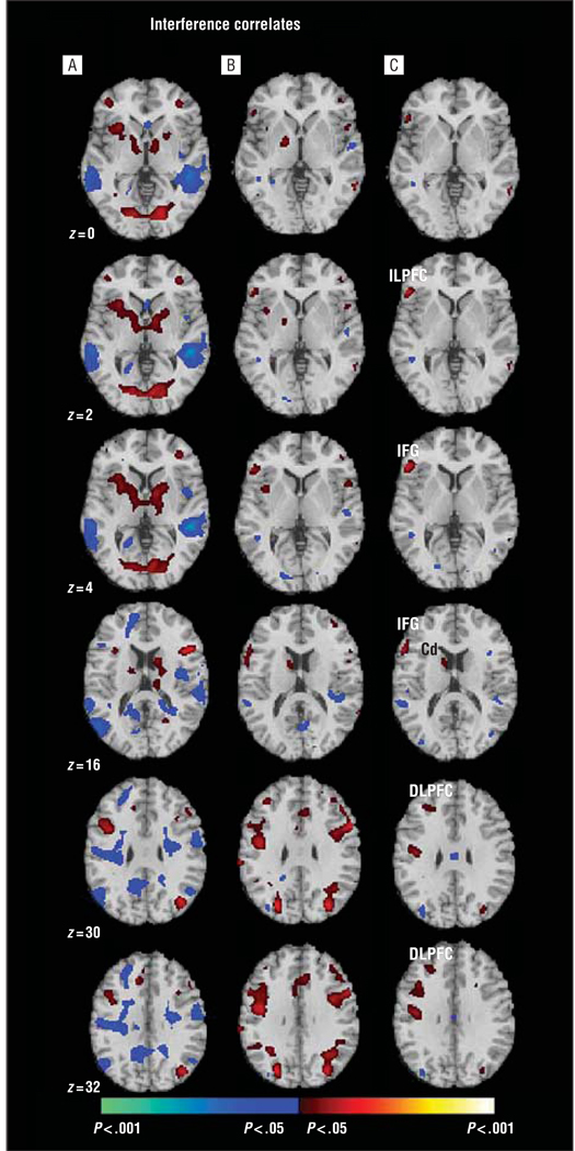
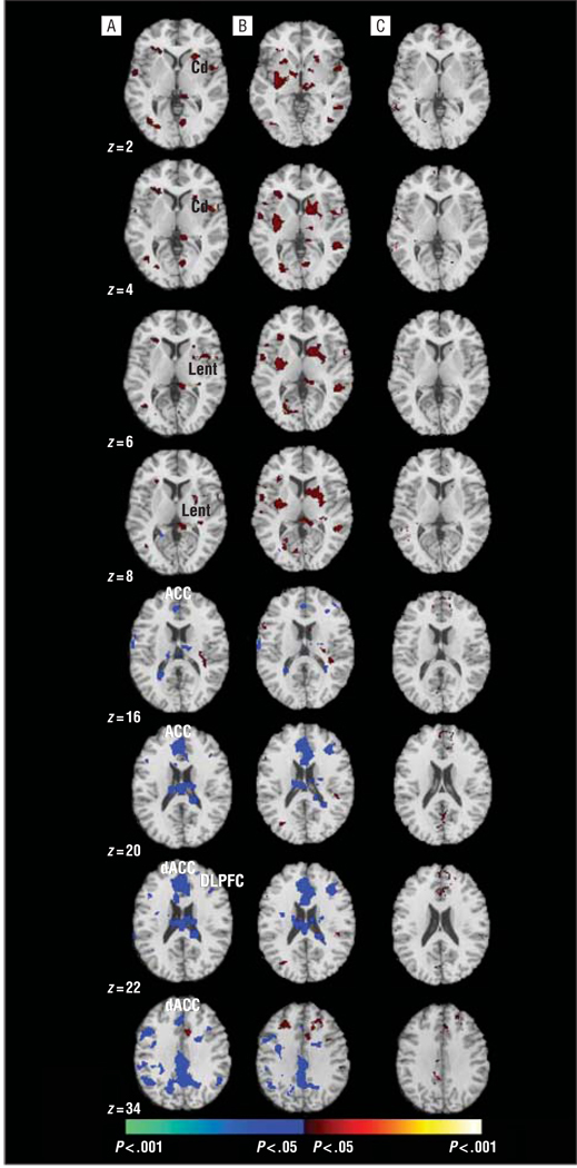
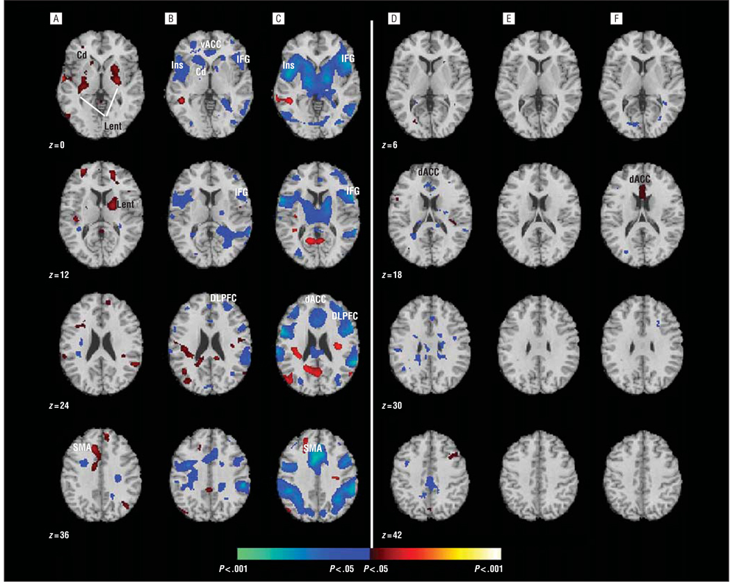
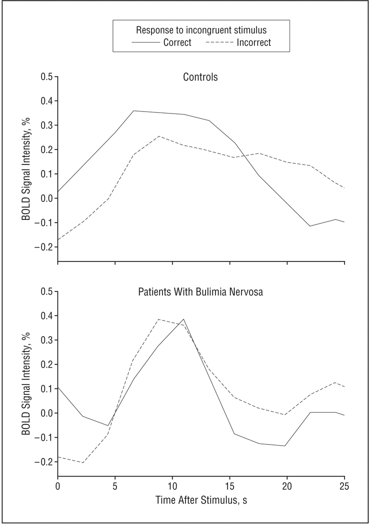
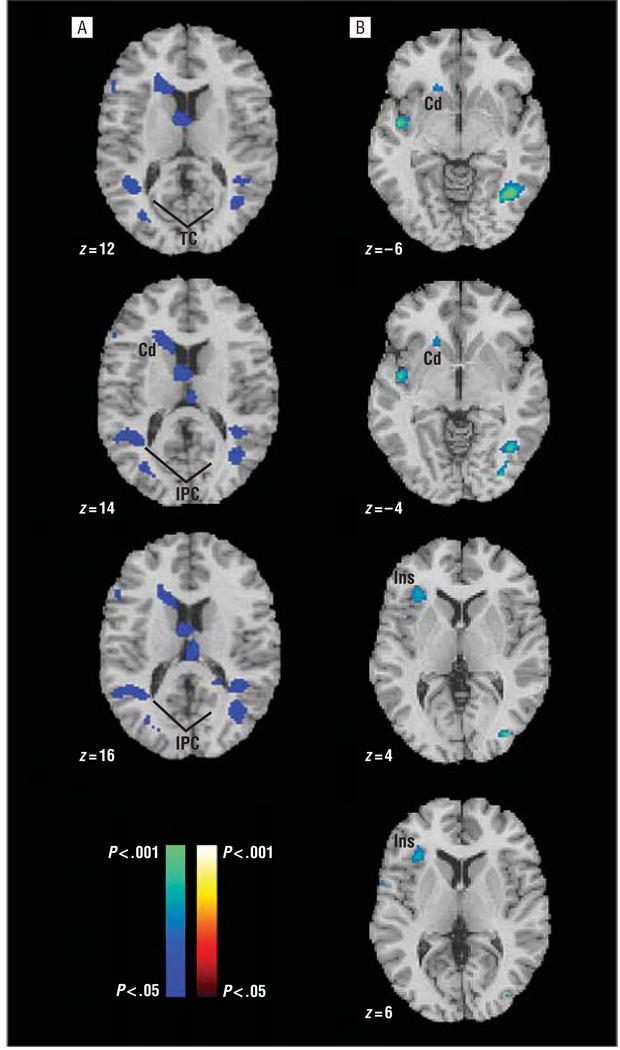
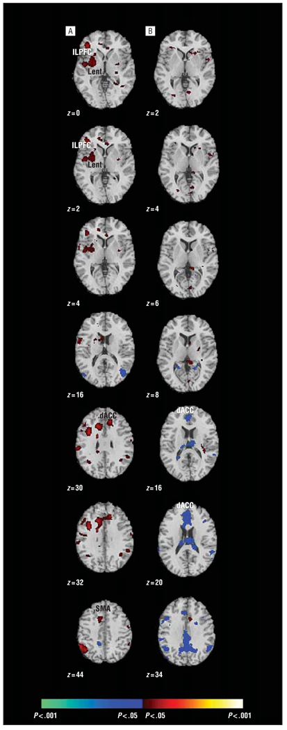
Results: Patients with BN responded more impulsively and made more errors on the task than did healthy controls; patients with the most severe symptoms made the most errors. During correct responding on incongruent trials, patients failed to activate frontostriatal circuits to the same degree as healthy controls in the left inferolateral prefrontal cortex (Brodmann area [BA] 45), bilateral inferior frontal gyrus (BA 44), lenticular and caudate nuclei, and anterior cingulate cortex (BA 24/32). Patients activated the dorsal anterior cingulate cortex (BA 32) more when making errors than when responding correctly. In contrast, healthy participants activated the anterior cingulate cortex more during correct than incorrect responses, and they activated the striatum more when responding incorrectly, likely reflecting an automatic response tendency that, in the absence of concomitant anterior cingulate cortex activity, produced incorrect responses.

Conclusions: Self-regulatory processes are impaired in women with BN, likely because of their failure to engage frontostriatal circuits appropriately. These findings enhance our understanding of the pathogenesis of BN by pointing to functional abnormalities within a neural system that subserves self-regulatory control, which may contribute to binge eating and other impulsive behaviors in women with BN.


Figures

Figure 1
Figure 1
Mean latency (A) and accuracy (B) of responses on the Simon Spatial Incompatibility task as a function of bulimia nervosa.
Figure 2
Figure 2
Axial slices showing group average brain activations during correct trials of the Simon Spatial Incompatibility task in healthy controls and patients with bulimia nervosa. A, Group × stimulus (congruent vs incongruent) interactions were detected in frontostriatal regions (red). Main effects of stimulus condition (congruent vs incongruent) are shown for the healthy participants (B) and those with bulimia nervosa (C) (P<.05, cluster>25 adjacent voxels; yielding a conjoint effective, P<.00000539). The combined application of a statistical threshold and cluster filter substantially reduces the false-positive identification of activated pixels at any given threshold. Increases in signal during correct incongruent trials relative to correct congruent trials are shown in red, and decreases are shown in blue. dACC indicates dorsal anterior cingulate cortex; GH, hippocampal gyrus; IFG, inferior frontal gyrus; ILPFC, inferolateral prefrontal cortex; Lent, lenticular nucleus; MTG, medial temporal gyrus; Put, putamen; SMA, supplementary motor area; STG, superior temporal gyrus; Thal, thalamus; vACC, ventral anterior cingulate cortex.
Figure 3
Figure 3
Correlations of mean reaction times with signal change during correct conflict trials of the Simon Spatial Incompatibility task in healthy controls (A) and patients with bulimia nervosa (B). Positive correlations are in red, and inverse correlations are in blue. C, Diagnosis × interference interactions in prefrontal regions and the caudate nucleus (Cd) stemming from the interference correlates in the patients (P<.05, cluster >25 adjacent voxels). DLPFC indicates dorsolateral prefrontal cortex; IFG, inferior frontal gyrus; and ILPFC, inferolateral prefrontal cortex.
Figure 4
Figure 4
Group average brain activity during the commission of errors. A, Group × response (incorrect vs correct) interactions were detected in frontostriatal brain areas (P<.05, cluster >25 adjacent voxels). Functional magnetic resonance imaging signal associated with incorrect vs correct responding on incongruent trials of the Simon Spatial Incompatibility task was greater in controls compared with patients with bulimia nervosa in the striatum (red). Increased activity in patients with bulimia nervosa was most prominent in the anterior cingulate cortex (ACC) (blue). Main effects of response are shown in the controls (B) and patients with bulimia nervosa (C). Increases in signal during incorrect incongruent trials relative to incorrect congruent trials are shown in red, and decreases are shown in blue. Cd indicates caudate nucleus; dACC, dorsal ACC; DLPFC, dorsolateral prefrontal cortex; and Lent, lenticular nucleus.
Figure 5
Figure 5
Correlations of activations during correct trials with the total number of errors made on the Simon Spatial Incompatibility task by healthy controls (A) and patients with bulimia nervosa (B). Positive correlations are shown in red, and inverse correlations are shown in blue. C, Diagnosis × error interactions were detected in prefrontal and striatal regions. Correlations of activations during the commission of errors with posterror adjustment scores in the controls (D) and patients with bulimia nervosa (E). These scores were calculated as the difference of the mean reaction times on trials immediately following erroneous responses and the mean reaction times on trials immediately following correct responses (these subsequent trials were always congruent stimuli because incongruent trials were never preceded by incongruent trials in our version of the Simon task). F, Diagnosis × posterror interactions in the dorsal anterior cingulate cortex (dACC) indicate that the controls who engaged this area the most during correct responses to conflict stimuli were those who slowed down most following correct conflict trials (all, P<.05, cluster >25 adjacent voxels). Cd indicates caudate nucleus; DLPFC, dorsolateral prefrontal cortex; IFG, inferior frontal gyrus; Ins, insula; Lent, lenticular nucleus; SMA, supplementary motor area; and vACC, ventral anterior cingulate cortex.
Figure 6
Figure 6
Temporal patterns of maximum dorsal anterior cingulate cortex (x=−4, y=34, z=26) activations following correct and incorrect responses to incongruent stimuli in healthy controls and patients with bulimia nervosa (extracted from Figure 3). Time courses were averaged across voxels in each region of interest for both groups. BOLD indicates blood oxygenation level–dependent.
Figure 7
Figure 7
Main effects of symptom severity in patients with bulimia nervosa. A, Inverse correlations of the number of objective bulimic episodes (from the Eating Disorders Examination) with the magnitude of activation during correct responding suggest that the patients with the most episodes of binge eating and purging engaged cortical areas (medial prefrontal cortex, temporal cortex [TC], and inferior parietal cortex [IPC]) and the head of the caudate the least. B, Inverse associations of ratings of preoccupation with weight and shape with task-related activations indicated that the most preoccupied patients engaged the caudate and insula (Ins) the least (all, P<.05, cluster >25 adjacent voxels). Cd indicates caudate nucleus.
Figure 8
Figure 8
Group differences in brain activations accounting for comorbid depression in patients with bulimia nervosa. A, Group × stimulus (congruent vs incongruent) interactions were detected in frontostriatal regions (red). Activations were still greater in healthy controls than in patients with bulimia nervosa when we accounted for Beck Depression Inventory II scores. B, Group × response (incorrect vs correct) interactions also remained the same when we accounted for Beck Depression Inventory II scores (P<.05, cluster >25 adjacent voxels). dACC indicates dorsal anterior cingulate cortex; ILPFC, inferolateral prefrontal cortex; Lent, lenticular nucleus; and SMA, supplementary motor area.

Similar articles

Cited by

References

    1. Klein DA, Walsh BT. Eating disorders. Int Rev Psychiatry. 2003;15(3):205–216. - PubMed
    1. Kaye W, Strober M, Jimerson DC. The neurobiology of eating disorders. In: Charney D, Nestler EJ, editors. The Neurobiology of Mental Illness. New York, NY: Oxford Press; 2004. pp. 1112–1128.
    1. Baumeister RF, Vohs KD. Handbook of Self Regulation. New York, NY: Guilford Press; 2004.
    1. Simon JR. Reactions toward the source of stimulation. J Exp Psychol. 1969;81(1):174–176. - PubMed
    1. Peterson BS, Kane MJ, Alexander GM, Lacadie C, Skudlarski P, Leung HC, May J, Gore JC. An event-related functional MRI study comparing interference effects in the Simon and Stroop tasks. Brain Res Cogn Brain Res. 2002;13(3):427–440. - PubMed

Publication types

MeSH terms