Comparative Study
. 2009 Jan 15;182(2):1192-201.
doi: 10.4049/jimmunol.182.2.1192.

Potent induction of IFN-alpha and chemokines by autoantibodies in the cerebrospinal fluid of patients with neuropsychiatric lupus

Affiliations
Comparative Study

Potent induction of IFN-alpha and chemokines by autoantibodies in the cerebrospinal fluid of patients with neuropsychiatric lupus

Deanna M Santer et al. J Immunol. .

Abstract

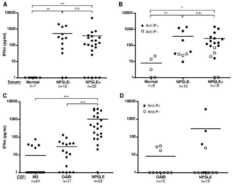
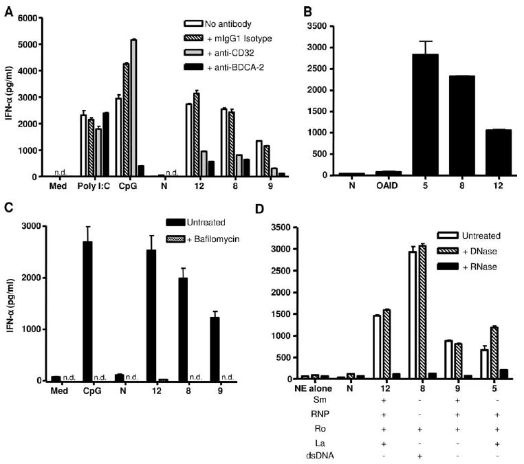
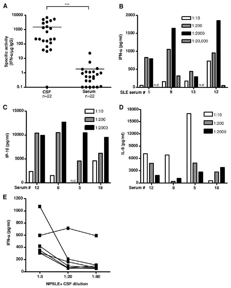
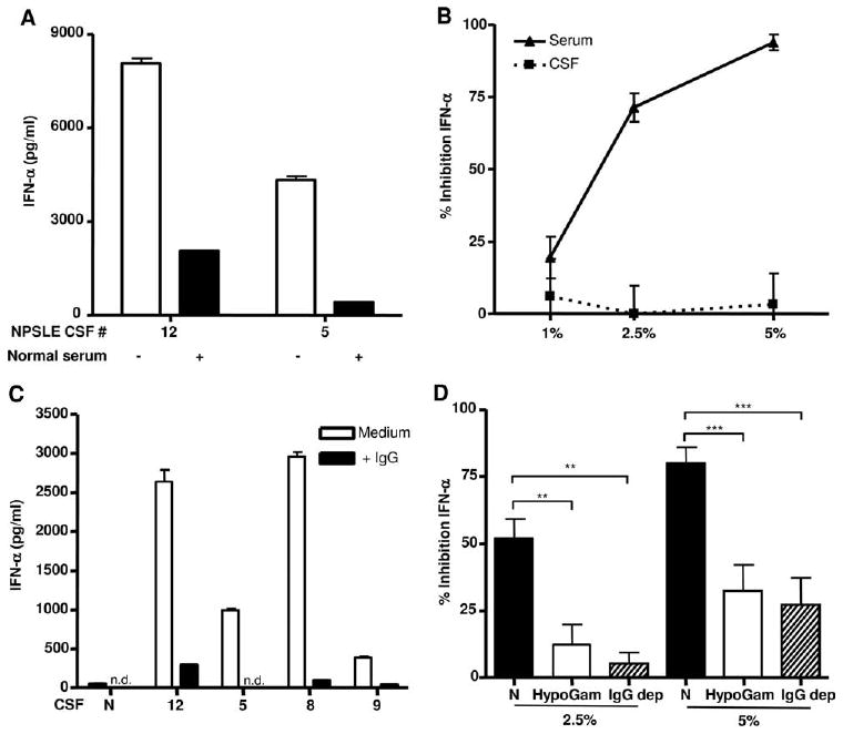
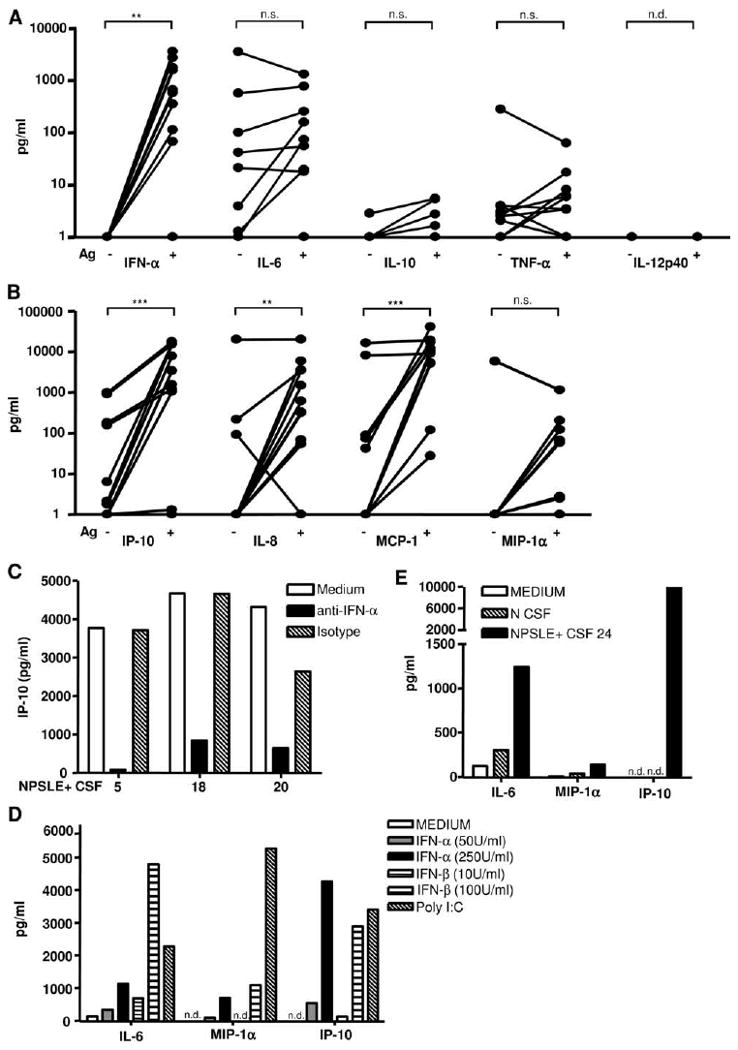
Neuropsychiatric disease in systemic lupus erythematosus (NPSLE) is a poorly understood, but potentially fatal, disease manifestation. A pathogenetic role for autoantibodies is suspected, but the mechanism is unclear. Since immune complexes in SLE can stimulate IFN-alpha and there is strong evidence in humans and in mice that IFN-alpha can cause neuropsychiatric manifestations, we asked whether NPSLE patient serum and/or cerebrospinal fluid (CSF) contain abnormally high IFN-alpha-inducing activity. In a bioassay containing plasmacytoid dendritic cells and a source of Ag, NPSLE CSF induced significantly higher IFN-alpha compared with CSF from patients with multiple sclerosis or other autoimmune disease controls. When normalized for IgG concentration, NPSLE CSF was 800-fold more potent at inducing IFN-alpha compared with paired serum due to inhibitors present in serum. Analysis of Ig-deficient patient serum, depletion of IgG from normal serum, as well as addition of purified IgG to NPSLE CSF and serum in the bioassays revealed that one inhibitor was contained within the IgG fraction itself. In addition to IFN-alpha, immune complexes formed by CSF autoantibodies produced significantly increased levels of IFN-gamma-inducible protein 10 (IP-10/CXCL), IL-8, and MCP-1, all of which have been reported to be elevated in CSF from NPSLE patients. Taken together, these findings are consistent with a two-step model of NPSLE whereby CSF autoantibodies bind to Ags released by neurocytotoxic Abs or other brain cell injury, and the resulting immune complexes stimulate IFN-alpha and proinflammatory cytokines and chemokines.


Conflict of interest statement

Disclosures: The authors have no financial conflicts of interest.

Figures

Figure 1
Figure 1
CSF from NPSLE+ patients induces high concentrations of IFN-α. A and B, Serum obtained from normal donors, NPSLE, or NPSLE+ patients was incubated at a 1/10 dilution with HeLa NE (80 μg/ml) (A) or ribosomes (50 μg/ml) (B) in the presence of type I IFN-primed PBMC. After 24 h, IFN-α concentrations in the supernatants were measured by ELISA. C, CSF (1/5 dilution) from patients with MS, OAID, or NPSLE+ were incubated with NE (80 μg/ml) and added to IFN-primed PBMC as above (standard bioassay). D, CSF from patients with OAID or NPSLE+ was incubated with affinity-purified ribosomal P (10 μg/ml) and IFN-α concentrations were tested as above. IFN-α levels below the level of detection (<10 pg/ml) were given a value of 1. Anti-P+ or anti-P− refers to whether serum contains anti-ribosomal P autoantibodies. Horizontal lines represent mean values. n.s., not significant; *, p < 0.05; **, p < 0.01; ***, p < 0.001; Mann-Whitney U test.
Figure 2
Figure 2
NPSLE+ CSF autoantibodies induce IFN-α by pDCs that is dependent on FcγRII engagement, endosomal transport, and RNA stimulation. A, Type I IFN-primed PBMC were preincubated for 1 h with 0.2 μg/ml anti-CD32, anti-BDCA-2, or mouse IgG1 before the addition of CSF (1/20) and Ag (80 μg/ml NE). Poly(I:C) (50 μg/ml) and CpG 2216 (0.5 μM) were used as controls. B, pDCs were isolated from PBMC by negative selection (>95% pure). Cells (1 × 10⁴) were primed with type I IFN and stimulated with CSF (1/20) and NE (80 μg/ml) as described in A. C, PBMC were preincubated for 1 h with 100 nM bafilomycin A1 before incubation with CSF (1/10) and NE Ag as in A. D, NE was pretreated with RNase (8 μg/ml) or DNase (500 U/ml) before addition with CSF (1/20) to PBMC. Serum anti-Sm, anti-RNP, anti-Ro, and anti-La levels were quantified as described in Materials and Methods. IFN-α was quantified as described in Fig. 1, and the results are shown as the mean ± SD of duplicates. n.d. indicates not detected; N, normal donor; numbers on the x-axis refer to NPSLE patients in Table I. The results shown are representative of two to three independent experiments.
Figure 3
Figure 3
IFN-α induction per μg IgG is >800-fold higher in CSF due to inhibitor(s) of IFN-α production in serum. A, The specific activity (concentration of IFN-α induced per microgram of IgG of test sample) for matched serum and CSF patient samples is shown. Lines represent mean values (***, p < 0.001; Mann-Whitney U test). B, NPSLE+ sera were diluted from 1/10 to 1/20,000 and incubated with NE in the presence of IFN-primed PBMC. IFN-α was quantified after 24 h as in Fig. 1. C and D, NPSLE patient sera were diluted 1/10, 1/200, and 1/2000 and added to unprimed PBMC with NE (80 μg/ml). IP-10 and IL-8 levels were measured in culture supernatants after 24 h by ELISA. Background readings from NE alone control were subtracted from values. E, Six different NPSLE+ patients' CSF was diluted as shown and added to IFN-primed PBMC with NE (80 μg/ml). IFN-α was quantified as in Fig. 1.
Figure 4
Figure 4
Normal serum and IgG are potent inhibitors of immune complex-stimulated IFN-α production. A, Equal amounts of NPSLE+ CSF and normal serum (both diluted 1/10) were incubated with NE and PBMC and IFN-α quantified as in Fig. 1. B, Normal CSF or serum (1%, 2.5%, or 5% (v/v)) was added to diluted NPSLE serum (1/2000), incubated with NE and PBMCs, and tested for IFN-α production in the bioassay after 24 h. Results are expressed as percentage inhibition of IFN-α production relative to cells that did not receive normal serum or CSF. C, Purified IgG (350 μg/ml) was added to 1/10 diluted normal or four different NPSLE+ patients' CSF, and IFN-α was quantified as above. The results in A and C are representative of two to three independent experiments, and in B, inhibition of IFN-α is expressed as the mean percentage ± SEM of three to six independent experiments. n.d. indicates not detected. D, Serum from normal individuals (N), four patients with hypogammaglobulinemia (HypoGam, associated with either X-linked agammaglobulinemia or common variable immunodeficiency), or three normal sera depleted of IgG (IgG dep) were incubated at 2.5% or 5% (v/v) with stimulatory NPSLE+ serum (1/2000), U937 freeze-thawed Ag (1% (v/v)), and primed PBMC. Results are shown as the mean percentage ± SEM of four independent experiments. **, p < 0.01; ***, p < 0.001; Mann-Whitney U test.
Figure 5
Figure 5
CSF from NPSLE patients induces other inflammatory mediators associated with NPSLE by activation of PBMC or microglia. A and B, NPSLE+ CSF (1/10) was incubated with unprimed PBMCs in the presence or absence of added nuclear Ag. Cytokines (A) and chemokines (B) were quantified after 24 h using a multiplex assay as described in Materials and Methods. IFN-α was measured by ELISA as in Fig. 1. Values below the level of detection were given a value of 1. Results with and without the addition of Ag were compared by the Wilcoxon signed-rank test (n.s., not significant; **, p < 0.01; ***, p < 0.001; n.d., not detected). C, NPSLE+ CSF (1/10) was incubated with NE and added to unprimed PBMC with or without a neutralizing mAb to IFN-α or mouse IgG1 isotype control (3 μg/ml), and IP-10 was measured as described above. D and E, Primary human microglia were plated as described in Materials and Methods and stimulated with IFN-α, IFN-β, orpoly(I:C) (50 μg/ml) (D) or normal (N) or NPSLE+ CSF (1/10) with nuclear Ag (80 μg/ml) (E). IL-6, IP-10, and MIP-1α were quantified as described above after 24 h.

References

    1. Johnson RT, Richardson EP. The neurological manifestations of systemic lupus erythematosus. Medicine. 1968;47:337–369. - PubMed
    1. Hanly JG. Neuropsychiatric lupus. Rheum Dis Clin North Am. 2005;31:273–298. - PubMed
    1. Zandman-Goddard G, Chapman J, Shoenfeld Y. Autoantibodies involved in neuropsychiatric SLE and antiphospholipid syndrome. Semin Arthritis Rheum. 2007;36:297–315. - PubMed
    1. Winfield JB, Shaw M, Silverman LM, Eisenberg RA, Wilson HA, 3rd, Koffler D. Intrathecal IgG synthesis and blood-brain barrier impairment in patients with systemic lupus erythematosus and central nervous system dysfunction. Am J Med. 1983;74:837–844. - PubMed
    1. Small P, Mass MF, Kohler PF, Harbeck RJ. Central nervous system involvement in SLE: diagnostic profile and clinical features. Arthritis Rheum. 1977;20:869–878. - PubMed

Publication types

MeSH terms