O6-methylguanine-induced cell death involves exonuclease 1 as well as DNA mismatch recognition in vivo
- PMID: 19124772
- PMCID: PMC2613938
- DOI: 10.1073/pnas.0811991106
O6-methylguanine-induced cell death involves exonuclease 1 as well as DNA mismatch recognition in vivo
Abstract
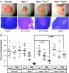
Alkylation-induced O(6)-methylguanine (O(6)MeG) DNA lesions can be mutagenic or cytotoxic if unrepaired by the O(6)MeG-DNA methyltransferase (Mgmt) protein. O(6)MeG pairs with T during DNA replication, and if the O(6)MeG:T mismatch persists, a G:C to A:T transition mutation is fixed at the next replication cycle. O(6)MeG:T mismatch detection by MutSalpha and MutLalpha leads to apoptotic cell death, but the mechanism by which this occurs has been elusive. To explore how mismatch repair mediates O(6)MeG-dependent apoptosis, we used an Mgmt-null mouse model combined with either the Msh6-null mutant (defective in mismatch recognition) or the Exo1-null mutant (impaired in the excision step of mismatch repair). Mouse embryonic fibroblasts and bone marrow cells derived from Mgmt-null mice were much more alkylation-sensitive than wild type, as expected. However, ablation of either Msh6 or Exo1 function rendered these Mgmt-null cells just as resistant to alkylation-induced cytotoxicity as wild-type cells. Rapidly proliferating tissues in Mgmt-null mice (bone marrow, thymus, and spleen) are extremely sensitive to apoptosis induced by O(6)MeG-producing agents. Here, we show that ablation of either Msh6 or Exo1 function in the Mgmt-null mouse renders these rapidly proliferating tissues alkylation-resistant. However, whereas the Msh6 defect confers total alkylation resistance, the Exo1 defect leads to a variable tissue-specific alkylation resistance phenotype. Our results indicate that Exo1 plays an important role in the induction of apoptosis by unrepaired O(6)MeGs.
Conflict of interest statement
The authors declare no conflict of interest.
Figures


Similar articles
-
Chromosomal instability, reproductive cell death and apoptosis induced by O6-methylguanine in Mex-, Mex+ and methylation-tolerant mismatch repair compromised cells: facts and models.Mutat Res. 1997 Nov 28;381(2):227-41. doi: 10.1016/s0027-5107(97)00187-5. Mutat Res. 1997. PMID: 9434879
-
Repair of O(6)-methylguanine is not affected by thymine base pairing and the presence of MMR proteins.Mutat Res. 2001 Nov 1;487(1-2):59-66. doi: 10.1016/s0921-8777(01)00105-7. Mutat Res. 2001. PMID: 11595409
-
Mutational-reporter transgenes rescued from mice lacking either Mgmt, or both Mgmt and Msh6 suggest that O6-alkylguanine-induced miscoding does not contribute to the spontaneous mutational spectrum.Oncogene. 2004 Aug 5;23(35):5931-40. doi: 10.1038/sj.onc.1207791. Oncogene. 2004. PMID: 15208683
-
MGMT: key node in the battle against genotoxicity, carcinogenicity and apoptosis induced by alkylating agents.DNA Repair (Amst). 2007 Aug 1;6(8):1079-99. doi: 10.1016/j.dnarep.2007.03.008. Epub 2007 May 7. DNA Repair (Amst). 2007. PMID: 17485253 Review.
-
BER, MGMT, and MMR in defense against alkylation-induced genotoxicity and apoptosis.Prog Nucleic Acid Res Mol Biol. 2001;68:41-54. doi: 10.1016/s0079-6603(01)68088-7. Prog Nucleic Acid Res Mol Biol. 2001. PMID: 11554312 Review.
Cited by
-
N-Methyl-N-nitrosourea Induced 3'-Glutathionylated DNA-Cleavage Products in Mammalian Cells.Anal Chem. 2022 Nov 15;94(45):15595-15603. doi: 10.1021/acs.analchem.2c02003. Epub 2022 Nov 4. Anal Chem. 2022. PMID: 36332130 Free PMC article.
-
Alkyltransferase-like protein (eATL) prevents mismatch repair-mediated toxicity induced by O6-alkylguanine adducts in Escherichia coli.Proc Natl Acad Sci U S A. 2010 Oct 19;107(42):18050-5. doi: 10.1073/pnas.1008635107. Epub 2010 Oct 4. Proc Natl Acad Sci U S A. 2010. PMID: 20921378 Free PMC article.
-
Integrated genomics of susceptibility to alkylator-induced leukemia in mice.BMC Genomics. 2010 Nov 17;11:638. doi: 10.1186/1471-2164-11-638. BMC Genomics. 2010. PMID: 21080971 Free PMC article.
-
Quaternary interactions and supercoiling modulate the cooperative DNA binding of AGT.Nucleic Acids Res. 2017 Jul 7;45(12):7226-7236. doi: 10.1093/nar/gkx223. Nucleic Acids Res. 2017. PMID: 28575445 Free PMC article.
-
Downregulation of both mismatch repair and non-homologous end-joining pathways in hypoxic brain tumour cell lines.PeerJ. 2021 Apr 30;9:e11275. doi: 10.7717/peerj.11275. eCollection 2021. PeerJ. 2021. PMID: 33986995 Free PMC article.
References
-
- Friedberg EC, Walker GC, Siede W. DNA Repair and Mutagenesis. Washington, DC: Am Soc Microbiol; 2006. p. 698.
-
- Jiricny J. The multifaceted mismatch-repair system. Nat Rev Mol Cell Biol. 2006;7:335–346. - PubMed
-
- Hickman MJ, Samson LD. Apoptotic signaling in response to a single type of DNA lesion, O6-methylguanine. Mol Cell. 2004;14:105–116. - PubMed
-
- Takagi Y, et al. Roles of MGMT and MLH1 proteins in alkylation-induced apoptosis and mutagenesis. DNA Repair. 2003;2:1135–1146. - PubMed
Publication types
MeSH terms
Substances
Grants and funding
LinkOut - more resources
Full Text Sources
Molecular Biology Databases
Research Materials
Miscellaneous

