. 2009 Jan 13;106(2):582-7.
doi: 10.1073/pnas.0805270106. Epub 2009 Jan 5.

Cryo-electron tomography of mouse hepatitis virus: Insights into the structure of the coronavirion

Affiliations

Cryo-electron tomography of mouse hepatitis virus: Insights into the structure of the coronavirion

Montserrat Bárcena et al. Proc Natl Acad Sci U S A. .

Abstract

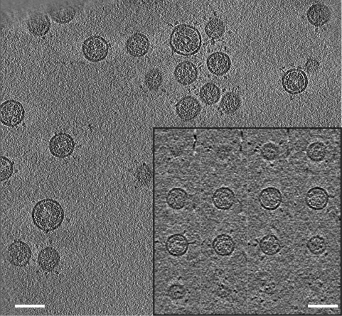
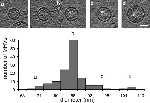
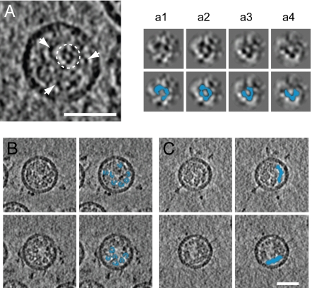
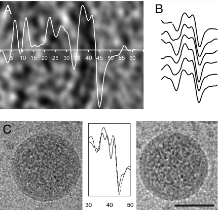
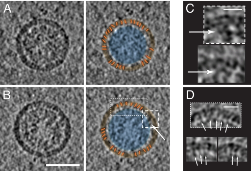
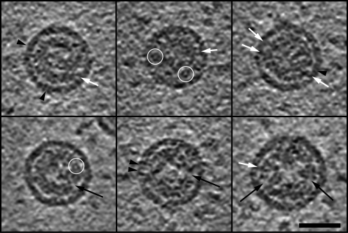
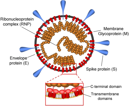
Coronaviruses are enveloped viruses containing the largest reported RNA genomes. As a result of their pleomorphic nature, our structural insight into the coronavirion is still rudimentary, and it is based mainly on 2D electron microscopy. Here we report the 3D virion structure of coronaviruses obtained by cryo-electron tomography. Our study focused primarily on the coronavirus prototype murine hepatitis virus (MHV). MHV particles have a distinctly spherical shape and a relatively homogenous size ( approximately 85 nm envelope diameter). The viral envelope exhibits an unusual thickness (7.8 +/- 0.7 nm), almost twice that of a typical biological membrane. Focal pairs revealed the existence of an extra internal layer, most likely formed by the C-terminal domains of the major envelope protein M. In the interior of the particles, coiled structures and tubular shapes are observed, consistent with a helical nucleocapsid model. Our reconstructions provide no evidence of a shelled core. Instead, the ribonucleoprotein seems to be extensively folded onto itself, assuming a compact structure that tends to closely follow the envelope at a distance of approximately 4 nm. Focal contact points and thread-like densities connecting the envelope and the ribonucleoprotein are revealed in the tomograms. Transmissible gastroenteritis coronavirion tomograms confirm all the general features and global architecture observed for MHV. We propose a general model for the structure of the coronavirion in which our own and published observations are combined.


Conflict of interest statement

The authors declare no conflict of interest.

Figures

Fig. 1.
Fig. 1.
Virtual slice (8.7 nm thick) in one of the reconstructed fields of purified MHV particles. Inset, Z-slices (5.8 nm thick) through one of the MHV cryo-tomograms. (Scale bars, 100 nm.)
Fig. 2.
Fig. 2.
Size histogram of MHV particles. Central sections (5.8 nm thick) of virions from the different regions of the histogram are shown in A–D. The arrows point at lower-density areas inside the virion. (Scale bar, 50 nm.)
Fig. 3.
Fig. 3.
(A) Slice through one of the reconstructed MHV particles (5.8 nm thick). The arrows point to some of the granular densities visible in the virion interior. These are likely to correspond to the N protein in complex with RNA. The dashed circle encloses a quasi-circular profile formed by these densities. (A). Serial sections (4 nm thick) of this region encompassing a total height of 11 nm. (B and C) Thick virtual slices (11 nm thick) of four different MHV particles displaying (B) circular profiles and (C) tubular fragments. These patterns are colored in blue. Assuming that they are favorable orientations of the RNP coil, a number of 4 to 5 granular densities per turn and a pitch of ≈10 nm were estimated. (Scale bars, 50 nm.)
Fig. 4.
Fig. 4.
MHV envelope. (A) Radial density profile of the rotationally averaged image superimposed to a central slice of the virion (5.8 nm thick). (B) Several density profiles of the envelope of reconstructed MHV particles. (C) Focal pairs of an MHV particle at 2 μm (Left) and 4 μm underfocus (Right), and radial density profiles (continuous line, 2 μm underfocus; dashed line, 4 μm underfocus). All measurements are in nanometers. (Scale bars, 50 nm.)
Fig. 5.
Fig. 5.
(A and B) Two 7.5-nm sections through two different MHV particles (Left) and highlighted features superimposed (Right). The striations in the envelope have been colored in orange. The RNP region is colored in blue and the envelope in orange to highlight the ≈4-nm low-density region in between. The arrow points to one of the focal contact points. (Scale bar, 50 nm.) (C and D) Close-ups of the focal contact points (C) and linking densities (D) between the RNP and envelope, indicated by arrows. The framed images are enlargements of the corresponding boxed areas in B. (Scale bar, 15 nm.)
Fig. 6.
Fig. 6.
Gallery of central virtual slices through some of the reconstructed TGEV particles (5.8 nm thick). TGEV virions show the same global architecture as MHV and analogous structural details (some of which are indicated). The RNP forms a complex arrangement with granular appearance in which some quasi-circular profiles (encircled in white) can be distinguished. The RNP closely follows the membrane at a distance of ≈4 nm, although infoldings (black arrows) and extensions contacting the envelope (white arrows) can be observed. Striations (black arrowheads) are visible in the TGEV virion envelope. (Scale bar, 50 nm.)
Fig. 7.
Fig. 7.
Structural model of the coronavirion based on our interpretation of the tomograms. Segments of unordered RNP alternate with coils in the interior of the virion. M monomers, dimers, trimers, and/or tetramers distribute all over the envelope and interact with each other at a local level. The C-terminal domains of the M proteins constitute an extra layer coating the interior of the lipid membrane (Inset). The focal contacts and string connections between the envelope and the RNP suggest anchor points for virion assembly and integrity.

References

    1. Peiris JS, et al. Coronavirus as a possible cause of severe acute respiratory syndrome. Lancet. 2003;361:1319–1325. - PMC - PubMed
    1. Ksiazek TG, et al. A novel coronavirus associated with severe acute respiratory syndrome. N Engl J Med. 2003;348:1953–1966. - PubMed
    1. van der Hoek L, et al. Identification of a new human coronavirus. Nat Med. 2004;10:368–373. - PMC - PubMed
    1. Woo PC, et al. Characterization and complete genome sequence of a novel coronavirus, coronavirus HKU1, from patients with pneumonia. J Virol. 2005;79:884–895. - PMC - PubMed
    1. de Haan CA, Rottier PJ. Molecular interactions in the assembly of coronavirus. Adv Virus Res. 2005;64:165–230. - PMC - PubMed

Publication types

Substances