. 2009;4(1):e4132.
doi: 10.1371/journal.pone.0004132. Epub 2009 Jan 6.

Ancestral vascular lumen formation via basal cell surfaces

Affiliations

Ancestral vascular lumen formation via basal cell surfaces

Tomás Kucera et al. PLoS One. 2009.

Abstract

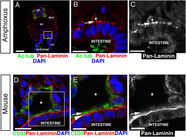
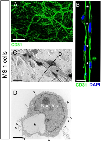
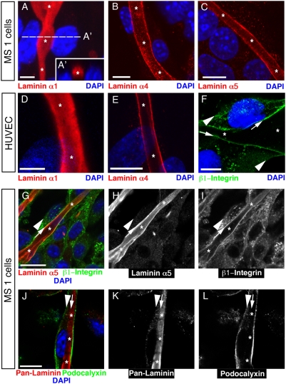
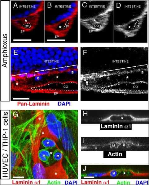
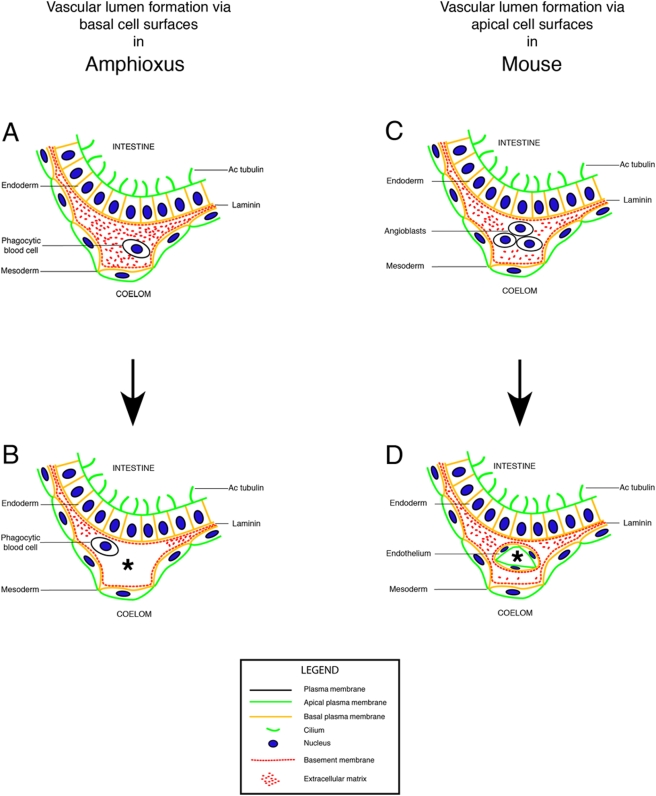
The cardiovascular system of bilaterians developed from a common ancestor. However, no endothelial cells exist in invertebrates demonstrating that primitive cardiovascular tubes do not require this vertebrate-specific cell type in order to form. This raises the question of how cardiovascular tubes form in invertebrates? Here we discovered that in the invertebrate cephalochordate amphioxus, the basement membranes of endoderm and mesoderm line the lumen of the major vessels, namely aorta and heart. During amphioxus development a laminin-containing extracellular matrix (ECM) was found to fill the space between the basal cell surfaces of endoderm and mesoderm along their anterior-posterior (A-P) axes. Blood cells appear in this ECM-filled tubular space, coincident with the development of a vascular lumen. To get insight into the underlying cellular mechanism, we induced vessels in vitro with a cell polarity similar to the vessels of amphioxus. We show that basal cell surfaces can form a vascular lumen filled with ECM, and that phagocytotic blood cells can clear this luminal ECM to generate a patent vascular lumen. Therefore, our experiments suggest a mechanism of blood vessel formation via basal cell surfaces in amphioxus and possibly in other invertebrates that do not have any endothelial cells. In addition, a comparison between amphioxus and mouse shows that endothelial cells physically separate the basement membranes from the vascular lumen, suggesting that endothelial cells create cardiovascular tubes with a cell polarity of epithelial tubes in vertebrates and mammals.


Conflict of interest statement

Competing Interests: The authors have declared that no competing interests exist.

Figures

Figure 1
Figure 1. Basal cell surfaces line the vascular lumen in amphioxus.
(A) Confocal image of a transverse section through an anterior part of juvenile amphioxus stained for laminin (red) to localize basement membranes, and acetylated tubulin (green) to localize apical cilia. Cell nuclei are stained with DAPI (blue). NT, neural tube. NC, notochord. MYO, myomere. CO, coelom. Scale bar, 20 µm. (B) Higher magnification of the boxed area outlined in (A) shows laminin (red) lining the lumen (asterisk) of the amphioxus aorta. The apical cilia (green) of the intestinal cells project into the intestinal lumen. Nuclei are labeled with DAPI (blue). Scale bar, 5 µm. (C) The red channel from (B) shows laminin staining (arrow) in the lumen (asterisk) of the amphioxus aorta. Scale bar, 5 µm. (D) Confocal image of a transverse section through an E9.0 mouse embryo shows a dorsal aorta with a lumen (asterisk). Endothelial cells stained for CD31 (green) line the vascular lumen and separate it from the laminin-containing basement membrane (red) of the intestinal epithelium. Nuclei are labeled with DAPI (blue). Scale bar, 20 µm. (E) Higher magnification of the boxed area outlined in (D) shows endothelial cells (green) lining the aortic lumen (asterisk) with their laminin-free luminal cell surface (arrow). The abluminal cell surface (arrowhead) is adjacent to the laminin-containing basement membrane of the intestinal epithelium (red). DAPI (blue). Scale bar, 10 µm. (F) The red channel from (E) shows laminin staining on the abluminal cell surface (arrowhead) of the mouse aorta. Scale bar, 10 µm.
Figure 2
Figure 2. Matrigel overlay induces an invertebrate vascular morphology in MS1 endothelial cells.
(A) Confocal image of a tubular network formed by Mile Sven 1 (MS1) cells upon Matrigel overlay. Vessels are stained for CD31 (green). Scale bar, 200 µm. (B) Confocal image at higher magnification of a vessel stained for CD31 (green) and cell nuclei (DAPI, blue). The lumen is labeled with asterisks. Scale bar, 10 µm. (C) Phase-contrast image of a hematoxylin-eosin stained vessel with a lumen (asterisks). Scale bar, 10 µm. (D) Electron micrograph showing a cross-section through a vessel with a lumen (asterisk). Microvilli (ˆ), characteristic of apical cell surfaces, are exclusively localized on the abluminal plasma membrane, whereas the luminal plasma membrane has a smooth appearance, characteristic of basal cell surfaces. Scale bar, 2 µm.
Figure 3
Figure 3. MS1 cells and HUVEC can use their basal cell surfaces to form a vascular lumen around basal ECM.
(A) Projection of a confocal z-stack through a vessel formed by MS1 cells shows that the lumen (asterisks) is filled with the Matrigel-derived laminin α1 chain (red). (A′) An optical cross-section is shown at the location indicated by a dashed line in (A). Nuclei (DAPI, blue). Scale bar, 5 µm. (B, C) Confocal images show the endothelial cell-specific (B) laminin α4 chain (red) and (C) laminin α5 chain (red) on the luminal cell surface of MS1 cells. Nuclei (DAPI, blue). Scale bars, 10 µm. (D–F) Confocal images of vascular tubes with a lumen (asterisk) formed by HUVEC 48 hrs after Matrigel overlay. (D) The lumen stains for Matrigel-derived laminin α1 chain (red) and (E) endothelial cell-derived laminin α4 chain (red). Nuclei are labeled with DAPI (blue). Scale bars, 5 µm in (D) and 10 µm in (E). (F) A HUVEC tube stained for β1-integrin (green), which is present on the luminal (arrows), rather than on the abluminal plasma membrane (arrowheads). Nuclei are labeled with DAPI (blue). Scale bar, 10 µm. (G) Confocal image of a tube formed by MS1 cells showing laminin α5 chain (red) and its β1-integrin receptor (green) on the luminal cell surface (arrow), but not on the abluminal cell surface (arrowhead). Nuclei (DAPI, blue). Scale bar, 20 µm. (H) The red channel from (G) shows laminin α5 chain on the luminal cell surface (arrow). (I) The green channel from (G) shows that β1-integrin is localized on the luminal cell surface (arrow). (J) Confocal image of a tube formed by MS1 cells shows that the apical marker podocalyxin (green) is localized on the abluminal cell surface (arrowhead), whereas the basement membrane protein laminin (red) is located inside the vascular lumen (asterisks). Nuclei (DAPI, blue). Scale bar, 10 µm. (K) The red channel from (J) shows laminin inside the lumen (asterisks). (L) The green channel from (J) shows localization of podocalyxin on the abluminal cell surface (arrowhead).
Figure 4
Figure 4. Phagocytotic blood cells generate a patent vascular lumen.
(A–F) Confocal images of 14-day old amphioxus larvae stained for laminin (red) and nuclei (DAPI, blue). Asterisks label the lumen of the subintestinal vessel. CO, coelom (dashed line); EP, epidermis. Scale bars, 10 µm. (A–D) Transverse sections. (A) An area of the vascular lumen (asterisk) that is filled with laminin (red). (B) An area of patent vascular lumen (asterisk) that is only lined by laminin. (C) Red channel from (A) showing an area filled with laminin (asterisk). (D) Red channel from (B) showing an area of a patent vascular lumen (asterisk). (E–F) Longitudinal sections. (E) Less laminin (red) is found in the lumen of the subintestinal vessel that contains blood cells (B) (dashed bracket), compared to the lumen that is without any blood cells (small bracket). (F) Red channel from (E) showing less laminin on the right side of the vessel where blood cells are found. (G) Confocal image of a vessel formed by HUVEC cells in the presence of a macrophage/monocyte cell line THP-1 (M). Macrophages (M) are found in a part of the vascular lumen that is without any laminin filling (red). Scale bar 5 µm. (H–J) A cross-section through a vessel shows a macrophage (M) inside a patent vascular lumen (asterisk) that is lined by, but not filled with laminin. (H) Laminin α1 chain is absent from the lumen (asterisk) that contains (I, J) a macrophage (M). (J) The composite image shows the localization of laminin α1 chain (red), actin (green) and nuclei (blue). Scale bar, 10 µm.
Figure 5
Figure 5. Formation of a basal vascular lumen in invertebrates and creation of an apical vascular lumen in vertebrates.
Hypothesis: In invertebrates, basal cell surfaces and basement membrane line the vascular lumen. In vertebrates, by contrast, evolution of endothelial cells led to the formation of blood vessels with apical cell surfaces lining the vascular lumen. This physically separated the basement membranes from blood. (A, B) The first vascular lumen (asterisk) in the invertebrate chordate amphioxus (Branchiostoma lanceolatum) forms between the basal cell surfaces of endoderm (e.g. intestinal epithelium) and mesoderm (e.g. coelomic mesothelium). (A) The blood vessel is filled with basally deposited ECM that contains laminin. (B) Phagocytotic blood cells remove this ECM to create a patent vascular lumen (asterisk) between the basal cell surfaces. (C, D) The first vascular lumen (asterisk) inside the mouse (Mus musculus) also forms between the basal cell surfaces of endoderm (e.g. intestinal epithelium) and mesoderm (e.g. coelomic mesothelium). (C) However, endothelial cells, or angioblasts, develop from the mesoderm and populate the ECM between the basal cell surfaces. (D) Finally, endothelial cells generate a blood vessel and separate the vascular lumen (asterisk) from the surrounding basement membranes.

References

    1. Bishopric NH. Evolution of the heart from bacteria to man. Ann N Y Acad Sci. 2005;1047:13–29. - PubMed
    1. Evans CJ, Hartenstein V, Banerjee U. Thicker than blood: conserved mechanisms in Drosophila and vertebrate hematopoiesis. Dev Cell. 2003;5:673–690. - PubMed
    1. Hartenstein V, Mandal L. The blood/vascular system in a phylogenetic perspective. Bioessays. 2006;28:1203–1210. - PubMed
    1. Mandal L, Banerjee U, Hartenstein V. Evidence for a fruit fly hemangioblast and similarities between lymph-gland hematopoiesis in fruit fly and mammal aorta-gonadal-mesonephros mesoderm. Nat Genet. 2004;36:1019–1023. - PubMed
    1. Olson EN. Gene regulatory networks in the evolution and development of the heart. Science. 2006;313:1922–1927. - PMC - PubMed

MeSH terms