. 2009;4(1):e4135.
doi: 10.1371/journal.pone.0004135. Epub 2009 Jan 6.

Protein palmitoylation regulates osteoblast differentiation through BMP-induced osterix expression

Affiliations

Protein palmitoylation regulates osteoblast differentiation through BMP-induced osterix expression

Wai Fook Leong et al. PLoS One. 2009.

Abstract

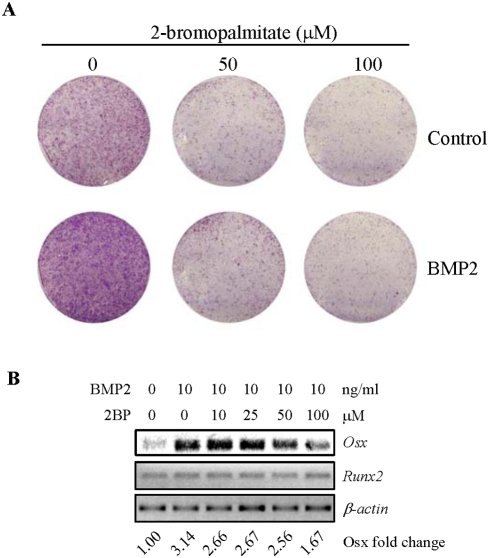
Osteoporosis is one of the most common diseases and can be treated by either anti-resorption drugs, anabolic drugs, or both. To search for anabolic drug targets for osteoporosis therapy, it is crucial to understand the biology of bone forming cells, osteoblasts, in terms of their proliferation, differentiation, and function. Here we found that protein palmitoylation participates in signaling pathways that control osterix expression and osteoblast differentiation. Mouse calvarial osteoblasts express most of the 24 palmitoyl transferases, with some being up-regulated during differentiation. Inhibition of protein palmitoylation, with a substrate-analog inhibitor, diminished osteoblast differentiation and mineralization, but not proliferation or survival. The decrease in differentiation capacity is associated with a reduction in osterix, but not Runx2 or Atf4. Inhibition of palmitoyl transferases had little effect in p53(-/-) osteoblasts that show accelerated differentiation due to overexpression of osterix, suggesting that osterix, at least partially, mediated the effect of inhibition of palmitoyl transferases on osteoblast differentiation. BMPs are the major driving force of osteoblast differentiation in the differentiation assays. We found that inhibition of palmitoyl transferases also compromised BMP2-induced osteoblast differentiation through down-regulating osterix induction. However, palmitoyl transferases inhibitor did not inhibit Smad1/5/8 activation. Instead, it compromised the activation of p38 MAPK, which are known positive regulators of osterix expression and differentiation. These results indicate that protein palmitoylation plays an important role in BMP-induced MAPK activation, osterix expression, and osteoblast differentiation.


Conflict of interest statement

Competing Interests: The authors have declared that no competing interests exist.

Figures

Figure 1
Figure 1. Expression of PATs and protein palmitoylation in osteoblasts.
A. RT-PCR analysis of the mRNA levels of the 24 known PATs during osteoblast differentiation. Primary osteoblasts were cultured in the differentiation medium for different periods of time. Total RNA was isolated from these cultures and was then used to carry out RT-PCR. These PATs were classified into unaltered expression (A), increased expression (B), and decreased expression (C), in comparison to the day 1 cultures. D. 2BP was able to inhibit protein palmitoylation in primary osteoblasts. Primary osteoblasts were treated with 100 µM of 2BP for 2 hrs and then 3H-palmitic acid was included in the culture medium for 24 hrs. The cells were harvested and the same amounts of total proteins were loaded for control and 2BP treated samples. The SDS-PAGE gel was dried and exposed to x-ray films. The palmitoylated proteins are radio-labeled. Actin was used as a loading control.
Figure 2
Figure 2. Inhibition of protein palmitoylation impeded osteoblast differentiation.
A. The effect of 2BP on ALP staining. Primary osteoblasts were treated with increasing amounts of 2BP for 4 days in differentiation medium and then stained for ALP. B. Quantitation of ALP activities that were normalized to the protein levels of the cells. C. The effect of 2BP on the expression of several osteoblast differentiation markers. The experiments were carried out like Fig. 2A and total RNA were isolated from these cells. RT-PCR was carried out to determine the mRNA levels of these markers. The value of control (lane 1) was set at 1.00. D. The effect of 2BP on bone nodule formation. The experiments were carried out like Fig. 2A. After 21 days in culture, the plates were stained using a Von Kossa method.
Figure 3
Figure 3. PAT inhibitor show minimal effect on proliferation/survival of osteoblast cultures.
A. Primary osteoblast cells were cultured in differentiation medium with different amounts of 2BP for 4 days and the cell numbers in each culture plates were determined by trypan blue exclusion methods. B. The experiments were done like Fig. 3A and the cells were collected and lysed in the same volume of lysis buffer and the protein concentrations were determined by the BioRad method. C. Recovery of 2BP treated cells in ALP expression. Primary osteoblasts were treated with different concentrations of 2BP for three days, washed off, and further cultured in differentiation medium for 4 more days before being stained for ALP (bottom panel), in comparison to the cells that were treated with 2BP all the time (upper panel).
Figure 4
Figure 4. Regulation of Osx by 2BP.
Primary osteoblasts were cultured in differentiation medium with or without 100 µM of 2BP for different periods of time. These cells were then collected to isolate total RNA. RT-PCR was carried out to determine the mRNA levels of these proteins. The value of control (lane 1) was set at 1.00.
Figure 5
Figure 5. Overexpression of Osx in p53−/− osteoblasts rendered resistance to the inhibitory effect of 2BP on differentiation.
A. 2BP treatment did not significantly down-regulate the mRNA levels of Osx. Primary osteoblasts were cultured in the presence or absence of 2BP for different periods of time and total RNA was isolated and RT-PCR was carried out to determine the mRNA levels of Osx. The value of control (lane 1) was set at 1.00. B. PAT inhibitor did not markedly affect ALP staining in p53−/− osteoblasts. p53−/− osteoblasts were treated with different amounts of 2BP for 7 days and then stained for ALP. C. PAT inhibitor did not markedly affect bone nodule formation in p53−/− osteoblasts. p53−/− osteoblasts were treated with different amounts of 2BP for 21 days and then stained for mineralization.
Figure 6
Figure 6. PAT inhibitor decreased BMP2-induced osteoblast differentiation, which was likely mediated by Osx.
A. 2BP diminished the ALP expression induced by BMP2. Primary osteoblasts were cultured in the presence or absence of 2BP for 1 day, and then were further cultured in the presence of 50 ng/ml of BMP2 for 4 days. The plates were then stained for ALP. B. 2BP down-regulated BMP2-induced Osx expression. Primary osteoblasts were cultured in the absence or presence of different concentrations of 2BP for a day, and then further cultured in the presence of 50 ng/ml of BMP2 for 16 hrs. The cells were collected and total RNA was isolated, which was used to perform RT-PCR to determine the mRNA levels of Osx and Runx2. The value of control (lane 1) was set at 1.00.
Figure 7
Figure 7. Inhibition of protein palmitoylation compromised p38 MAPK.
A. 2BP showed an inhibitory effect on p38 MAPK activation in differentiation assays, without a significant effect on Erk1/2 activation or Smad1/5/8 activation. Primary osteoblasts were cultured in differentiation medium with or without 2BP for different periods of time. Cells were collected for Western blot analysis of the activation of p38, Erk1/2, and Smad1/5/8. B. 2BP also compromised p38 MAPK activation in response to BMP2. Primary osteoblasts were pretreated with PAT inhibitor overnight, and then stimulated with 100 ng/ml of BMP2. Cells were collected at different time points for analysis of p38 and Erk1/2 activation.

References

    1. Krane SM. Identifying genes that regulate bone remodeling as potential therapeutic targets. J Exp Med. 2005;201:841–843. - PMC - PubMed
    1. Manolagas SC, Jilka RL. Bone marrow, cytokines, and bone remodeling. Emerging insights into the pathophysiology of osteoporosis. N Engl J Med. 1995;332:305–311. - PubMed
    1. Martin TJ, Sims NA. Osteoclast-derived activity in the coupling of bone formation to resorption. Trends Mol Med. 2005;11:76–81. - PubMed
    1. Canalis E, Economides AN, Gazzerro E. Bone morphogenetic proteins, their antagonists, and the skeleton. Endocr Rev. 2003;24:218–235. - PubMed
    1. Zaidi M, Blair HC, Moonga BS, Abe E, Huang CL. Osteoclastogenesis, bone resorption, and osteoclast-based therapeutics. J Bone Miner Res. 2003;18:599–609. - PubMed

Publication types

MeSH terms