. 2009 May 15;87(7):1610-9.
doi: 10.1002/jnr.21980.

Expression of estrogen receptor GPR30 in the rat spinal cord and in autonomic and sensory ganglia

Affiliations

Expression of estrogen receptor GPR30 in the rat spinal cord and in autonomic and sensory ganglia

Siok L Dun et al. J Neurosci Res. .

Abstract

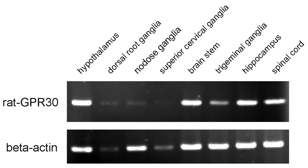
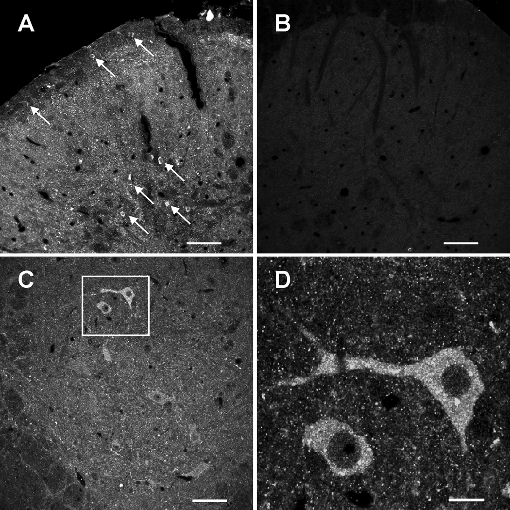
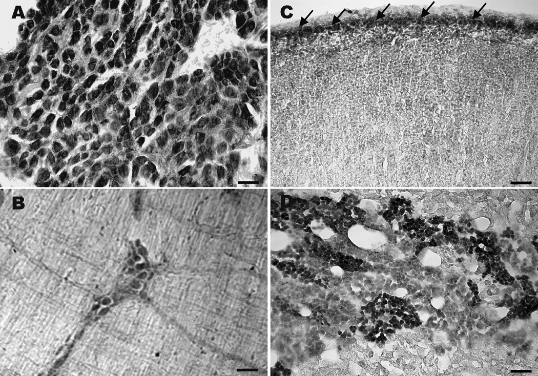
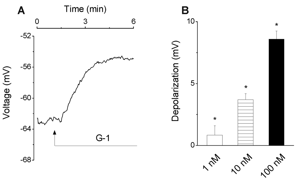
The G protein-coupled receptor GPR30 has recently been identified as a nonnuclear estrogen receptor. Reverse transcriptase-polymerase chain reaction revealed expression of GPR30 mRNA in varying quantities in the rat spinal cord, dorsal root ganglia, nodose ganglia, trigeminal ganglia, hippocampus, brain stem, and hypothalamus. Immunohistochemical studies that used a rabbit polyclonal antiserum against the human GPR30 C-terminus revealed a fine network of GPR30-immunoreactive (irGPR30) cell processes in the superficial layers of the spinal cord; some of which extended into deeper laminae. A population of neurons in the dorsal horn and ventral horn were irGPR30. Dorsal root, nodose, and trigeminal ganglionic neurons displayed varying intensities of irGPR30. Positively labeled neurons were detected in the major pelvic ganglion, but not in the superior cervical ganglion. A population of chromaffin cells in the adrenal medulla was irGPR30, so were cells of the zona glomerulosa. Double-labeling the adrenal medulla with GPR30 antiserum and tyrosine hydroxylase antibody or phenylethanolamine-N-methyltransferase antiserum revealed that irGPR30 is expressed in the majority of tyrosine hydroxylase-positive chromaffin cells. Last, some of the myenteric ganglion cells were irGPR30. Tissues processed with preimmune serum resulted in no staining. Voltage-sensitive dye imaging studies showed that the selective GPR30 agonist G-1 (1, 10, and 100 nM) depolarized cultured spinal neurons in a concentration-dependent manner. Collectively, our result provides the first evidence that GPR30 is expressed in neurons of the dorsal and ventral horn as well as in sensory and autonomic neurons, and activation of GPR30 by the selective agonist G-1 depolarizes cultured spinal neurons.


Figures

Fig. 1
Fig. 1
RT-PCR results of GPR30 mRNA in various tissues. The total RNA was extracted by Trizol reagent. 3 µg of total RNA was used for RT-PCR as described under "Experimental Procedures." Housekeeping gene β-actin was used as an internal control. The product size of rat GPR30 was 987 bp and that of β-actin was 876 bp. The figure is the representative of three experiments with similar results.
Fig. 2
Fig. 2
Fluorescence images of rat spinal cord sections labeled with GPR30 antiserum or pre immune serum. A, lumbar spinal section where irGPR30 is detected in terminals and neurons of small diameter (arrows) in the dorsal horn. B, lumbar spinal cord section processed with pre immune serum, where immunoreactivity is not detected. C, few intensely and many moderately labeled ventral horn cells are noted. D, higher magnification of the boxed ventral horn area shown in C; irGPR30 is detected in the cytoplasm. Scale bar: A– C, 50 µm; D, 10 µm.
Fig. 3
Fig. 3
Images of rat trigeminal, nodose, dorsal root and superior cervical ganglion sections labeled with GPR30 antiserum. A, section of trigeminal ganglion where numerous ganglion cells are irGPR30. B, section of nodose ganglion where ganglion cells of varying intensities are detected. C, numerous irGPR30 cells of large and small diameter are noted in this lumbar dorsal root ganglion section. D, section of superior cervical ganglion where little or no irGPR30 is detected. Scale bar: A and D, 25 µm; B and C, 50 µm.
Fig. 4
Fig. 4
Sections of rat major pelvic ganglion, myenteric plexus and adrenal gland labeled with GPR30 antiserum. A, numerous irGPR30 cells are noted in the major pelvic ganglion. B, several irGPR30 cells in a myenteric ganglion; moderately labeled cell processes extend from some of these cells. C, section of adrenal cortex where cells of the zona glomerulosa (arrows) located underneath the adrenal capsule, are labeled. D, section of adrenal medulla where groups of irGPR30 chromaffin cells intermix with non labeled or lightly labeled chromaffin cells. Scale bar: A, B and D, 50 µm; C, 100 µm.
Fig. 5
Fig. 5
Confocal images of rat adrenal medulla sections double-labeled with GPR30 antiserum and tyrosine hydroxylase (TH) or phenylethanolamine-N-methyltransferase (PNMT) antiserum and major pelvic ganglion and spinal cord sections labeled with GPR30 and choline acetyltransferase (ChAT) antisera. A and B, section double-labeled withGPR30 and TH antiserum; some irGPR30 chromaffin cells (green fluorescence) are also TH-positive (red fluorescence). C, a merged image of A and B, where double-labeled cells appear orange/yellow. D and E, section double-labeled with GPR30 (green fluorescence) and PNMT antiserum (red fluorescence). F, a merged image of D and E where there is a clear distinction between irGPR30 and irPNMT cells. G and H, pelvic ganglion section double-labeled with ChAT (green fluorescence) and GPR30 (red fluorescence) antiserum. irGPR30 was present in some cholinergic cells (solid arrows); while other irGPR30 cells (open arrows) do not express ChAT, but are surrounded by ChAT immunoreactive fibers. I, a merged image of G and H; several double-labeled cells (solid arrows) are irGPR30 and ChAT positive, and several irGPR30 cells are ChAT negative (open arrows). J and K, spinal cord sections double-labeled with ChAT (green fluorescence) and GPR30 (red fluorescence). L, merged image of J and K; few double-labeled cells are irGPR30 and ChAT positive. Scale bar: A–F, 20 µm, G–L, 50 µm
Fig. 6
Fig. 6
Depolarization of cultured spinal neurons by G-1. A, administration of G-1 (100 nM) produced a depolarization with the amplitude of 8 mV in this neuron. B, G-1-induced depolarizations were concentration-dependent; G-1 (1 nM, 10 nM, 100 nM) depolarized cultured spinal neurons by 0.84 ± 0.76 mV (n=14), 3.7 ± 0.48 mV (n=19), and 8.6 ± 0.64 mV (n=23). One asterisk indicates statistically significant difference as compared to control (P<0.05).

References

    1. Abraham I, Han HJ, Todman MG, Korach KS, Herbison AE. Estrogen receptor-β mediates rapid estrogen actions on gonadotropin-releasing hormone neurons in vitro. J Neurosci. 2003;23:5771–5777. - PMC - PubMed
    1. Ahmed Y, Lin DL, Ferguson C, Esparza N, Damaser MS. Effect of estrogen on urethral function and nerve regeneration following pudendal nerve crush in the female rat. J Urol. 2006;175:1948–1952. - PubMed
    1. Baquedano MS, Berensztein E, Saraco N, Dorn GV, de Davila MT, Rivarola MA, Belgorosky A. Expression of the IGF system in human adrenal tissues from early infancy to late puberty: implications for the development of adrenarche. Pediatr Res. 2005;58:451–458. - PubMed
    1. Baquedano MS, Saraco N, Berensztein E, Pepe C, Bianchini M, Levy E, Goñi J, Rivarola MA, Belgorosky A. Identification and developmental changes of aromatase and estrogen receptor expression in prepubertal and pubertal human adrenal tissues. J Clin Endocrinol Metab. 2007;92:2215–2222. - PubMed
    1. Bechi N, Ietta F, Romagnoli R, Focardi S, Corsi I, Buffi C, Paulesu L. Estrogen-like response to p-Nonyphenol in human first trimester placenta and BeWo Choriocarcinoma cells. Toxicol Sci. 2006;93:75–81. - PubMed

Publication types

MeSH terms

Substances