

doi: 10.1002/ajmg.a.32596.
Elements of morphology: standard terminology for the hands and feet
Affiliations
- PMID: 19125433
- PMCID: PMC3224990
- DOI: 10.1002/ajmg.a.32596
Item in Clipboard
Elements of morphology: standard terminology for the hands and feet
Am J Med Genet A.
2009 Jan.
Abstract
An international group of clinicians working in the field of dysmorphology has initiated the standardization of terms used to describe human morphology. The goals are to standardize these terms and reach consensus regarding their definitions. In this way, we will increase the utility of descriptions of the human phenotype and facilitate reliable comparisons of findings among patients. Discussions with other workers in dysmorphology and related fields, such as developmental biology and molecular genetics, will become more precise. Here we introduce the anatomy of the hands and feet and define and illustrate the terms that describe the major characteristics of the hands and feet.
Figures
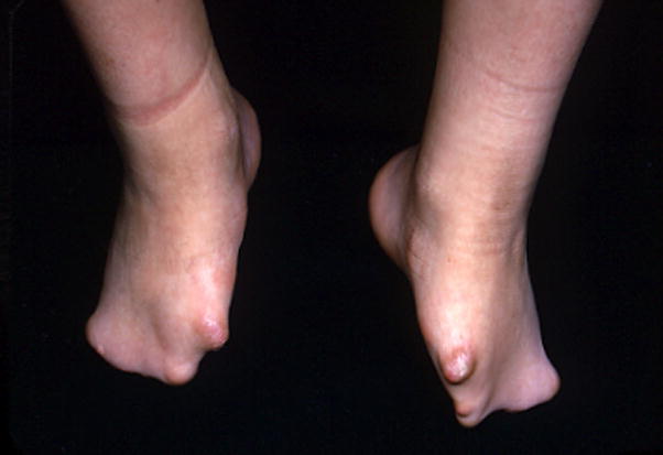
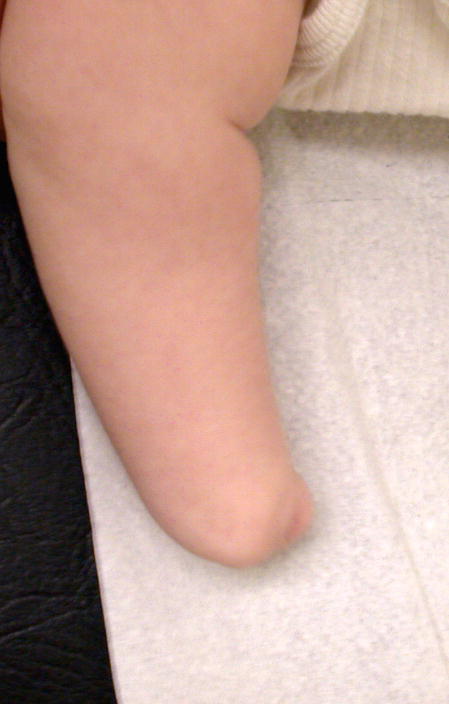
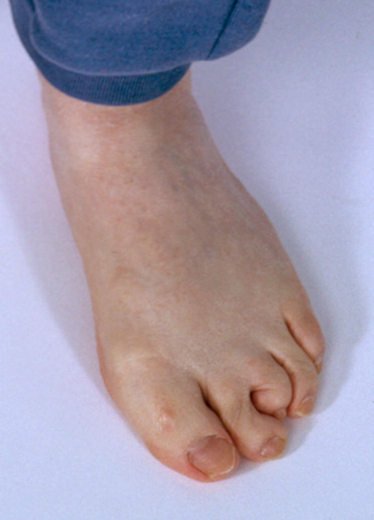
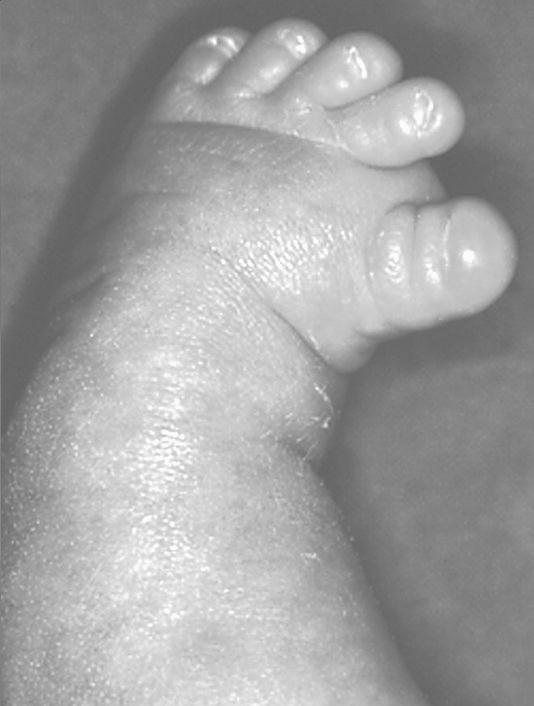
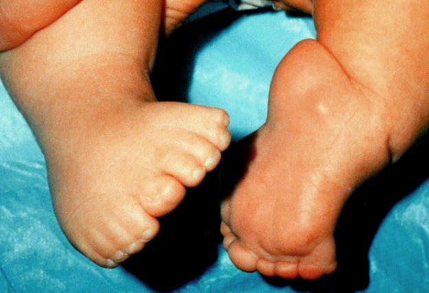
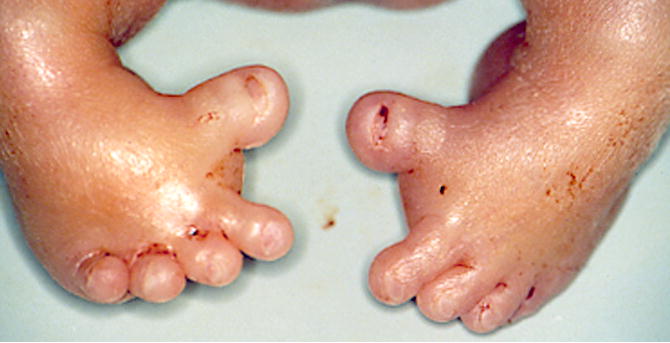
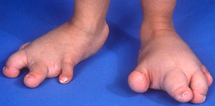
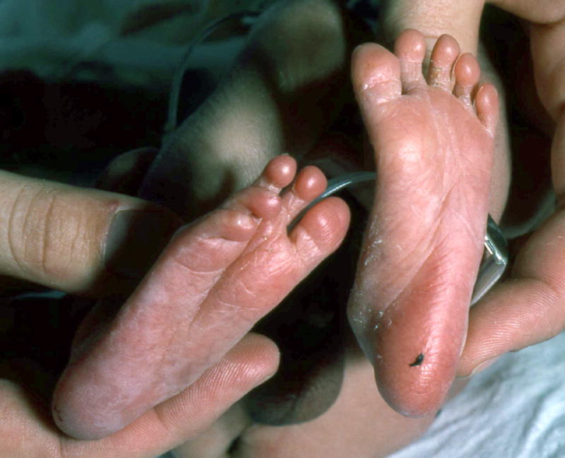
Adactyly of the feet, bilateral.
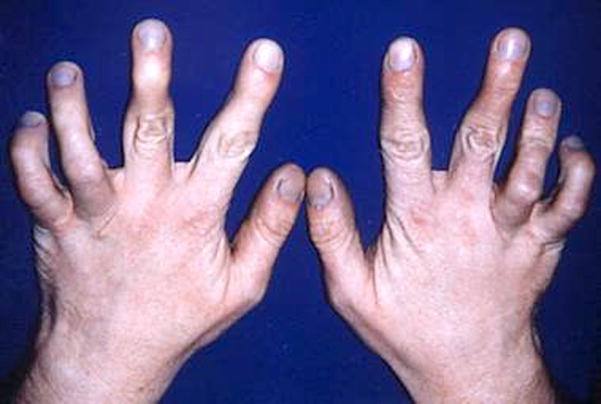
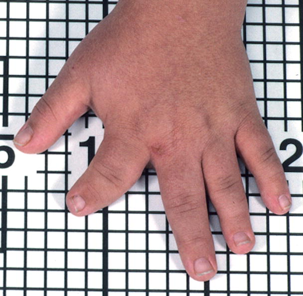
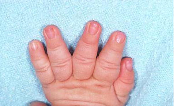
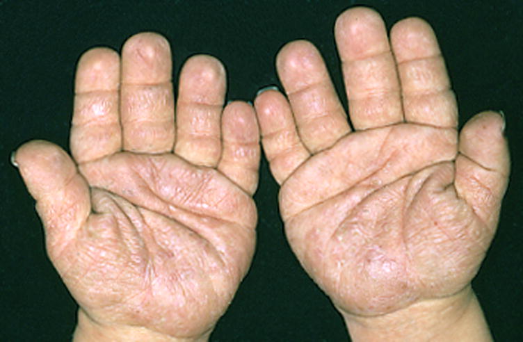
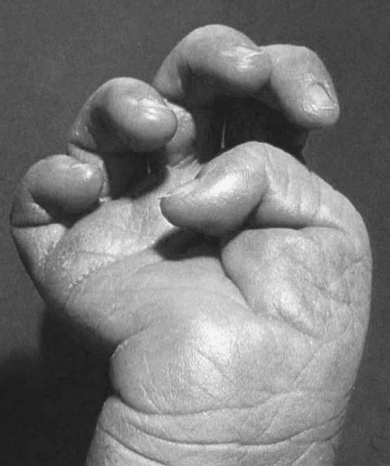
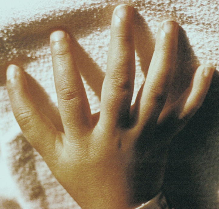
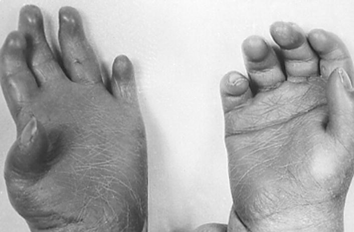
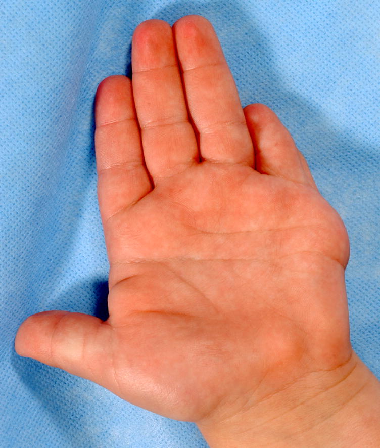
Camptodactyly of F45, bilateral.
This person also has Short finger, F5, bilateral, subjective. See also Figs. 13, 20.
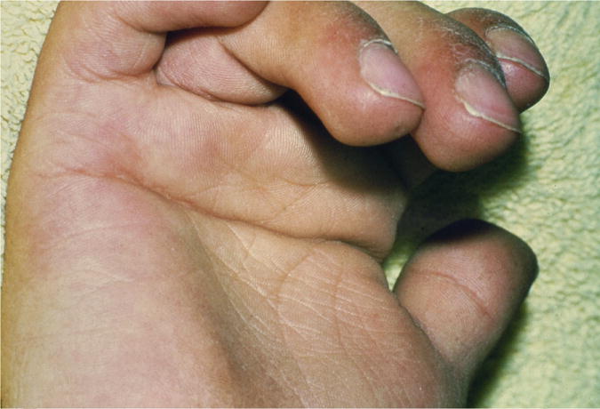
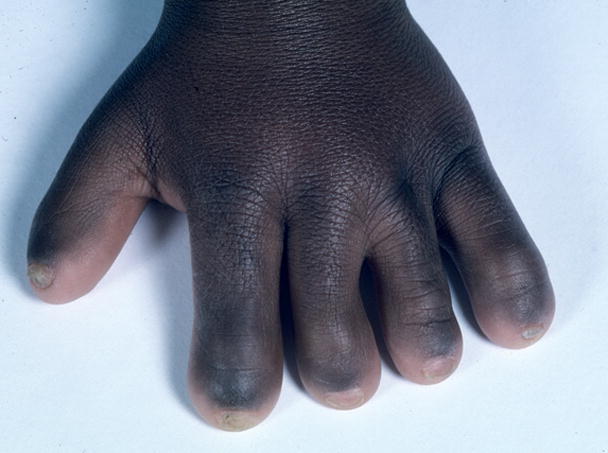
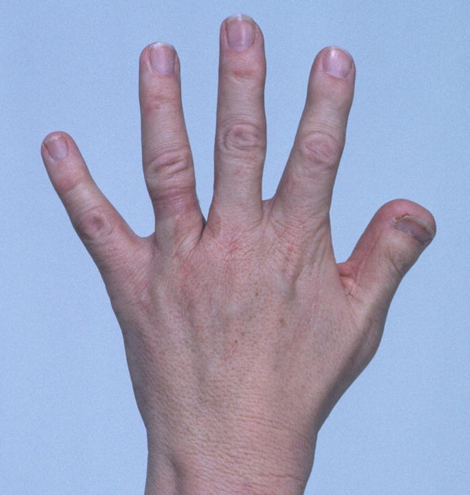
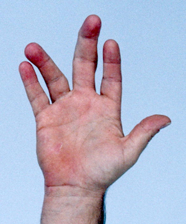
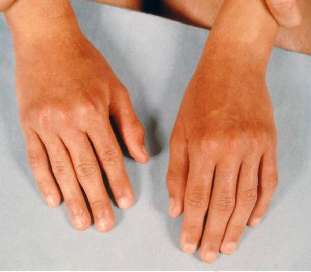
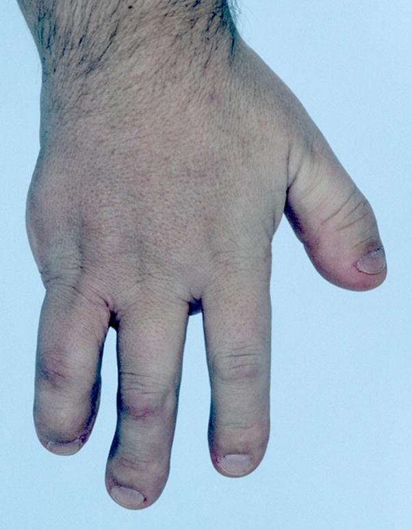
Note that the clubbing is best viewed laterally (e.g., the thumb) whereas it is difficult to appreciate in the other digits viewed dorsally. See also Figs. 5 and 63.
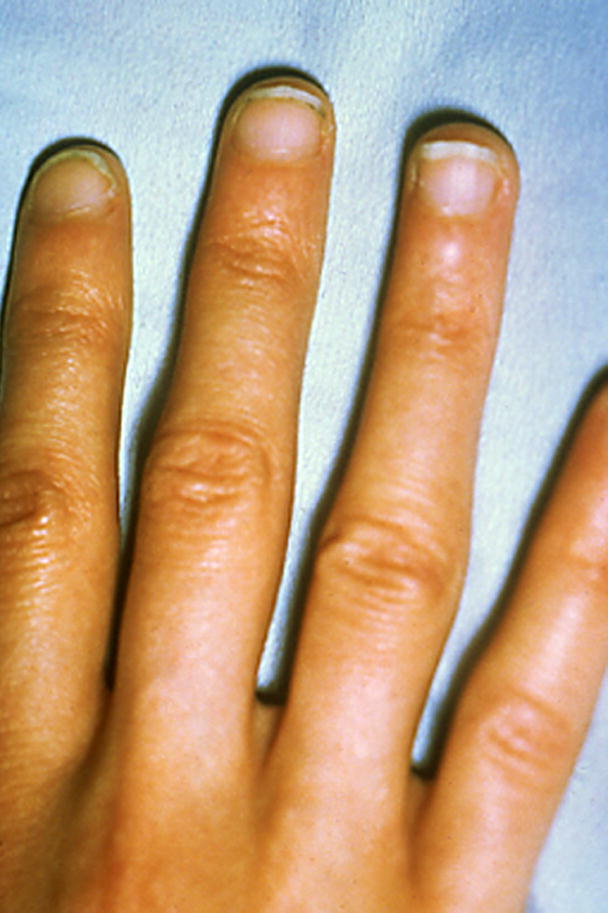
Note that this patient also has Clubbing and a Single transverse palmar crease.
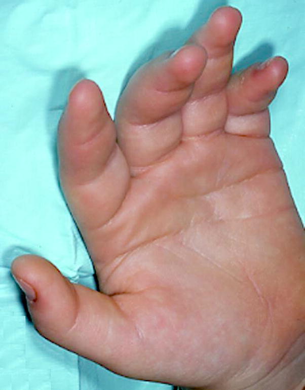
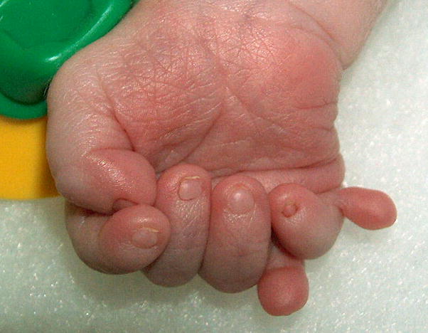
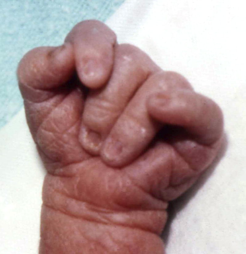
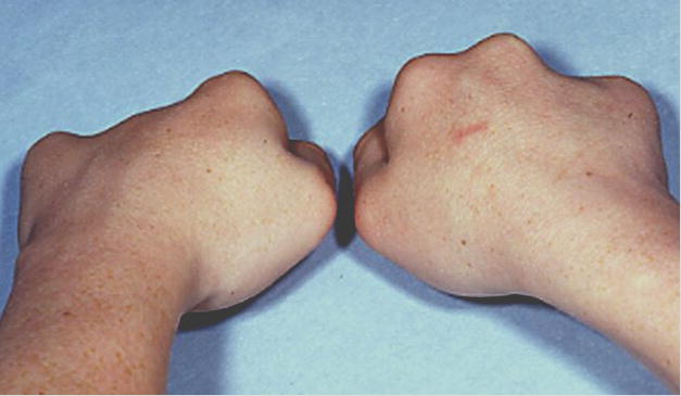
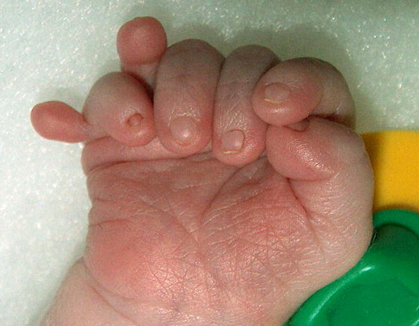
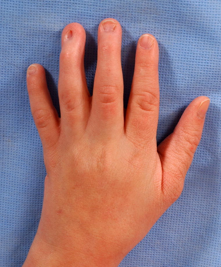
Digital constriction rings, left hand, F1-5, near MCPJ.
Note here that the identity of the missing digit is not specified, as there are no clinical data to allow this to be determined. See also Fig. 68.
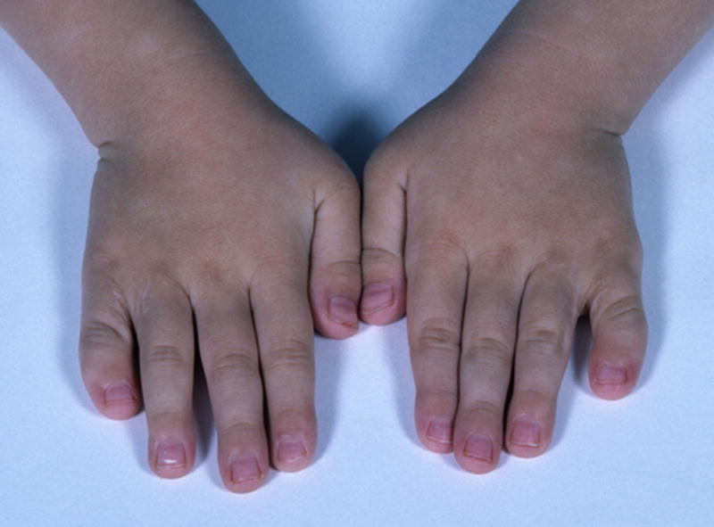
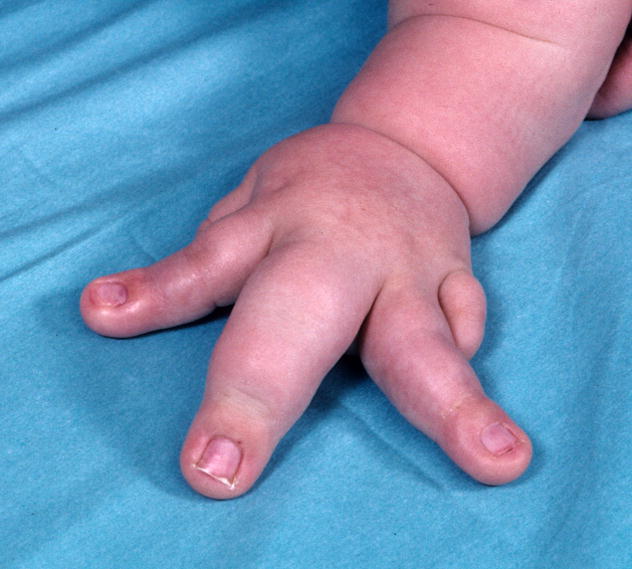
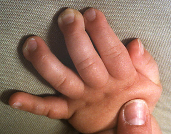
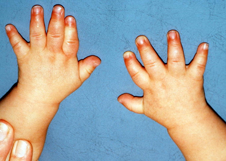
Appreciate that the dorsal-ventral dimension of these digits is not increased, whereas the lateral (proximo-distal) dimension is increased. See also Fig. 99.
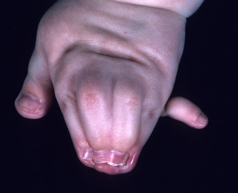
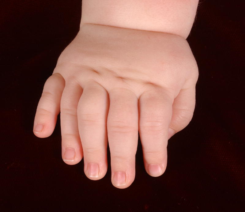
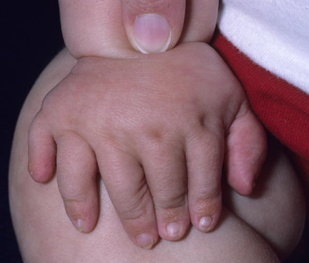
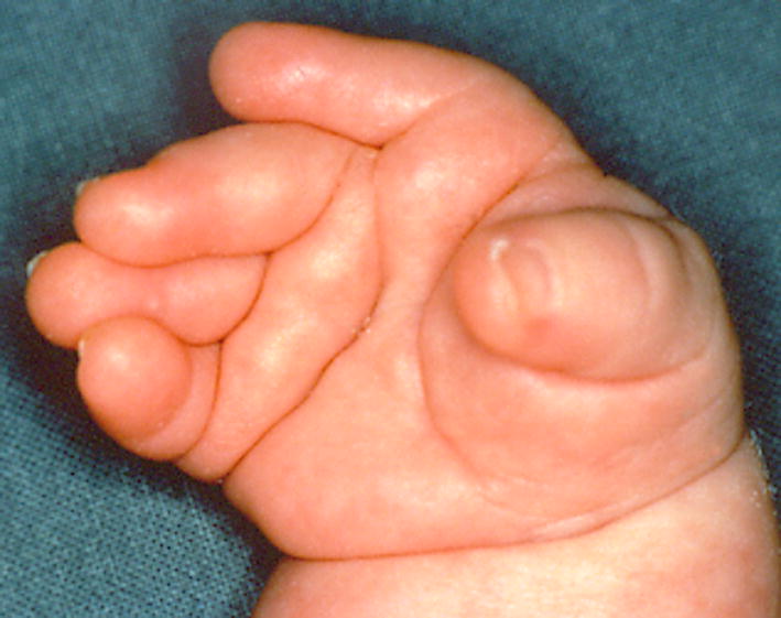
A. Cutaneous syndactyly of F2-5, left hand, complete, objective. Note that this patient also has a Broad thumb, and Postaxial polydactyly of the hand, type B and Fused nails F2- 5. B. This patient has Cutaneous syndactyly of F2-4, partial, subjective. See also Fig. 14.
A. Cutaneous syndactyly of F2-5, left hand, complete, objective. Note that this patient also has a Broad thumb, and Postaxial polydactyly of the hand, type B and Fused nails F2- 5. B. This patient has Cutaneous syndactyly of F2-4, partial, subjective. See also Fig. 14.
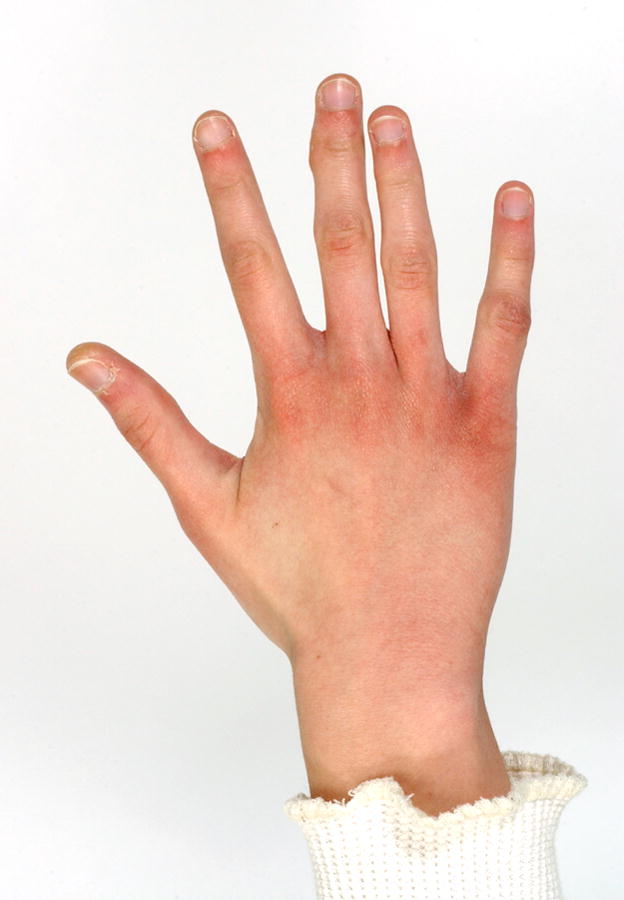
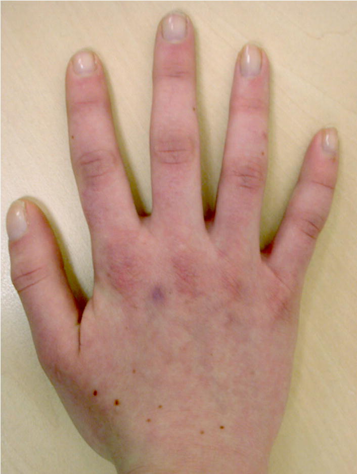
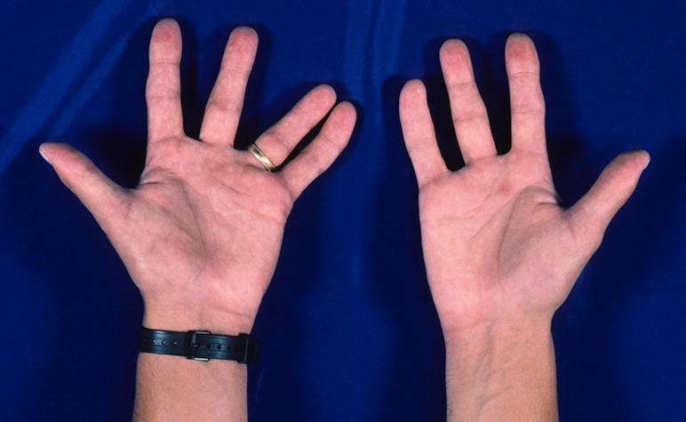
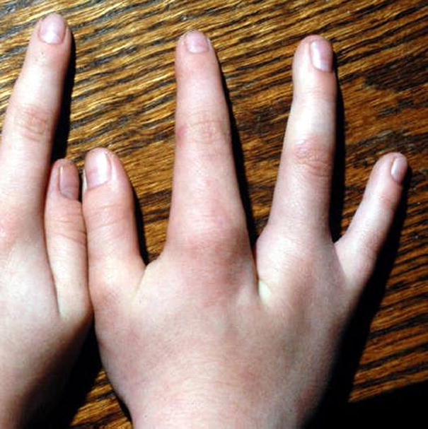
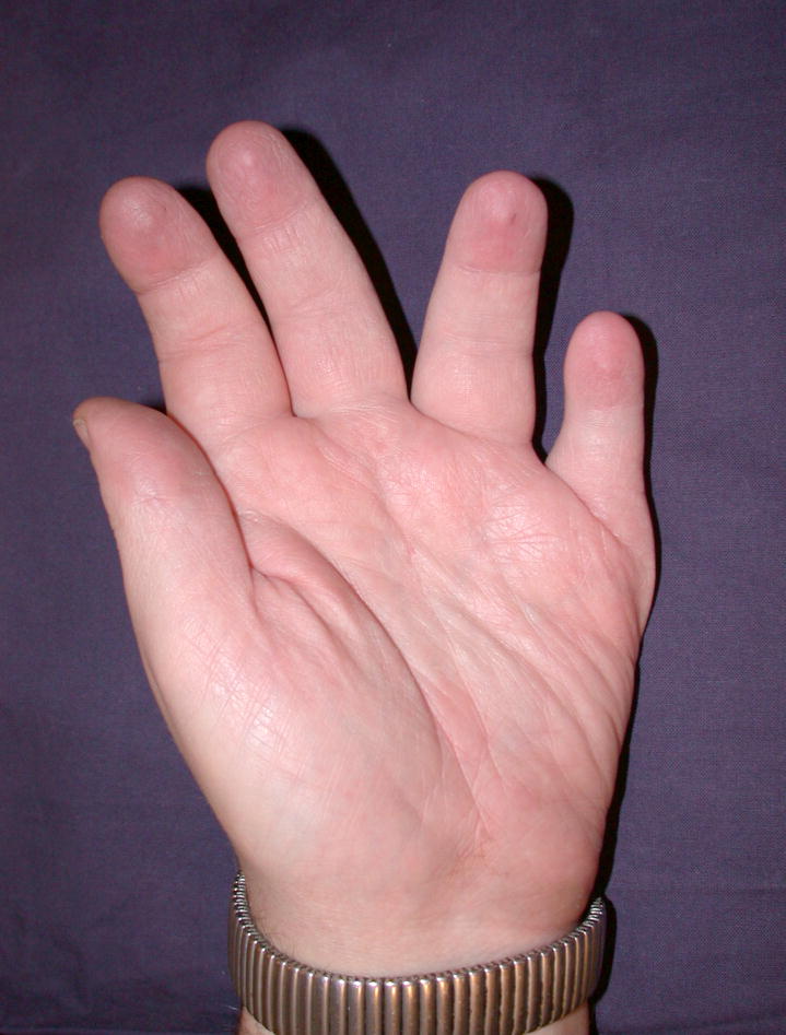
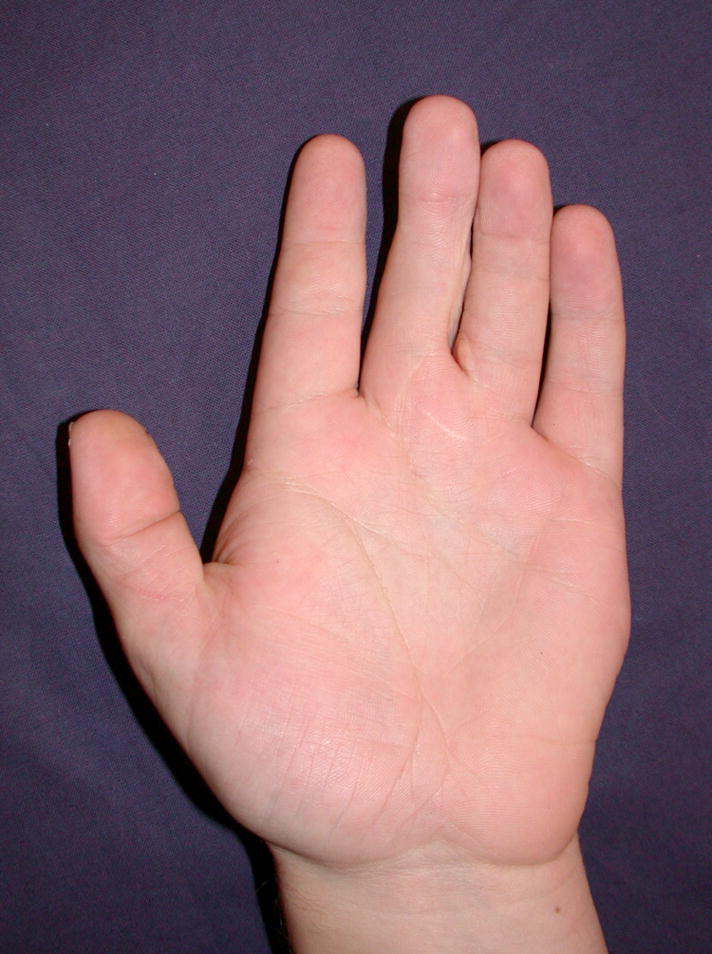
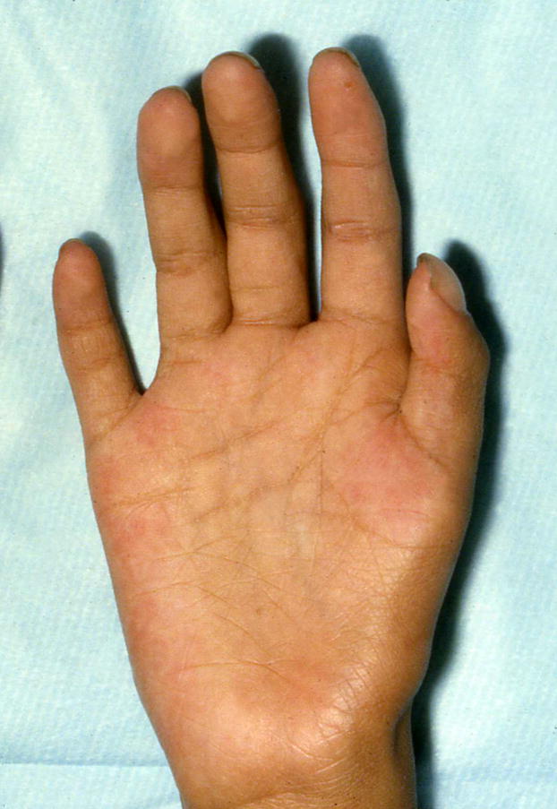
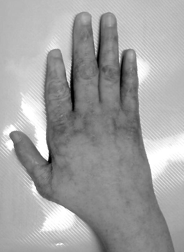
Long fingers, right hand, subjective.
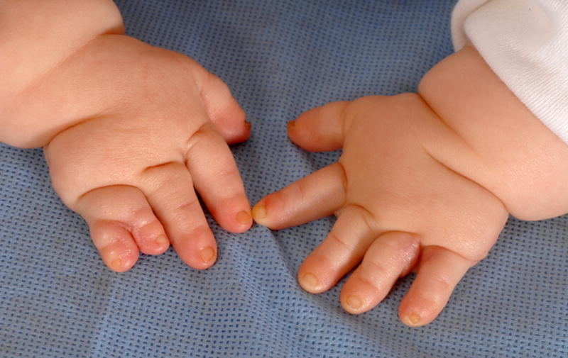
A. Overlapping fingers, right hand, F54, F56. Note that this patient also has Mesoaxial polydactyly and Postaxial polydactyly, type B. B. Overlapping fingers, right hand, F23, F54. See also Figs. 37, 45, and 58A.
A. Overlapping fingers, right hand, F54, F56. Note that this patient also has Mesoaxial polydactyly and Postaxial polydactyly, type B. B. Overlapping fingers, right hand, F23, F54. See also Figs. 37, 45, and 58A.
Note that this patient also has Small nails, F1-5.
Note that this patient also has Clinodactyly, F2, radial and that these two findings are distinct. See also Fig. 47.
See also Figs. 3, 47, 69, and 99. Note that this patient also has Short palms, subjective and cutaneous syndactyly of F4,5, right hand, objective.
Note that this patient also has a Short nail, F1. See also Fig. 60A.
Note that only some of the digits are clearly shown in this figure.
This patient also has Deep palmar creases.
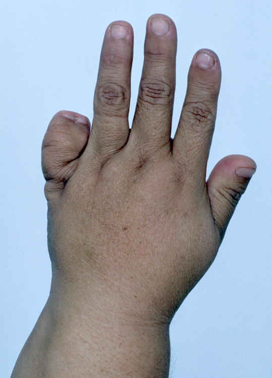
Note that this patient also has Macrodactyly F2-4
A. Tapered finger, left hand, F4. B. Tapered fingers, right hand, F2-5. See also Figs. 42, 44, and 93.
A. Tapered finger, left hand, F4. B. Tapered fingers, right hand, F2-5. See also Figs. 42, 44, and 93.
Note that this patient also has Clinodactyly of F4, ulnar. Note that his middle finger manifests Clinodactyly radial, F3, but that this finger is not deviated, demonstrating the distinction of these two features. See also Fig. 45.
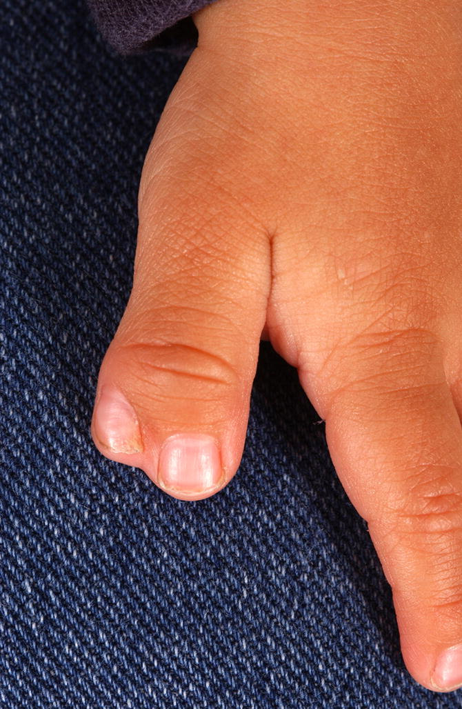
A. Broad fingertip, right hand, F1. Note how the digit widens at the IPJ. B. Broad fingertips, left hand, F3,4. See also Fig. 93.
A. Broad fingertip, right hand, F1. Note how the digit widens at the IPJ. B. Broad fingertips, left hand, F3,4. See also Fig. 93.
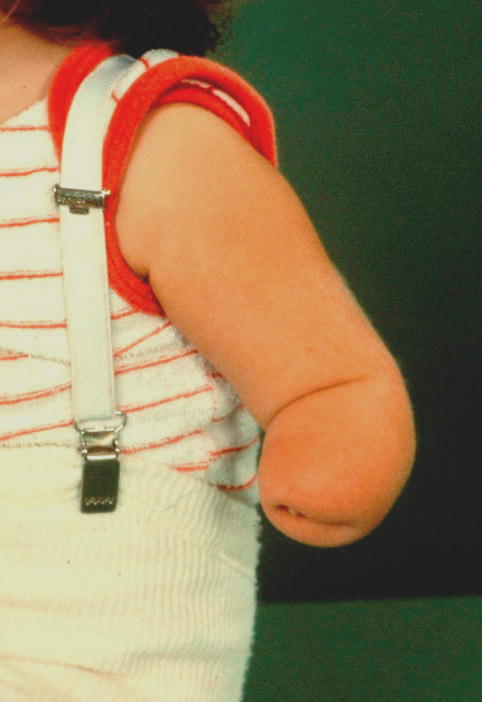
Note that this is the same patient as is shown in Fig. 27. One limb has partial absence of the foot and the other complete.
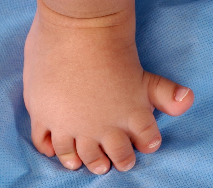
In this example, the forefoot appears more broad than does the midfoot, but this distinction is not noted in the nomenclature.
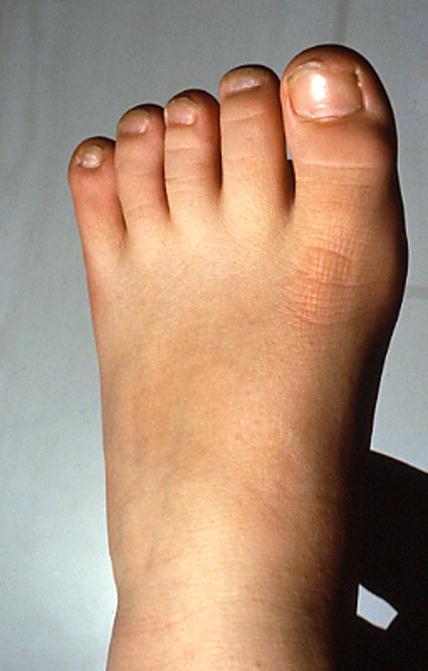
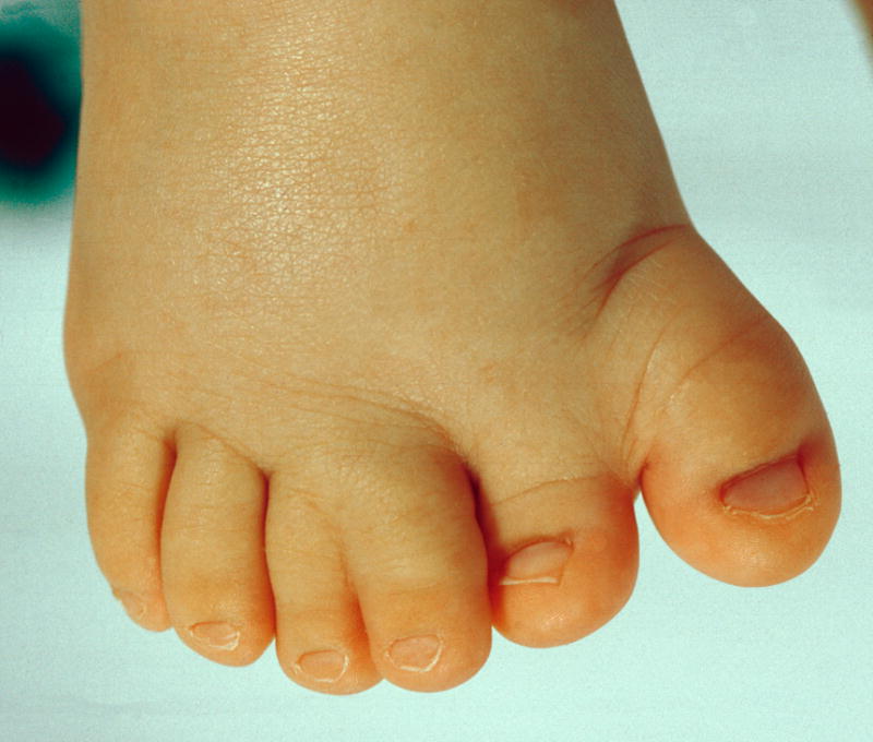
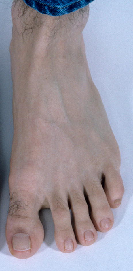
Note that this patient also has Long toes.
Note that this is the same patient as is shown in Fig. 22. One limb has complete absence of the foot and the other partial.
Note that this patient also has Small nails.
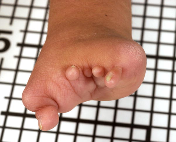
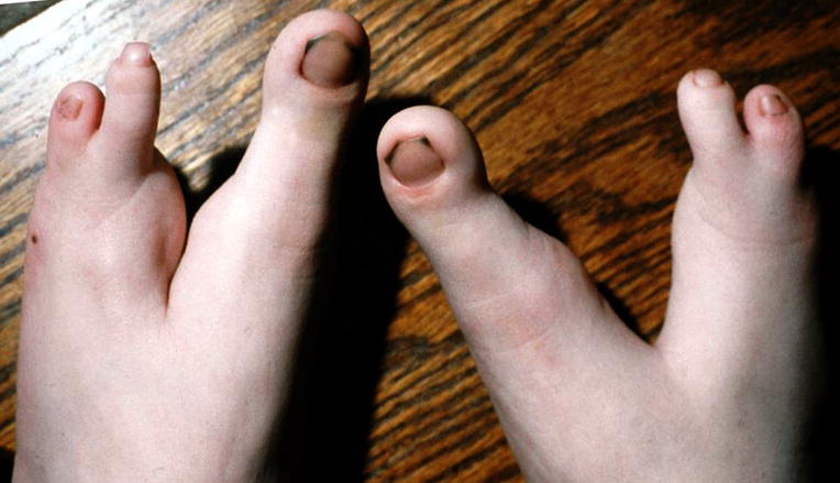
A. Preaxial polydactyly of the right foot. Note that this duplication is nearly complete. B. Preaxial polydactyly of the right foot. This patient has the same finding, but the duplicated digits are more separated than in Fig. 29A.
A. Preaxial polydactyly of the right foot. Note that this duplication is nearly complete. B. Preaxial polydactyly of the right foot. This patient has the same finding, but the duplicated digits are more separated than in Fig. 29A.
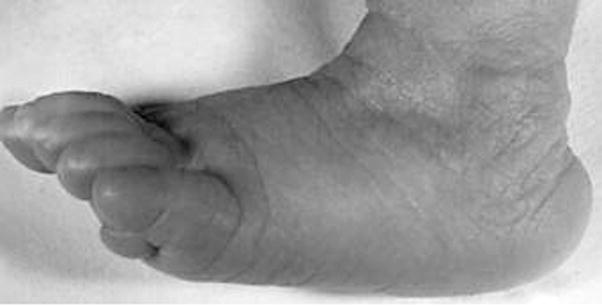
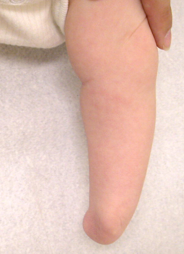
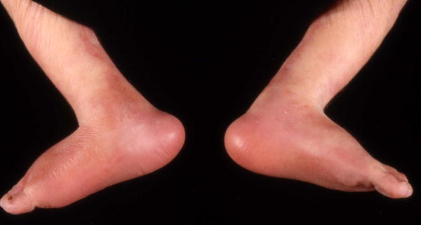
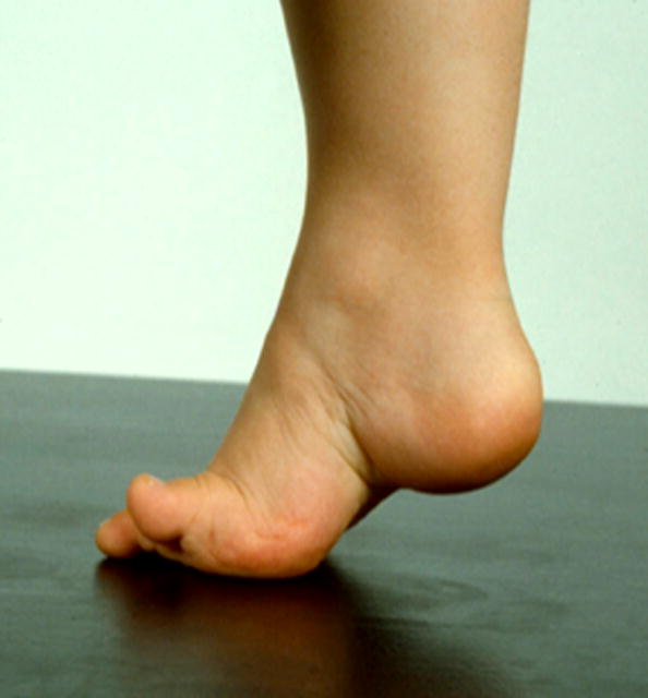
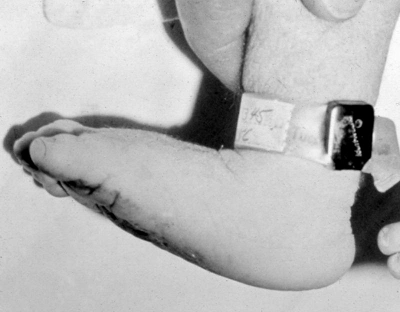
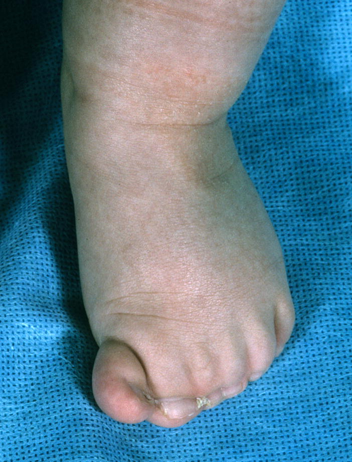
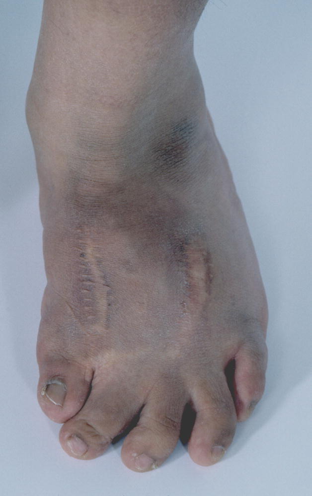
Rocker bottom foot, left.
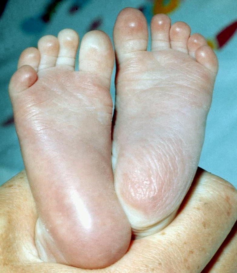
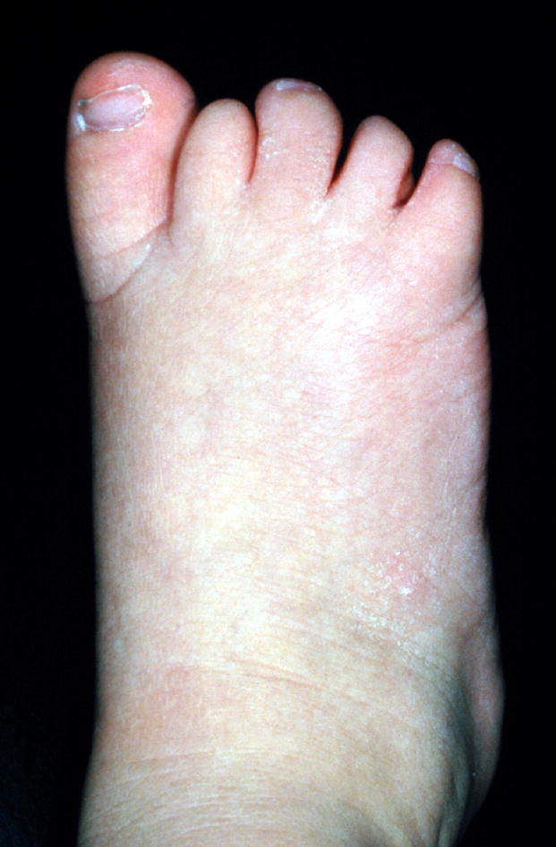
Short feet, subjective.
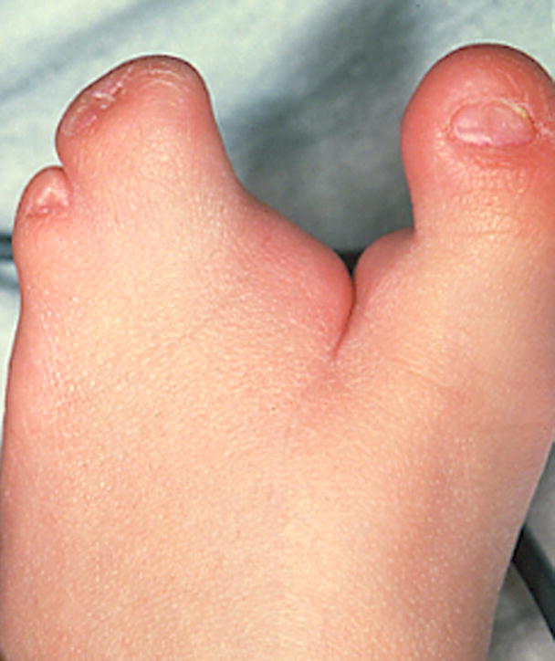
A. Split foot, left. B. Split feet. Note that this patient has a more severe, deeper notch in the feet than does the patient in A. The shape and numbers of other affected digits is highly variable.
A. Split foot, left. B. Split feet. Note that this patient has a more severe, deeper notch in the feet than does the patient in A. The shape and numbers of other affected digits is highly variable.
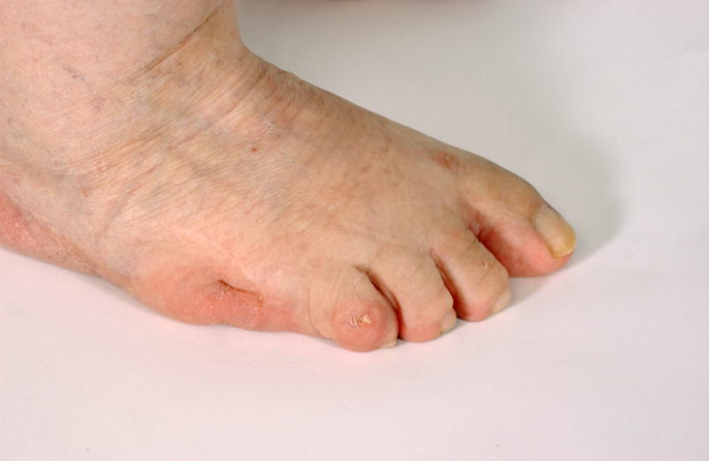
Absent hallux, right.
See also Fig. 60B.
See also Fig. 57.
Note that this patient also has a shortened radius and ulna, although that finding is not required for this finding.
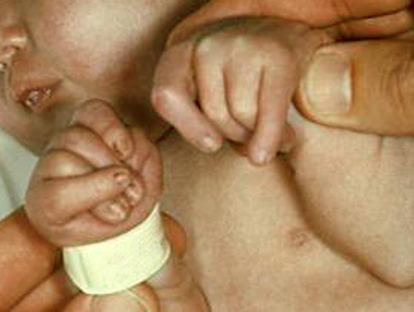
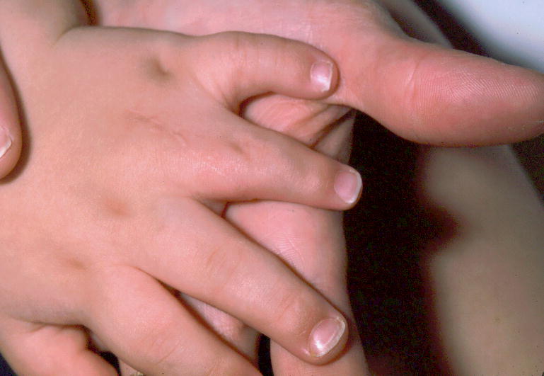
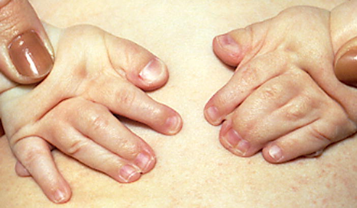
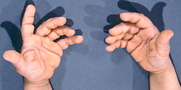
Note that the left hand does not warrant the term because not all of the digits are completely flexed at the MCPJ and IPJ. The left hand would warrant the finding of Overlapping fingers F23, F54, left.
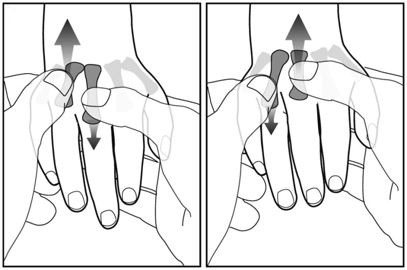
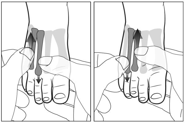
This figure shows the maneuver used to detect this finding. The abnormal finding is not shown. The examiner grasps two adjacent metacarpals and alternately moves them to determine if they are fused or independent.
A. Postaxial polydactyly of the right hand, type A. B. Postaxial polydactyly of the right hand. Note that this patient has a digit that is intermediate between type A and type B, so that is not specified. See also Figs. 49A, 58A, and 73A. See Figs. 9A and 11A for examples of Postaxial polydactyly, type B.
A. Postaxial polydactyly of the right hand, type A. B. Postaxial polydactyly of the right hand. Note that this patient has a digit that is intermediate between type A and type B, so that is not specified. See also Figs. 49A, 58A, and 73A. See Figs. 9A and 11A for examples of Postaxial polydactyly, type B.
A Preaxial polydactyly of the left hand, partial. B. Preaxial polydactyly of the right hand
A Preaxial polydactyly of the left hand, partial. B. Preaxial polydactyly of the right hand
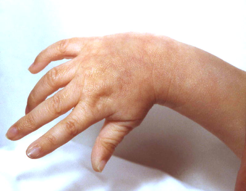
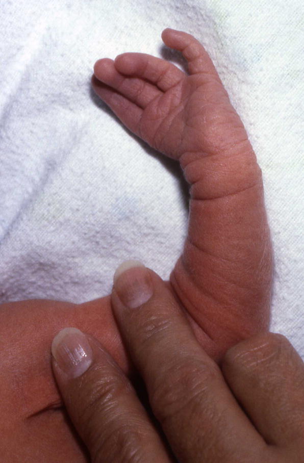
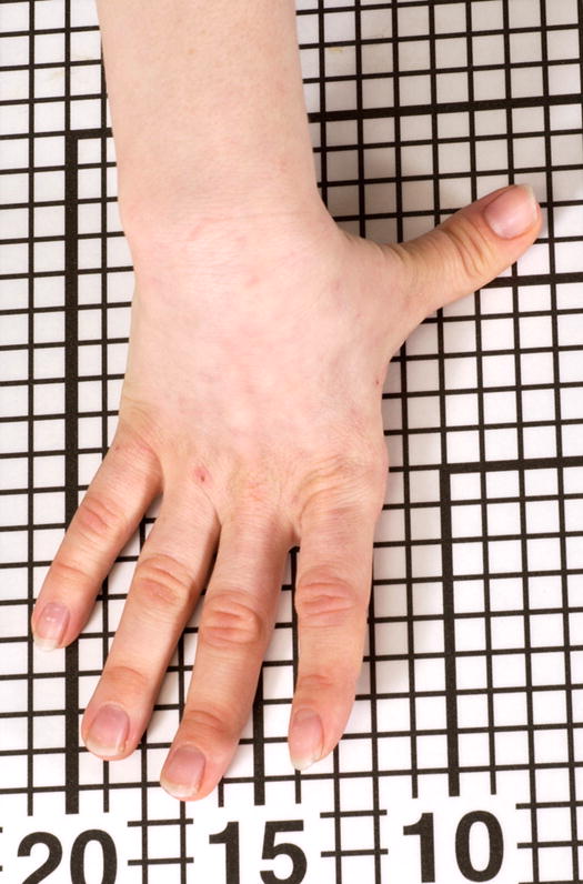
Radial deviation of the hand, right.
Note that this patient also has Tapered fingers.
Note that this patient also has Tapered fingers, but that is not required for the finding.
Note that this patient also has Ulnar deviation of the fingers, F2-3 and Overlapping fingers F45.
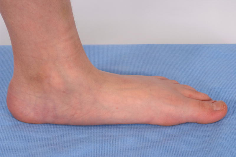
Prominent heels.
Note that this patient also has Short fingers F2-5 and Radial deviation of fingers F2-3.
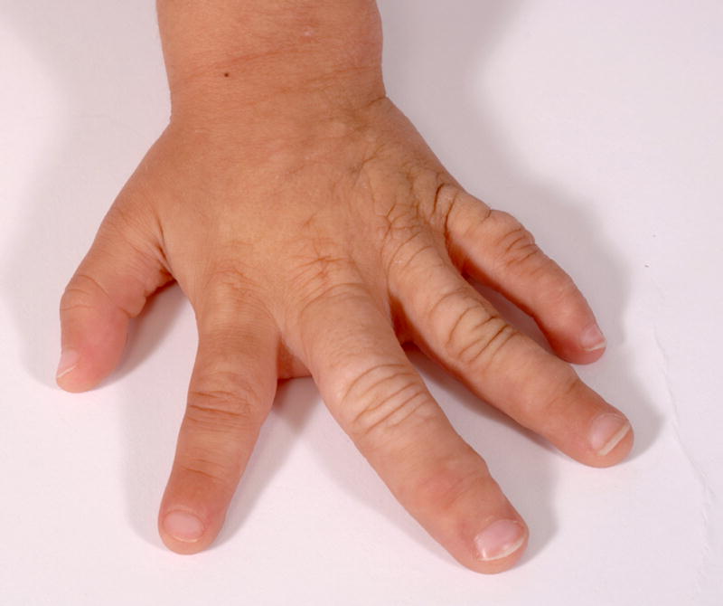
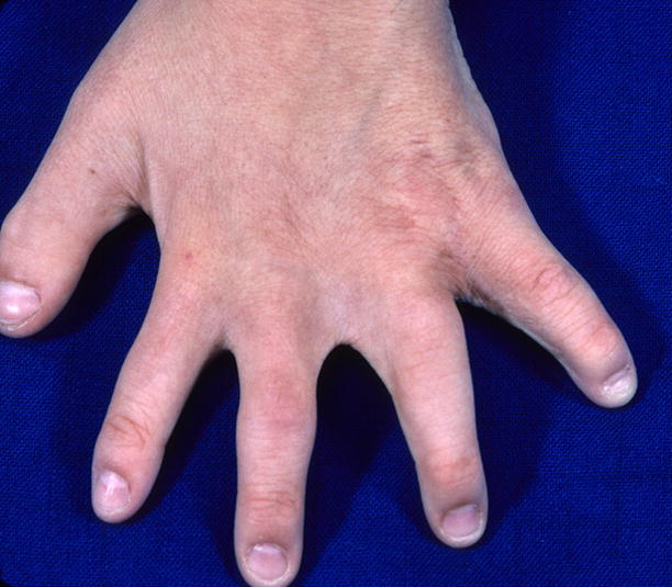
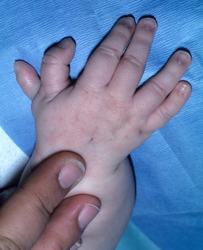
A. Macrodactyly of F2-3, left hand. Note that this person also has Clinodactyly, but that finding should be coded separately. B. Macrodactyly of T1-2, right foot. This patient also has a Sandal gap. See also Figs. 18, 81, and 83.
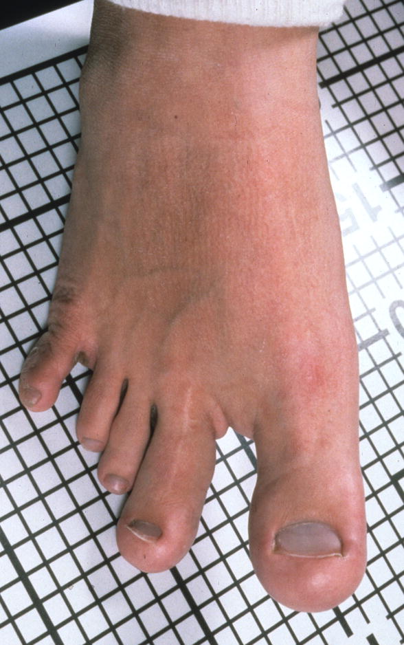
A. Macrodactyly of F2-3, left hand. Note that this person also has Clinodactyly, but that finding should be coded separately. B. Macrodactyly of T1-2, right foot. This patient also has a Sandal gap. See also Figs. 18, 81, and 83.
A. Short metacarpal, F5, left hand. Note that this patient also has a Postaxial polydactyly, partial. B. Short metacarpals, F34, left hand F4, right hand. Note that this patient’s hands are shown in dorsal view, with the fingers flexed, which can facilitate the recognition of this finding.
A. Short metacarpal, F5, left hand. Note that this patient also has a Postaxial polydactyly, partial. B. Short metacarpals, F34, left hand F4, right hand. Note that this patient’s hands are shown in dorsal view, with the fingers flexed, which can facilitate the recognition of this finding.
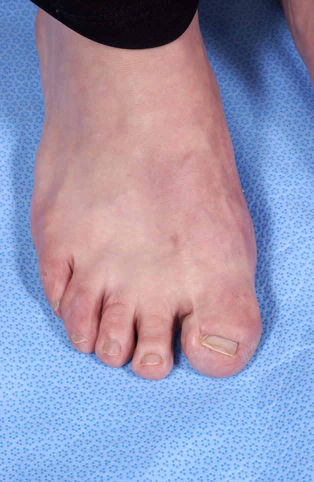
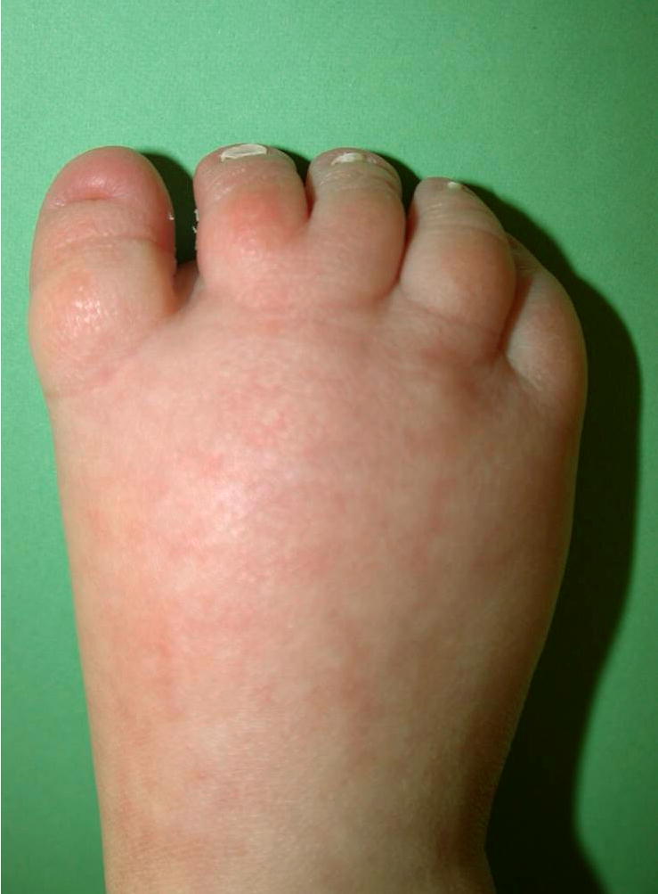
Short metatarsals, T3,4, bilateral.
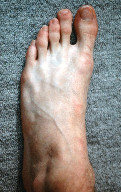
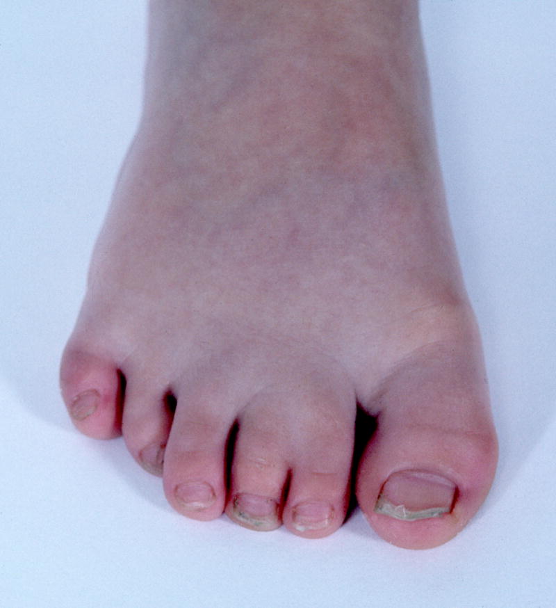
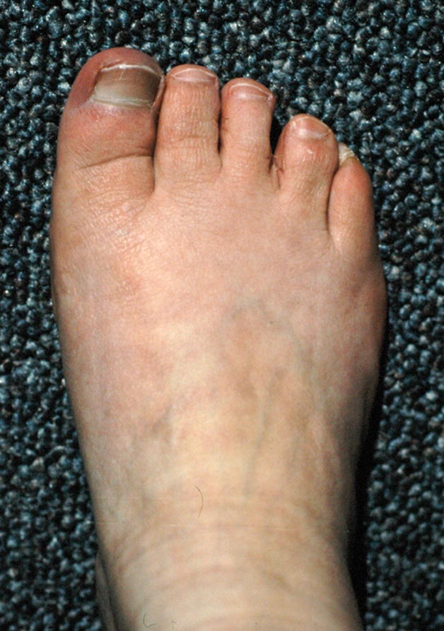
Note that this patient also has a Sandal gap.
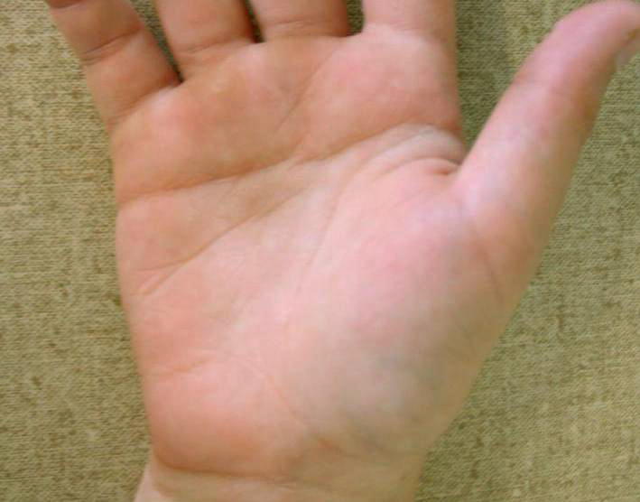
Note that this patient had normal palm length, so that this is not an example of a short palm with only an apparently broad palm.
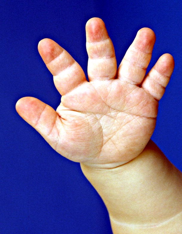
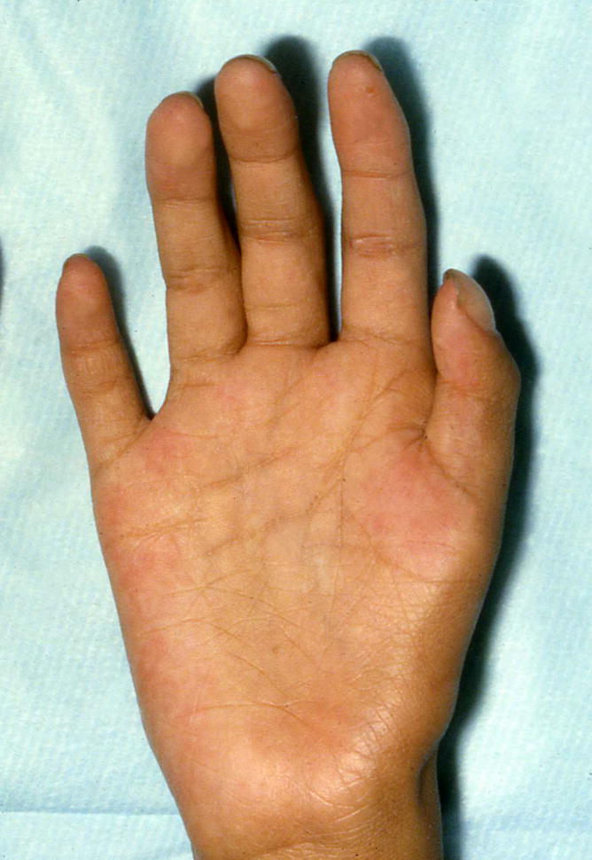
Note that this patient had normal palm width, so that this is not an example of a narrow palm with only an apparently broad palm. Note that this image illustrates a number of other features, which are not delineated. See also Figs. 63 and 85.
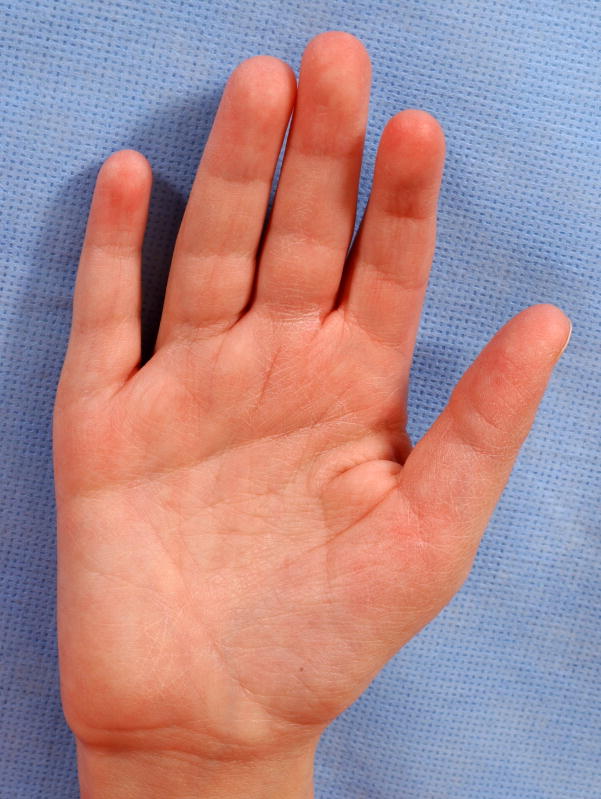
Note that this patient had normal palm width, so that this is not an example of a long palms with only apparently narrow palms.
Note that this patient also has Short fingers. This patient does not warrant a finding of Small hands, because the fingers are of normal caliber. See also Fig. 14.
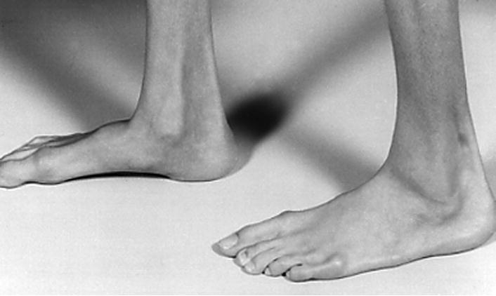
Note that this patient could also be said to have pes equinus, but that term is not included in the current terminology set.
Note that this patient also has Hammertoes bilateral T2.
A. Mesoaxial polydactyly of the hand, right. Note that this patient has heptadactyly, manifesting as well Postaxial polydactyly, type B. The figure also shows Small nail, F6 and Overlapping fingers F56. See also Fig. 11A. B. Mesoaxial polydactyly of the right foot. This patient also has Cutaneous syndactyly of the toes, partial, T23.
A. Mesoaxial polydactyly of the hand, right. Note that this patient has heptadactyly, manifesting as well Postaxial polydactyly, type B. The figure also shows Small nail, F6 and Overlapping fingers F56. See also Fig. 11A. B. Mesoaxial polydactyly of the right foot. This patient also has Cutaneous syndactyly of the toes, partial, T23.
Note that this patient also has Cutaneous syndactyly of numerous digits, notable on the right foot.
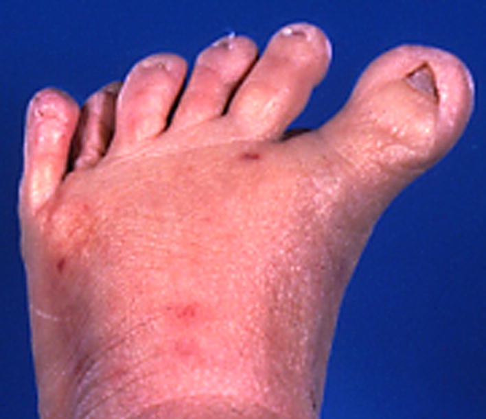
A. Absent ray, right hand. Note that this patient also has Short distal phalanges of the fingers and Short nails. B. Absent ray, left foot. Note that this patient also manifests a Broad hallux, which should be coded separately.
A. Absent ray, right hand. Note that this patient also has Short distal phalanges of the fingers and Short nails. B. Absent ray, left foot. Note that this patient also manifests a Broad hallux, which should be coded separately.
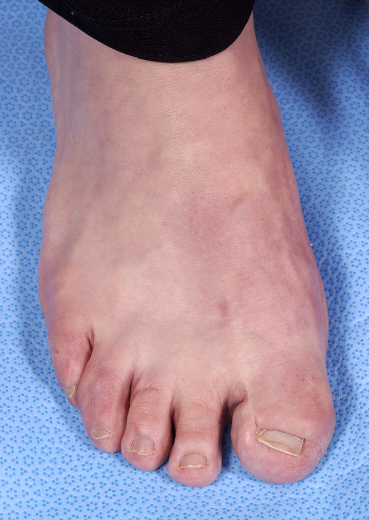
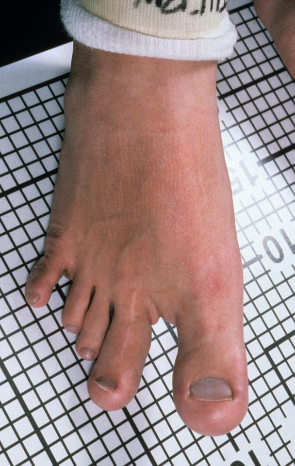
A. Sandal gap, right foot, subjective. B. Sandal gaps, objective. See also Figs. 48 and 51.

A. Sandal gap, right foot, subjective. B. Sandal gaps, objective. See also Figs. 48 and 51.
Note the heel in this patient is not sufficiently prominent to warrant the descriptor of Rocker bottom foot.
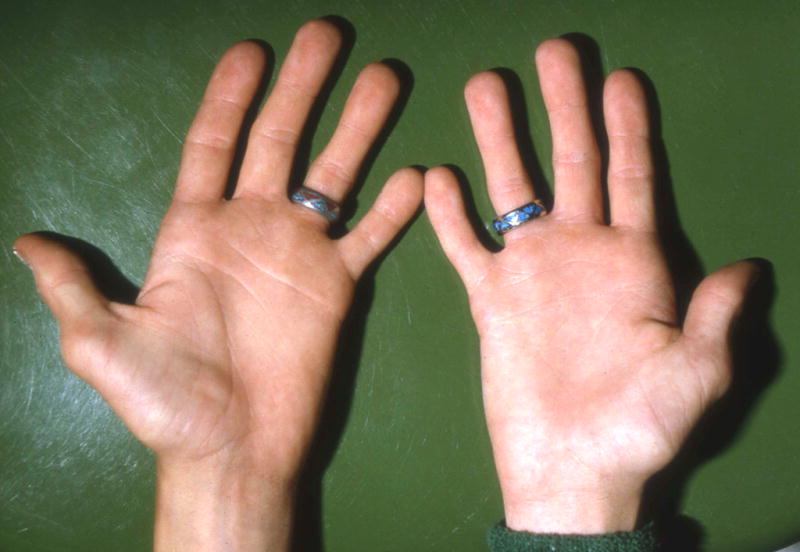
Note that this person also has Clubbing (visible in the thumb only). This patient also has Long palm, right hand, subjective.
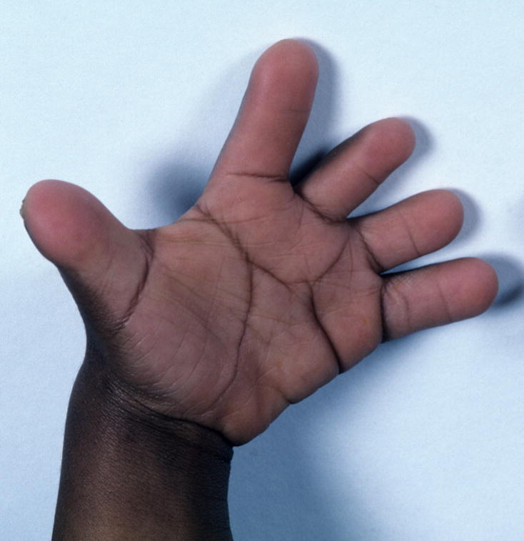
Absent thumb, left.
Thumb, adducted, right.
See also Fig. 9A.
Hitchhiker thumb, right.
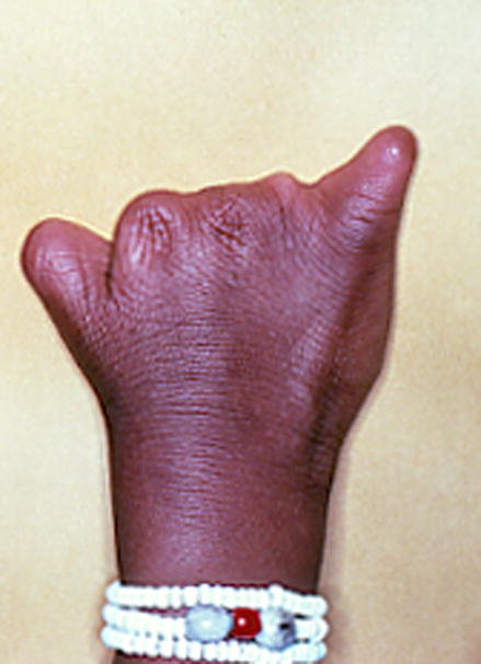
Note that this patient also has Absent fingers, F2-4, and Absent finger F5, partial.
Note that this patient also has Short finger, F2.
Triphalangeal thumb, right.
Absent toe, right.
See also Figs 92, 94, and 101.
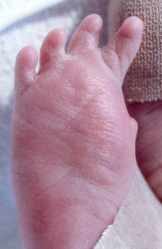
A. Cutaneous syndactyly of the toes, complete, TT1-6, left, objective. See also Figs. 58B, 59, 78, 92, 94, and 101. Note that his patient also has Preaxial polydactyly of the foot, partial, left. B. Cutaneous syndactyly of the toes, partial, T2-4, bilateral, objective. As specified in the definitions, this finding is objective if the syndactyly extends more than half the proximo-distal length of the digits.
A. Cutaneous syndactyly of the toes, complete, TT1-6, left, objective. See also Figs. 58B, 59, 78, 92, 94, and 101. Note that his patient also has Preaxial polydactyly of the foot, partial, left. B. Cutaneous syndactyly of the toes, partial, T2-4, bilateral, objective. As specified in the definitions, this finding is objective if the syndactyly extends more than half the proximo-distal length of the digits.
See also Figs. 24 and 79.
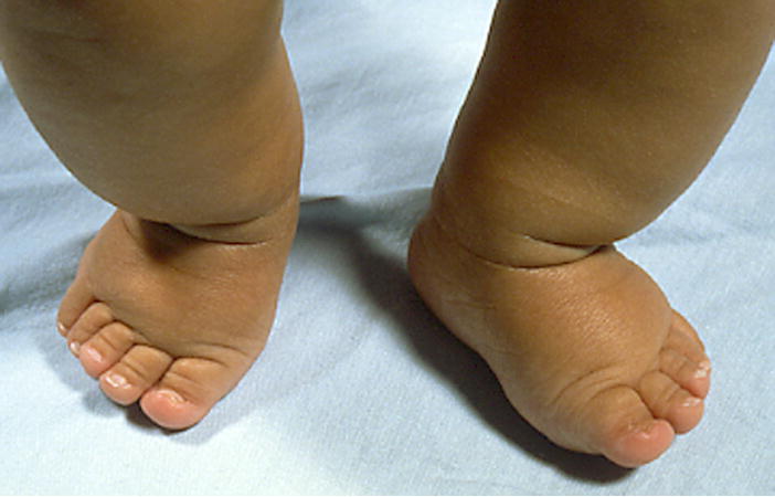
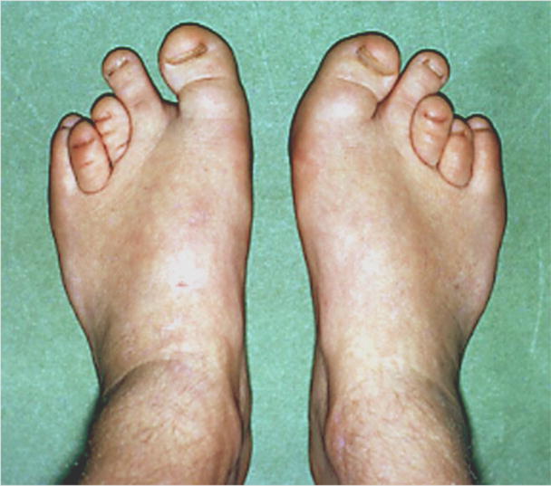
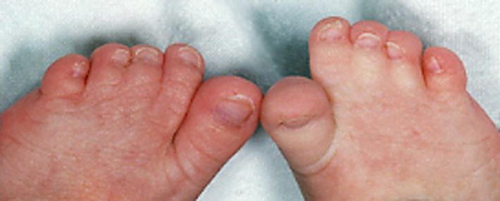
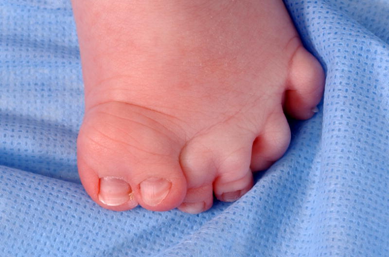
Overlapping toes T45, bilateral.
Note that this is the same image as in Fig. 82, Toe, tapered.
Note that this patient also has Short distal phalanges of toes T2,3.
Note that this patient also has Cutaneous syndactyly of toes T23, subjective. See also Fig. 77.

Note that this patient also has Long toes T1-5.
See also Fig. 81.
Note that this finding differs from Toes, widely spaced, because in splayed toes the toes have a divergent axis of orientation. This patient also has Small toe, T1, right and Macrodactyly of T2,3, right, and T1,2, left.

Note that this is the same image as in Fig. 76, Toe, partial absence.
Note that this patient also has Macrodactyly of T1,2.
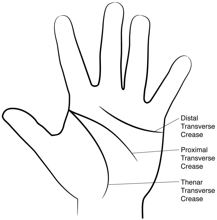
Normal palmar creases. See text for explanatory material.
Note that this patient also has Long palm, subjective, left and Single transverse crease, right hand.
Bridged palmar crease, right hand.
Palmar creases, decreased, right hand.
See also Fig. 17.
See also Figs. 5 and 85.
Deep longitudinal plantar crease, right foot.
Sydney crease, left hand.
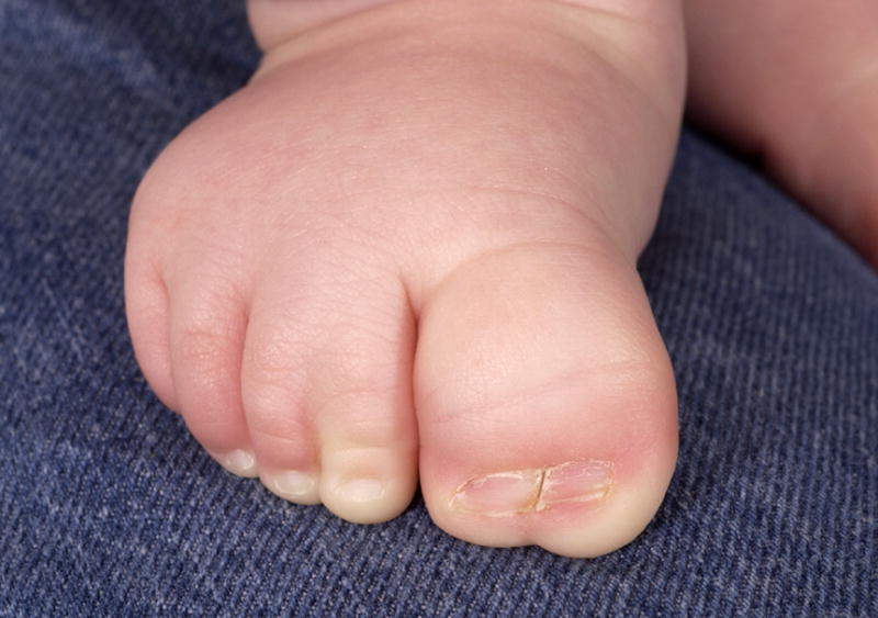
See also Fig. 99. Note that this patient also has a Broad toe, T1 and Cutaneous syndactyly, partial, T23.
See also Fig. 103. Note that this image also shows Tapered fingers, left, F23, and Broad fingertip, left, F4.
See also Fig. 9A. This patient also has a Broad toe, left, T1 and Cutaneous syndactyly, partial, left, T23.
See also Fig. 102.
Note that the length of this nail is normal, so it is coded as narrow, not small.
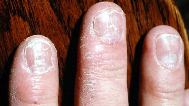
Nails, pitted, F2-4, right hand.
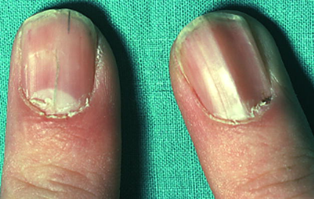
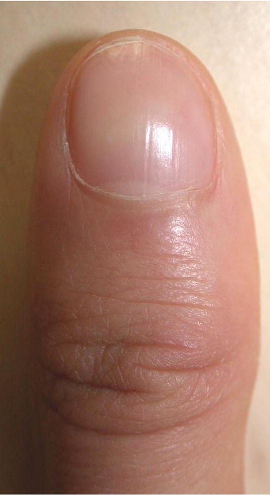
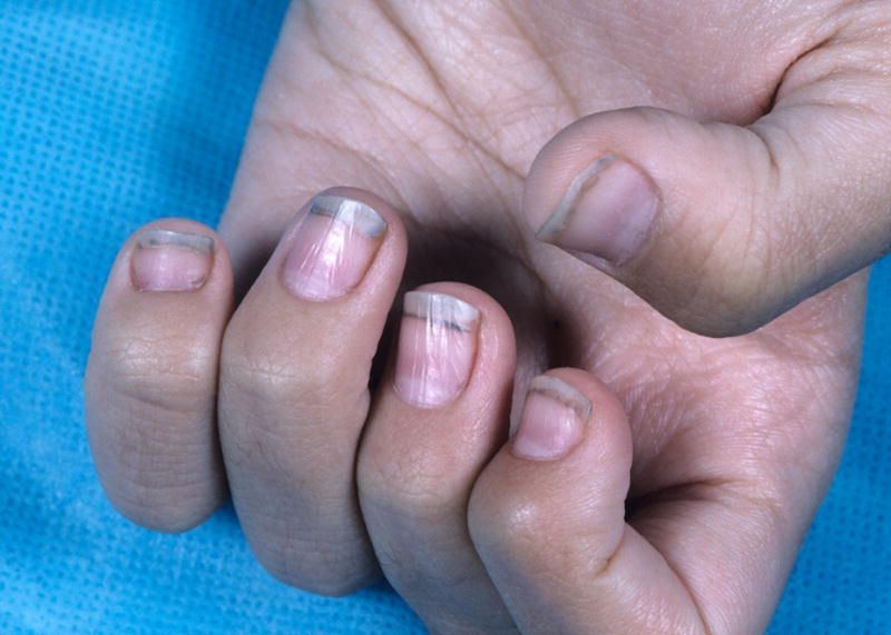
Nails, ridged, F1, bilateral.
See also Fig. 15 and 60A. Note that this patient also has Bifid nail, F5 left hand; Broad finger, F5, left hand; and Short finger, left hand, F1.
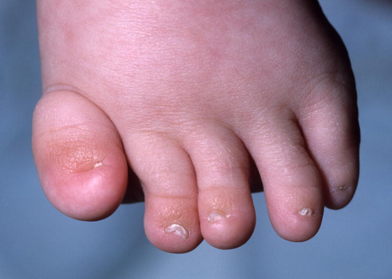
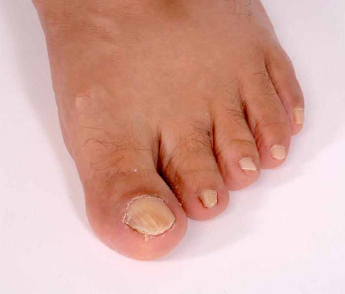
Nails, small, left foot. See also Figs. 12, 28, and 58A.
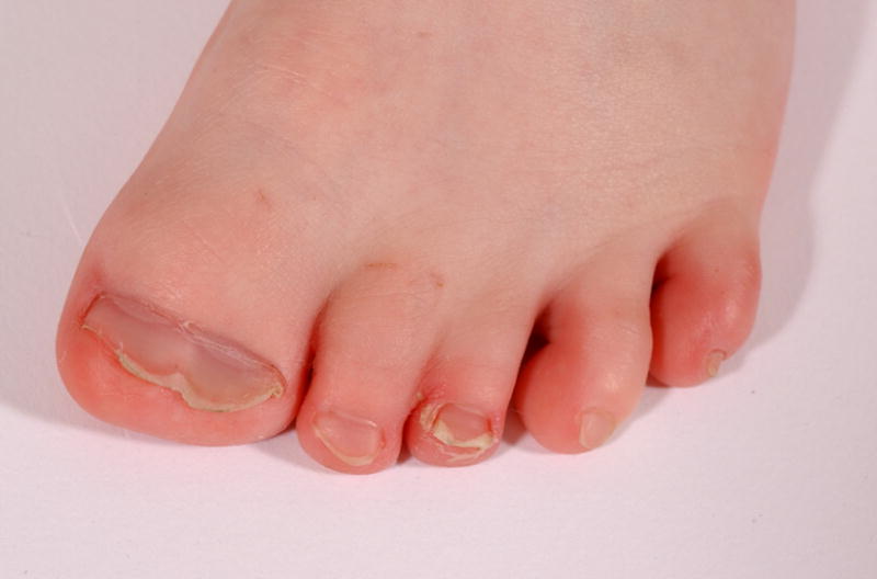
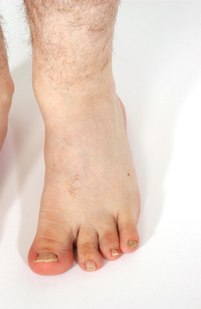
Nail, split, T1, left foot. Note that this image also shows a Broad toe, T1, left foot and Cutaneous syndactyly, partial, T23, left foot.
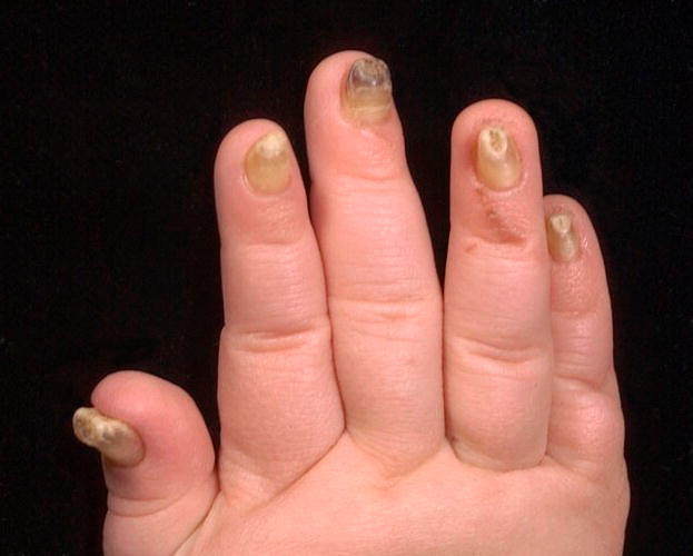
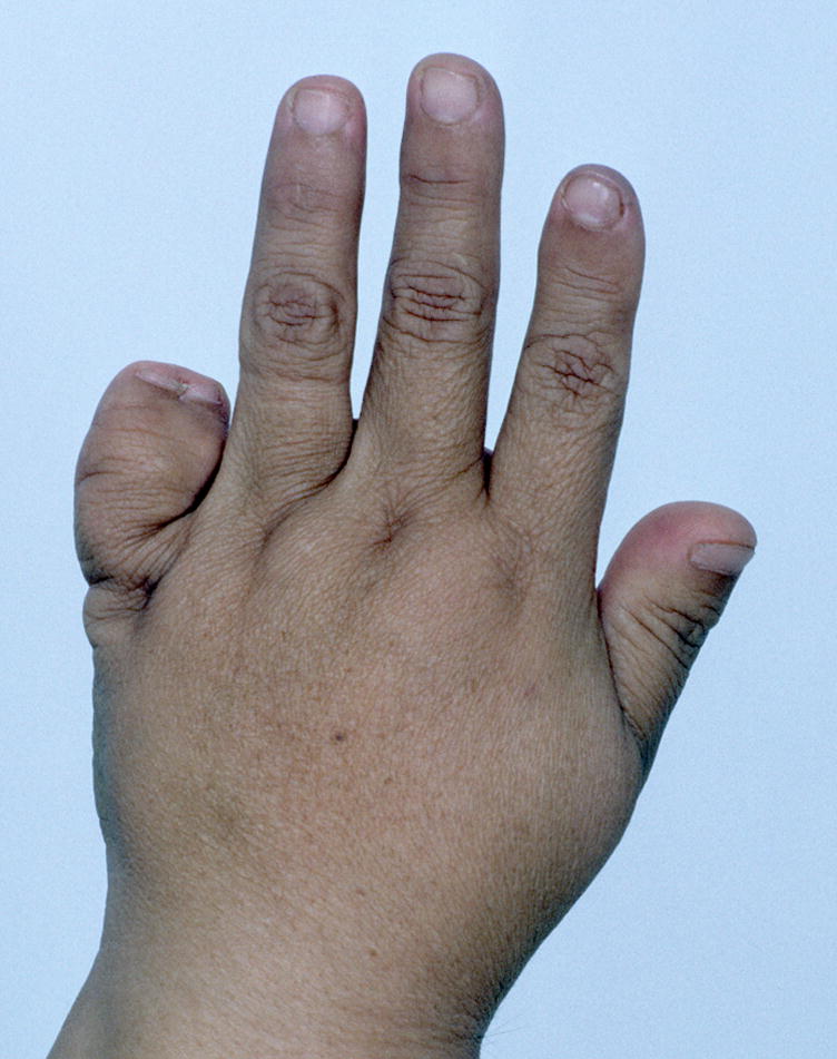
Nails, thick, right hand, F1-5. Note that this image also shows Hyperconvex nails.
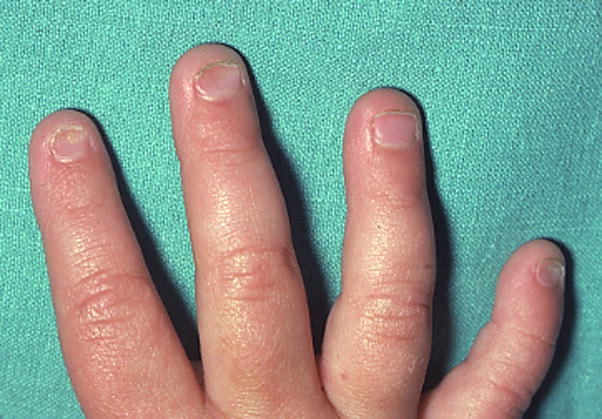
Nails, thin, F2,5, right hand. This image also shows Concave nail, F2, left hand.
Comment in
-
John Locke and a case of macrodactyly.Am J Med Genet A. 2009 Jun;149A(6):1364. doi: 10.1002/ajmg.a.32879. Am J Med Genet A. 2009. PMID: 19449427 No abstract available.
References
-
- Aase JM. Diagnostic dysmorphology. New York: Plenum Medical Book Co; 1990. p. 299.
-
- Anderson M, Blais MM, Green WT. Lengths of the growing foot. J Bone Joint Surg Am. 1956;38-A:998–1000. - PubMed
-
- Biesecker LG. A maneuver to assess the presence of metacarpal or metatarsal osseous syndactyly: A physical finding useful for the differential diagnosis of polydactyly. Am J Med Genet A. 2007;143:1788–1789. - PubMed
Publication types
MeSH terms
Grants and funding
LinkOut - more resources
Full Text Sources
Other Literature Sources
Medical
