

doi: 10.1002/ajmg.a.32612.
Elements of morphology: standard terminology for the head and face
Affiliations
- PMID: 19125436
- PMCID: PMC2778021
- DOI: 10.1002/ajmg.a.32612
Item in Clipboard
Elements of morphology: standard terminology for the head and face
Am J Med Genet A.
2009 Jan.
Abstract
An international group of clinicians working in the field of dysmorphology has initiated the standardization of terms used to describe human morphology. The goals are to standardize these terms and reach consensus regarding their definitions. In this way, we will increase the utility of descriptions of the human phenotype and facilitate reliable comparisons of findings among patients. Discussions with other workers in dysmorphology and related fields, such as developmental biology and molecular genetics, will become more precise. Here we introduce the anatomy of the craniofacies and define and illustrate the terms that describe the major characteristics of the cranium and face.
Figures
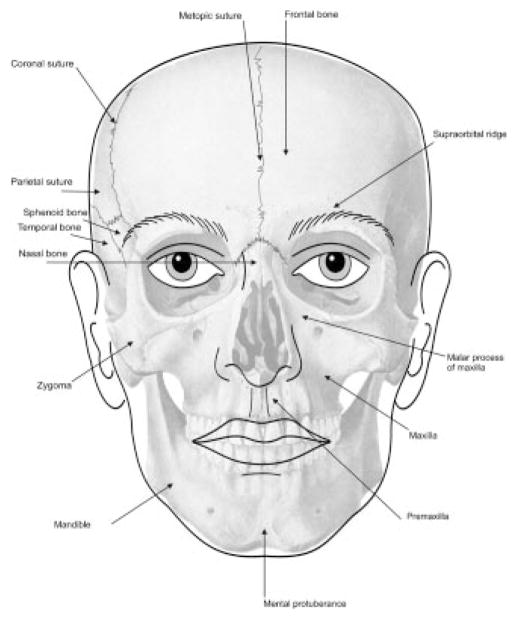
An antero-posterior view of the cranium and face shows bony landmarks.
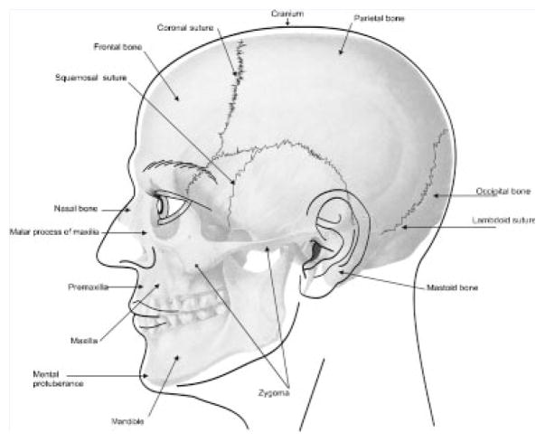
A lateral view of the cranium and face shows bony landmarks.
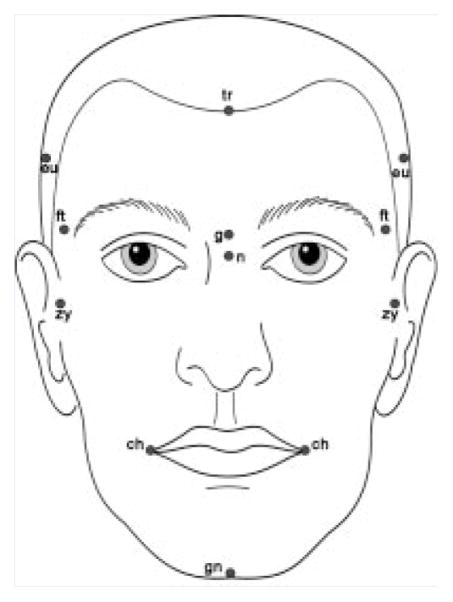
Anthropological landmarks of the face, frontal view, which are described in this paper.
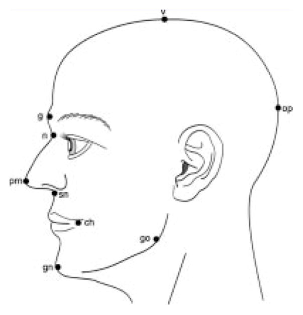
Anthropological landmarks of the face, lateral view, which are described in this paper.
Brachycephaly: The skull has a reduced antero-posterior dimension with the back of the head appearing to have reduced convexity.
Dolichocephaly. The skull has an increased antero-posterior dimension. Scaphocephaly is demonstrated on the right. Note that this subtype of dolichocephaly is “boat-shaped” with pointed anterior and posterior aspects of the cranial vault.
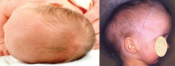
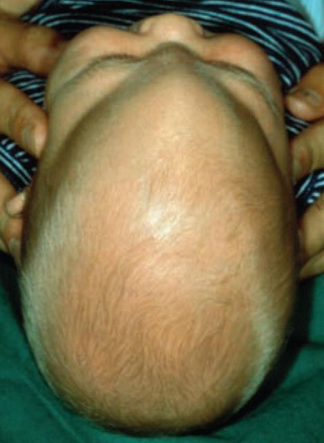
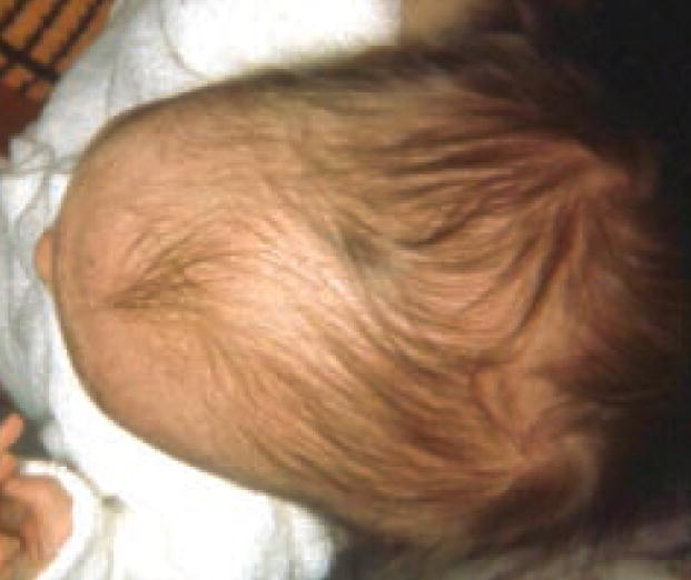
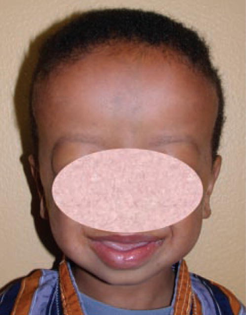
Macrocephaly. Note the increased size of the cranium. Differences in size are difficult to appreciate but increased head size in this child is notable because of comparison with the smaller face.
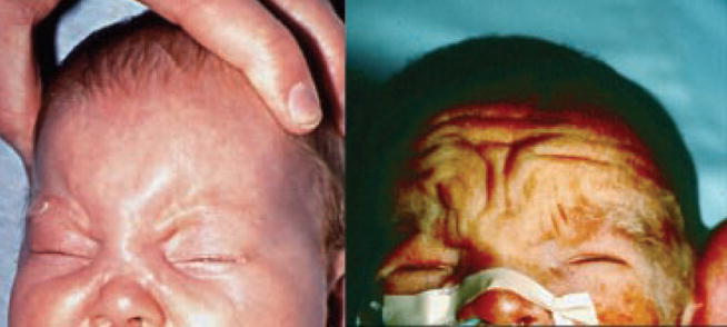
Microcephaly. Decreased size of the cranium is accompanied by marked posterior sloping of the forehead.
There is reduced convexity of the occiput giving an appearance of flattening of the back of the skull.
The posterior part of the skull shows increased convexity.
Plagiocephaly. There is asymmetry of head shape: Note that one can see a combination of unilateral occipital flattening with ipsilateral frontal prominence, leading to rhomboid cranial shape or asymmetry of the posterior skull alone. These figures are kindly provided by John Graham Jr.
The skull has a trilobar configuration when viewed from the front or behind.
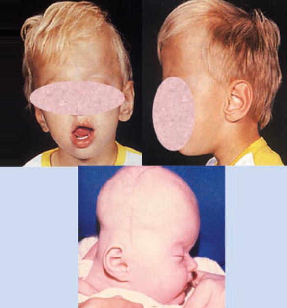
Trigonocephaly. Note the wedge-shaped, or triangular head, with the apex of the triangle at the midline of the forehead and the base of the triangle at the occiput.
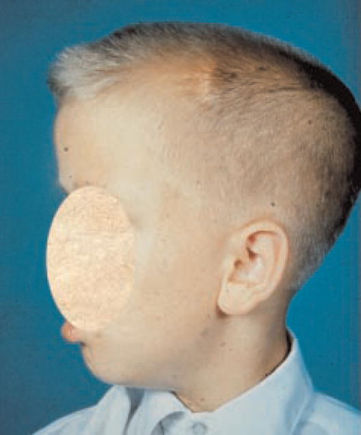
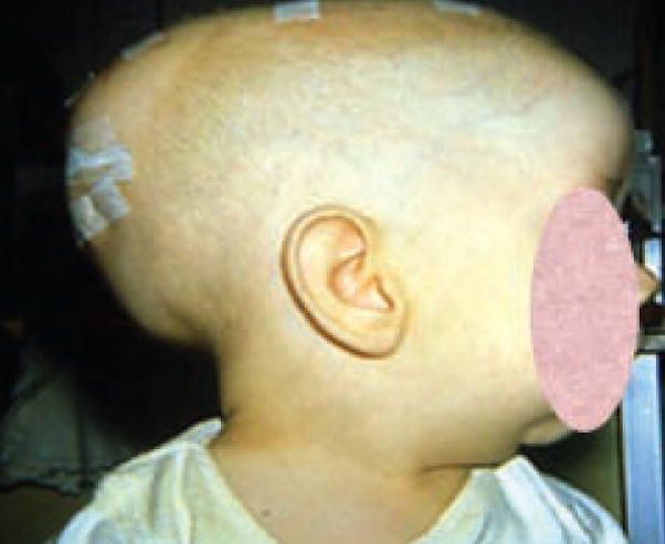
Turricephaly. The head is tall head relative to its width and length. The terms acrocephaly or oxycephaly are used when there is turricephaly and the top of the skull assumes a cone shape (lower image).
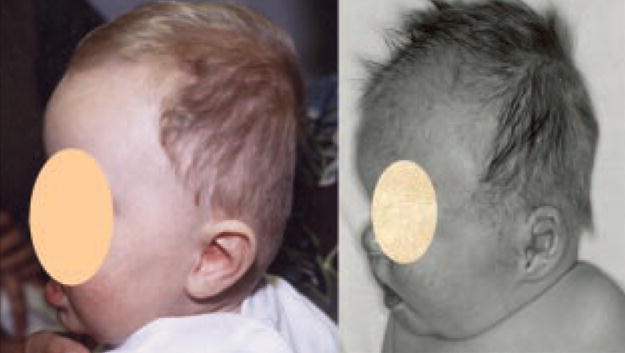
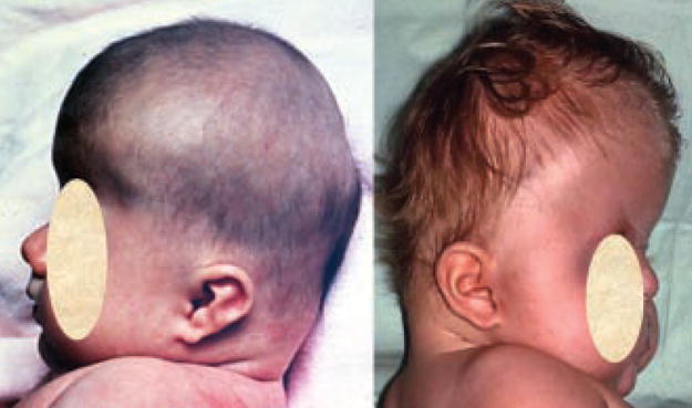
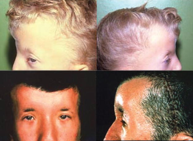
Note the absence of hair in the anterior midline and/or parietal areas.
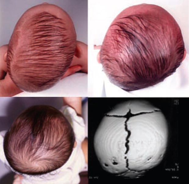
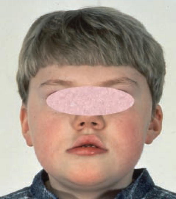
Note the pattern of upward and sideward growth of anterior hair.
Hair whorl, double.
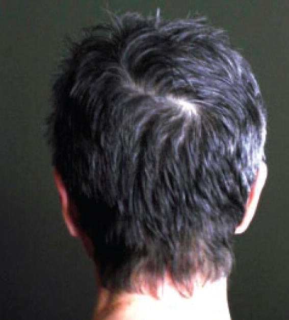
The hair whorl is positioned postero-inferiorly than its usual location lateral to the midline and close to the vertex of the skull.
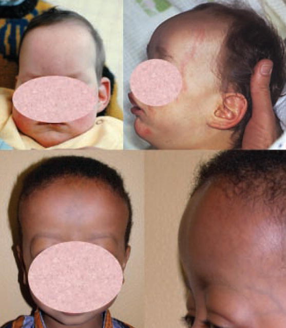
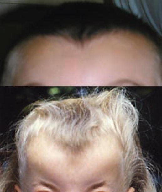
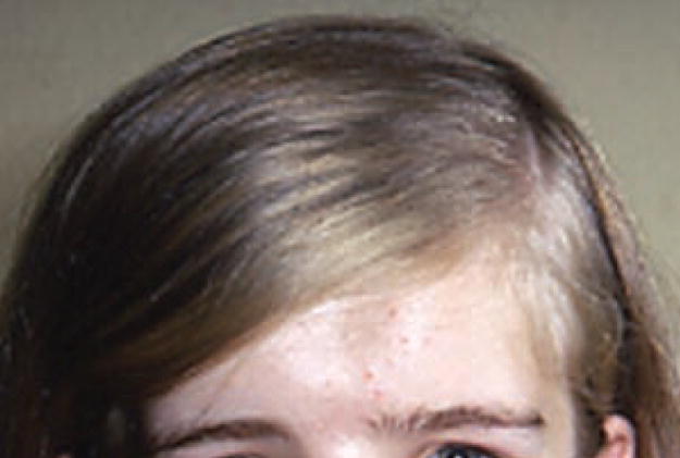
The high anterior hairline contributes to an appearance of tall forehead.
The low anterior hairline contributes to an appearance of short forehead.
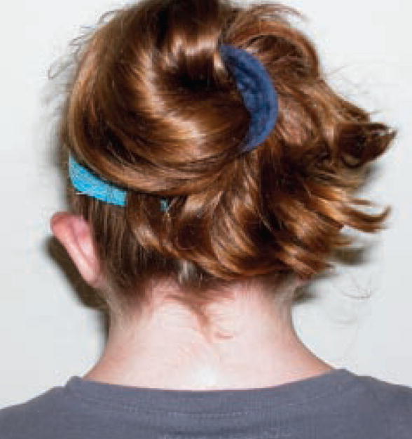
Hair on the neck extends more inferiorly than usual, particularly in the lateral aspects.
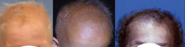
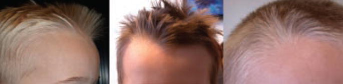
Hair density is reduced giving a thinned appearance.
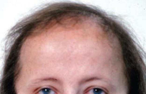
Frontal hairline shows bilateral arcs to a low point in the midline of the forehead.
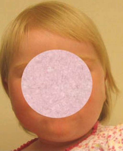
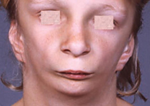
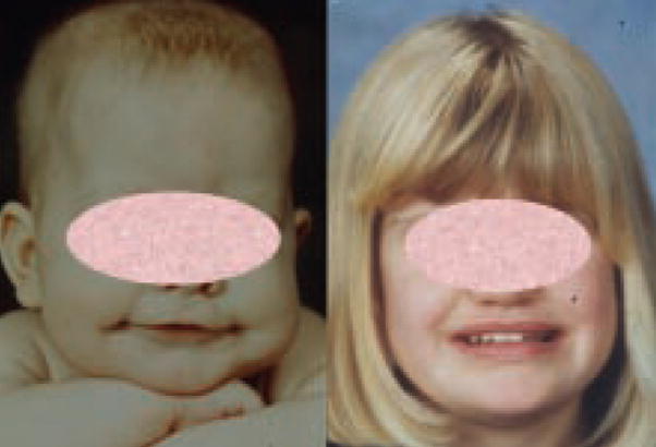
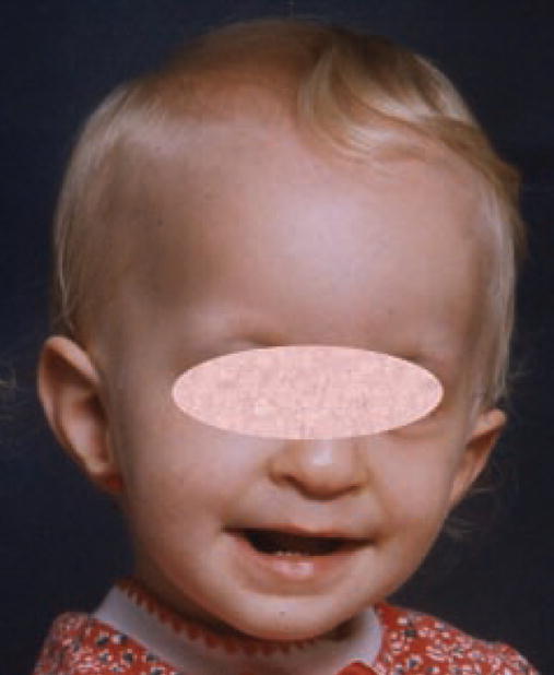
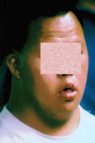
An increased width of the upper and lower face.
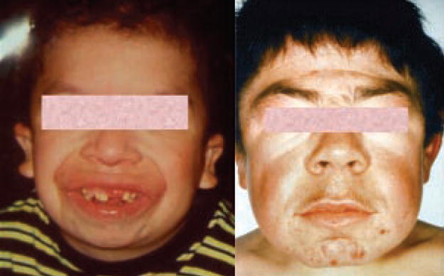
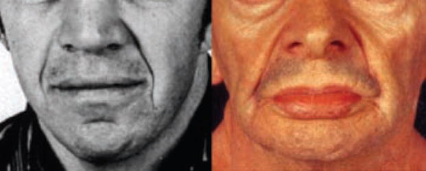
Facial features lack the usual fine and sharp appearance and are rounded and heavy with thickened skin, subcutaneous or bony tissues.
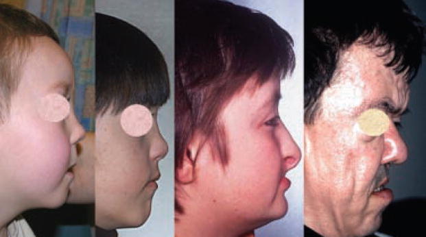
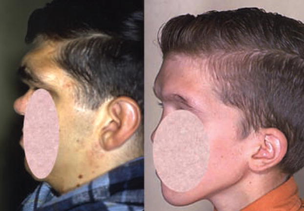
The profile of the face is flat with no concavity or convexity.
Height (length) of the face is increased in comparison to face width. Without actual measurement it can be difficult to decide whether increased height or reduced width is real.
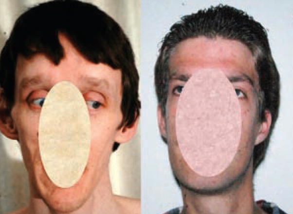
There is reduction in width of the upper and lower face. Without actual measurement it can be difficult to decide whether increased height or reduced width is real.
Facial appearance is more circular than usual.
Decreased height (length) of the face is usually appreciated in comparison to face width and it may be difficult to decide whether reduced height or increased width is present without measurement.
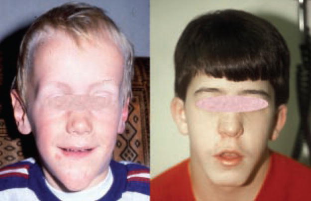
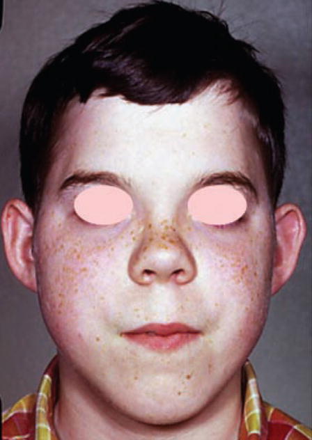
The upper face/cranium and lower face/mandible are both broad, creating a square appearance.
Facial contours are triangular in shape, with breadth at the temples tapering to a narrow chin.
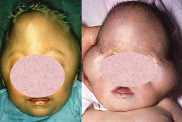
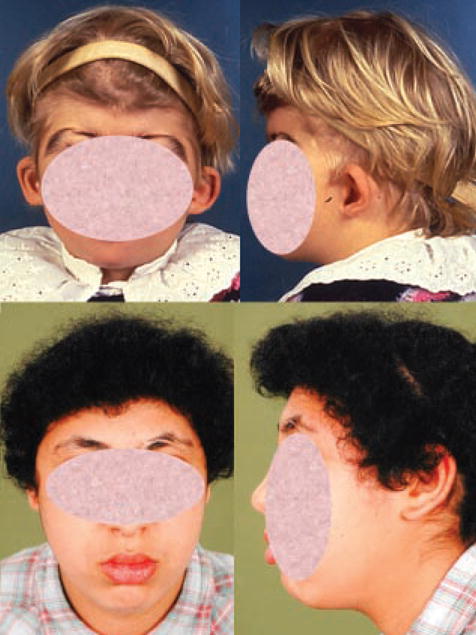
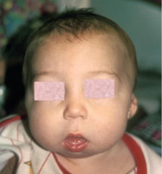
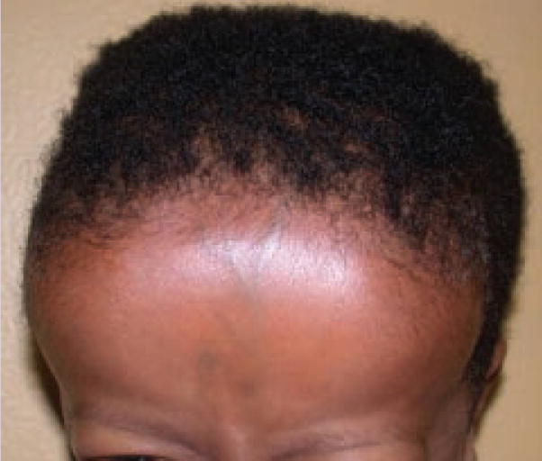
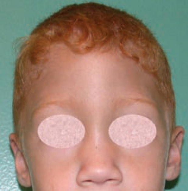
Note the increased distance between the two sides of the forehead.
Note the decreased distance between the two sides of the forehead with narrowing at the temples.
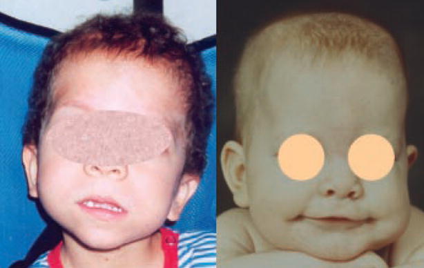
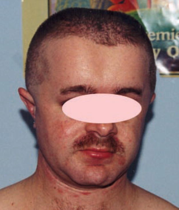
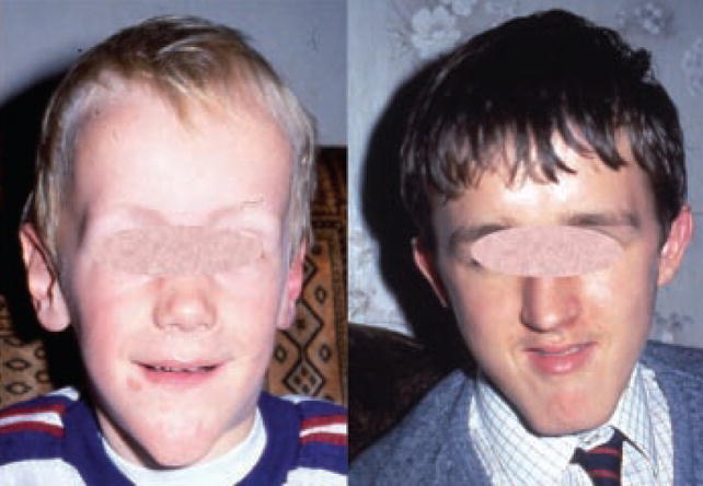
The entire forehead is prominent due to protrusion of the frontal bone.
The anterior surface of the forehead slopes posteriorly in an excessive manner.
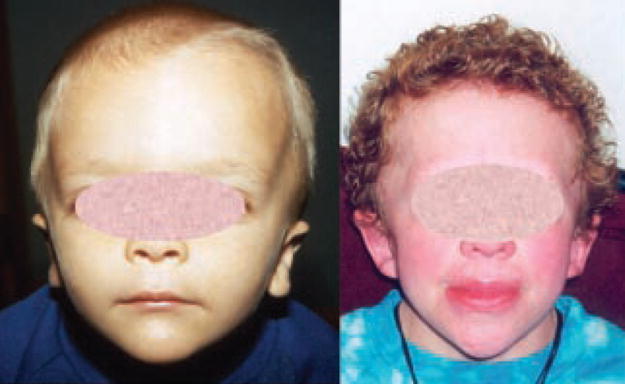
Vertical soft tissue creases are noted in the midline of the forehead. These often extending from the hairline to the brow.
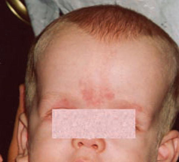
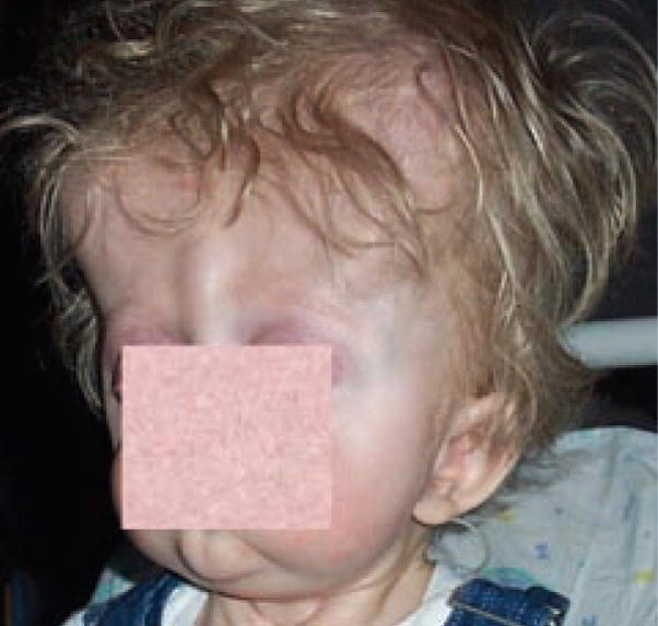
There is bilateral bulging of the lateral aspects of the forehead with relative sparing of the midline.
Note the depression of the midline forehead between the supraorbital ridges.
Note prominence of the glabella, the area of the forehead in the midline between the supraorbital ridges, just above the nasal root.
There is a linear vertical groove in the midline of the forehead, extending from hairline to glabella.
Note the vertical bony ridge in the midline of the forehead.
The supraorbital portion of the frontal bones protrudes forward and laterally.
The supraorbital portion of the frontal bones is less prominent than usual.
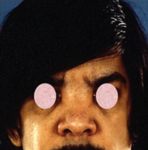
The cheekbones overlying the zygoma of the temporal bone of the skull are more prominent than usual.
The cheekbones overlying the zygoma of the temporal bone of the skull are less prominent than usual.
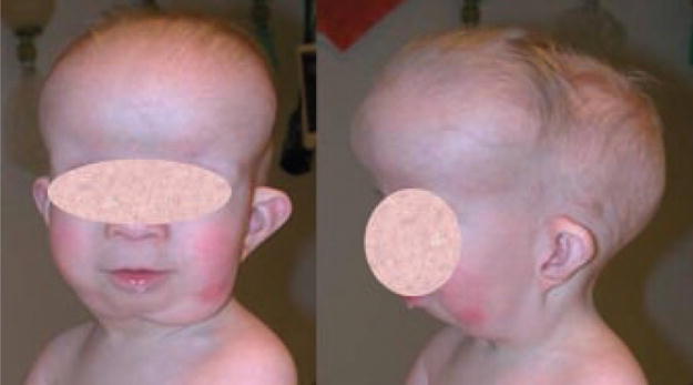
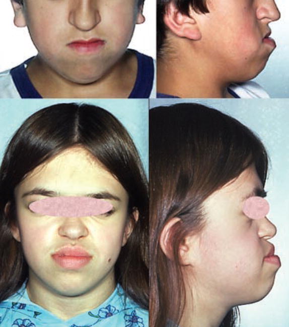
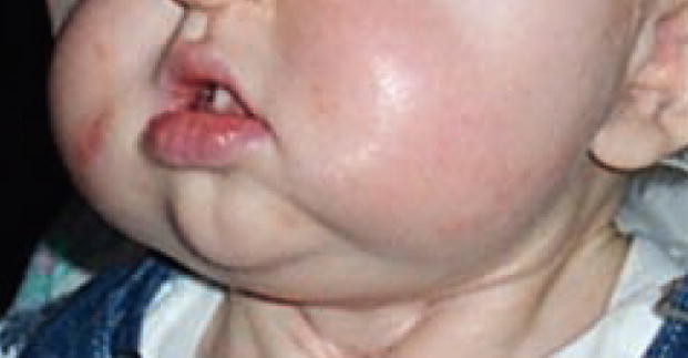
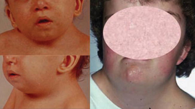
Note the increased prominence or roundness of the soft tissues between the cheekbones and mandible.
Note the reduced prominence or fullness of the soft tissues between the cheekbones and mandible.
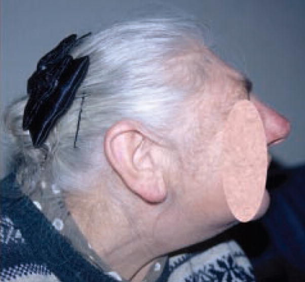
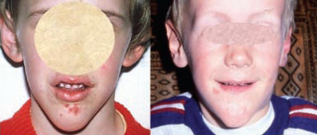
Note the underdevelopment of bony tissues lateral to the nasal bridge extending from the inner corner of the eye to the medial aspect of the cheekbone.
Note the prominence of bony tissues lateral to the nasal bridge extending from the inner corner of the eye to the medial aspect of the cheekbone.
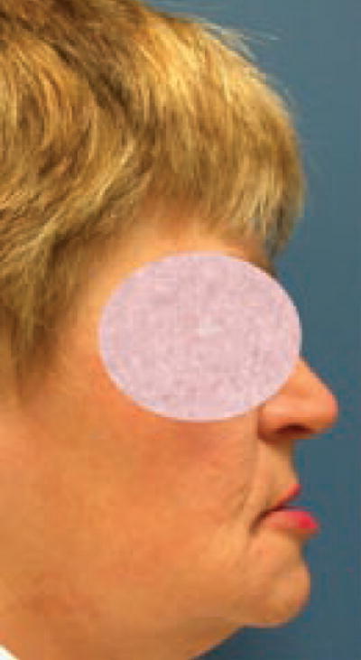
Note prominence of the infraorbital and perialar regions leading to more pronounced convexity of the face and increased nasolabial angle.
Note underdevelopment of the infra-orbital and peri-alar regions leading to more pronounced concavity of the face and reduced nasolabial angle. This gives the appearance of prognathia.
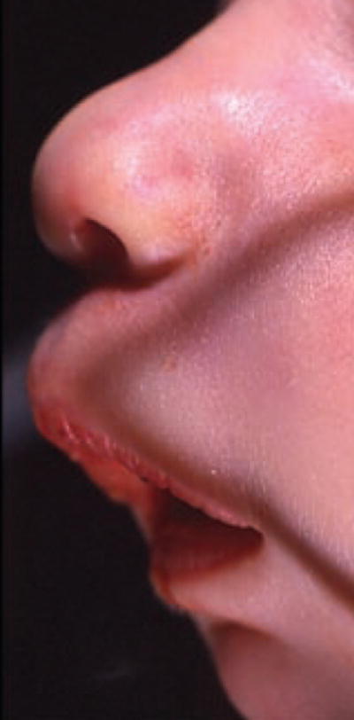
The crease or fold of skin running from the lateral margin of the nose, where nasal base meets the skin of the face, to a point just lateral to the corner of the mouth is more prominent than usual.
The crease or fold of skin running from the lateral margin of the nose, where nasal base meets the skin of the face, to a point just lateral to the corner of the mouth is less prominent than usual.
Note increased convexity of the face and an increased nasolabial angle giving the impression of retrognathia.
Note decreased convexity of the face and nasolabial angle giving the impression of prognathia.
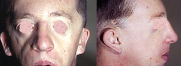
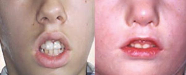
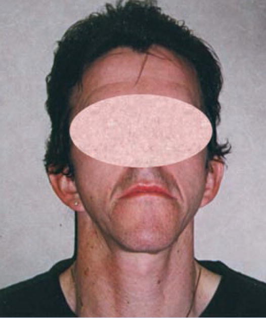
Note increased width of the lower jaw (mandible).
Note decreased width of the lower jaw (mandible).
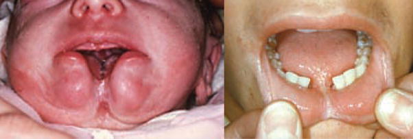
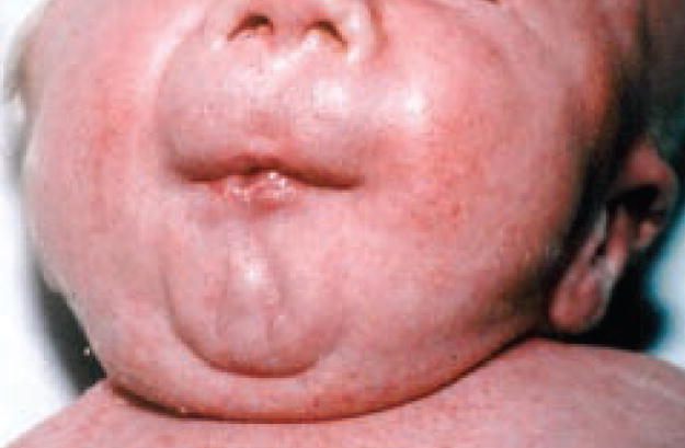
There is a complete midline deficiency of the mandible on the left and deficiency of overlying tissues on the right.
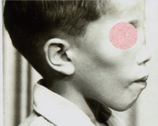
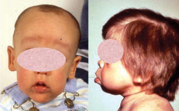
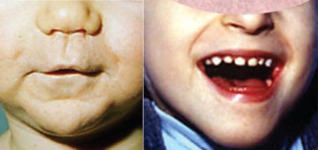
Micrognathia. There is shortening and narrowing of the mandible and chin.
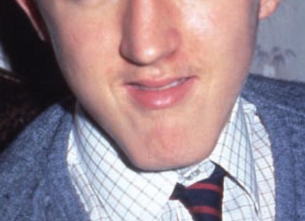
Prognathism. There is anterior protrusion of the mandible such that the alveolar ridge extends beyond the vertical plane of the maxillary alveolar ridge.
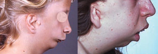
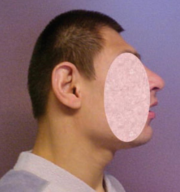
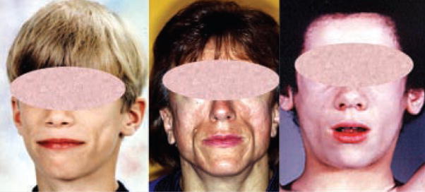
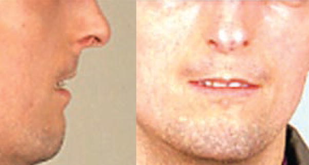
Retrognathia. The lower jaw is set back from the plane of the face.
The midpoint of the mandible (mental protuberance) and overlying soft tissue is broader than usual.
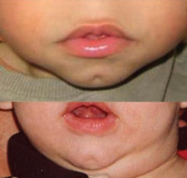
Note the midline depression of the skin over the fat pad of the chin.

Note the horizontal crease or fold situated below the vermilion border of the lower lip and above the fatty pad of the chin.
Note the H-shaped crease in the fat pad of the chin.
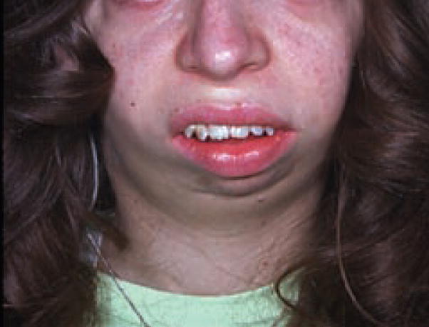
Note the marked tapering of the lower face to the chin with the two sides of the mandible meeting at an acute angle.
Note the reduced vertical distance from the vermilion border of the lower lip to the inferior-most point of the chin.
Note the increased vertical distance from the vermillion border of the lower lip to the inferior-most point of the chin.
Note the vertical crease in the fat pad of the chin.
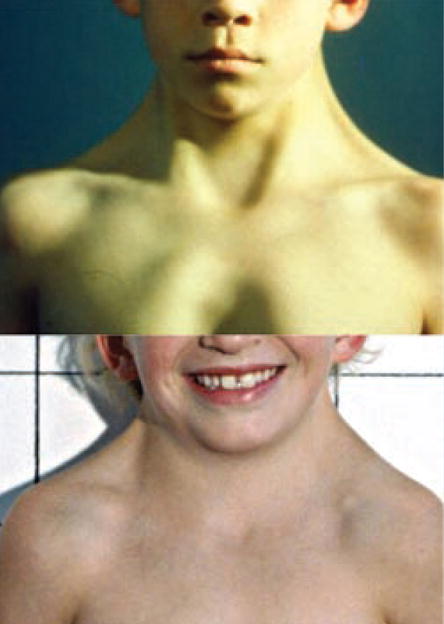
Note the increased width of the neck.
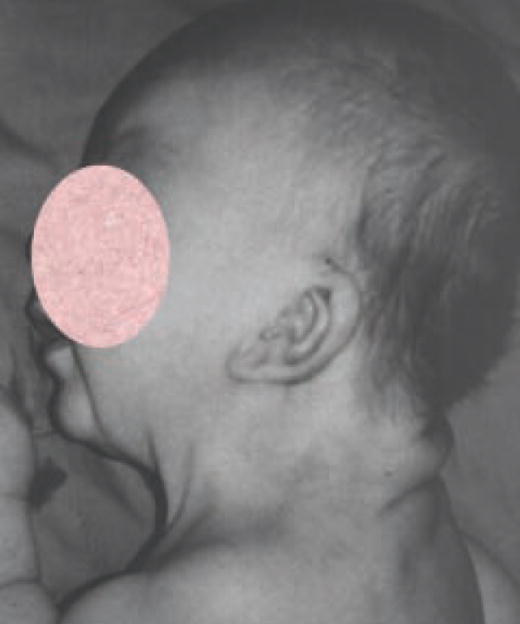
Note the increased distance from the point where neck and shoulders meet to the inferior margin of the occipital bone.
Note the decreased distance from the point where neck and shoulders meet to the inferior margin of the occipital bone.
Note the bilateral folds of skin on the posterolateral aspect of the neck.
Note the excess skin around the neck.
References
-
- Carey JC, Cohen MM, Jr, Curry C, Devriendt K, Holmes L, Verloes A. Elements of morphology: Standard terminology for the lips, mouth, and oral region. Am J Med Genet Part A. 2008;149A:77–92. - PubMed
-
- Farkas LG. Anthropometry of the head and face in medicine. New York: Elsevier; 1981.
-
- Hall JG, Allanson JE, Gripp K, Slavotinek A. Handbook of normal physical measurements. 2. New York: Oxford University Press; 2007.
MeSH terms
Grants and funding
LinkOut - more resources
Full Text Sources
Other Literature Sources
Medical
Molecular Biology Databases
