In vitro examination of ontogenesis of developing neuronal cells in vagal nuclei in medulla oblongata in newborns
- PMID: 19125713
- PMCID: PMC5677285
- DOI: 10.17305/bjbms.2008.2904
In vitro examination of ontogenesis of developing neuronal cells in vagal nuclei in medulla oblongata in newborns
Abstract
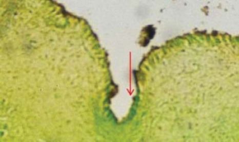
The development of neuron cells in vagal nerve nuclei in medulla oblongata was studied in vitro in live newborns and stillborns from different cases. Morphological changes were studied in respiratory nuclei of dorsal motor centre (DMNV) and nucleus tractus solitarius (NTS) in medulla oblongata. The material from medulla oblongata was fixated in 10 micro buffered formalin solution. Fixated material was cut in series of 10mu thickness, with starting point from obex in +/- 4 mm thickness. Special histochemical and histoenzymatic methods for central nervous system were used: cresyl echt violet coloring, tolyidin blue, Sevier-Munger modification and Grimelius coloring. In immature newborns (abortions and immature) in dorsal motor nucleus of the vagus (DMNV) population stages S1, S2, S3 are dominant. In neuron population in vagal sensory nuclei (NTS) stages S1, S2 are dominant. In more advanced stages of development of newborns (premature), in DMNV stages S3 and S4 are seen and in NTS stages S2 and S3 are dominant. In mature phase of newborns (maturity) in vagal nucleus DMNV stages S5 and S6 are dominant, while in sensory nucleus NTS stages S4 and S5 are dominant. These data suggest that neuron population in dorsal motor nucleus of the vagus (DMNV) are more advanced in neuronal maturity in comparison with sensory neuron population of vagal sensory nucleus NTS. This occurrence shows that phylogenetic development of motor complex is more advanced than the sensory one, which is expected to take new information's from the extra uterine life after birth (extra uterine vagal phenotype).
Figures


References
-
- Erlichmana JS. Nicotinic receptors mediate spontaneous GABA release in the rat dorsal motor nucleus of the vagus. Neuroscience Aug. 1997;79:671–681. - PubMed
-
- Goodwin BP, Anderson GF, Barraco RA. Characterization of muscarinic receptors in the rat NTS. Neurosci Lett May. 1995;19:131–135. - PubMed
-
- Miguel MP. Neuropathologic correlates of perinatal asphyxia. Int. Pediatr. 2000;15(4):221–228.
-
- Kenney HC, Armstrong DD. Perinatal Neuropathology. In: Graham DI, Lantos PL, editors. Greenfield’s Neuropathology. London: Arnold Publushers; 1997.
-
- Fitzgerald JT. Basic and Applied. Tindal. Vol. 122. London: 1985. Neuroanatomy; pp. 146–148.