Review
. 2009 Feb;126(2):147-64.
doi: 10.1111/j.1365-2567.2008.02848.x.

Tricks with tetramers: how to get the most from multimeric peptide-MHC

Affiliations
Review

Tricks with tetramers: how to get the most from multimeric peptide-MHC

Linda Wooldridge et al. Immunology. 2009 Feb.

Erratum in

  • Immunology. 2009 Mar;126(3):447

Abstract

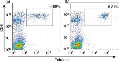
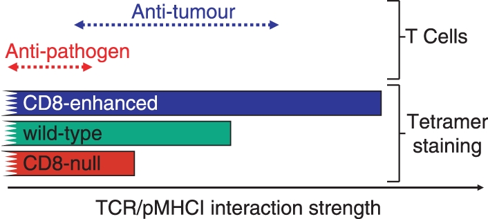
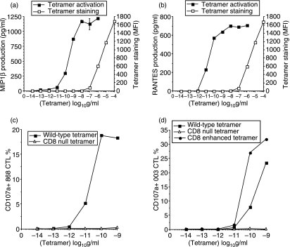
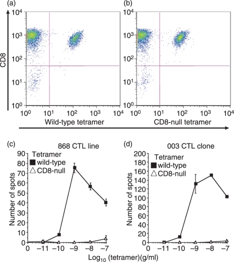
The development of fluorochrome-conjugated peptide-major histocompatibility complex (pMHC) multimers in conjunction with continuing advances in flow cytometry has transformed the study of antigen-specific T cells by enabling their visualization, enumeration, phenotypic characterization and isolation from ex vivo samples. Here, we bring together and discuss some of the 'tricks' that can be used to get the most out of pMHC multimers. These include: (1) simple procedures that can substantially enhance the staining intensity of cognate T cells with pMHC multimers; (2) the use of pMHC multimers to stain T cells with very-low-affinity T-cell receptor (TCR)/pMHC interactions, such as those that typically predominate in tumour-specific responses; and (3) the physical grading and clonotypic dissection of antigen-specific T cells based on the affinity of their cognate TCR using mutant pMHC multimers in conjunction with new approaches to the molecular analysis of TCR gene expression. We also examine how soluble pMHC can be used to examine T-cell activation, manipulate T-cell responses and study allogeneic and superantigen interactions with TCRs. Finally, we discuss the problems that arise with pMHC class II (pMHCII) multimers because of the low affinity of TCR/pMHCII interactions and lack of 'coreceptor help'.


Figures

Figure 1
Figure 1
Multivalent T-cell receptor/peptide–major histocompatibility complex (TCR/pMHC) binding leads to a considerable avidity ‘bonus effect’ as the result of cooperate interactivity and extends the interaction half-life. Streptavidin was linked to a BIAcore™ (GE Healthcare Ltd) CM-5 chip by amine coupling, biotin-tagged pMHCI was loaded onto each flow cell, and data were collected at 25° with a flow rate of 5 μl/min. Five microlitres of biotinylated TCR monomer at 1 mg/ml and 25 μl of TCR tetramer at 50 μg/ml were flowed over all flow cells. Negligible responses were observed to non-cognate pMHCI for both TCR monomers and tetramers. To facilitate visual comparison of monomer and tetramer binding events, the much larger monomer response values were normalized to the peak values for the tetramers. Kinetic binding parameters for the tetramers were estimated using BIAEvaluation™ software as described in ref [1]. There are two apparent off-rates for the TCR tetramers: (1) a minority fast off-rate thought to correspond to those tetramers binding less than three antigens; and, (2) a slow (true) off-rate for those tetramers probably binding three pMHCI molecules. The latter half-life is shown. Some irreversible binding of biotinylated TCR monomers is observed owing to incomplete blocking of the streptavidin-coated chip surface with soluble biotin. Representative data are shown. Curves are the best fit of the model described in. Data reproduced from with permission.
Figure 2
Figure 2
Tetrahedral avidin–biotin-based peptide–major histocompatibility complex class I (pMHCI) tetramers engage three T-cell receptors (TCRs) and three CD8 molecules at the cell surface.
Figure 3
Figure 3
The T-cell receptor/peptide–major histocompatibility complex (TCR/pMHC) interaction threshold for tetramer staining varies with CD8 engagement and temperature. Staining of cytotoxic T-lymphocyte (CTL) clone ILA1 with seven different human telomerase reverse transcriptase (hTERT540–548) variants, as indicated, refolded with wild-type human leucocyte antigen (HLA) A2 (a) or CD8-null HLA A2 D227K/T228A (b) at 37°. The mean fluorescence intensity (MFI) values observed with pMHCI tetramer staining are plotted against the TCR/pMHCI interaction KD values for experiments conducted at 37° (c) and 4° (d) with wild-type HLA A2 and CD8-null HLA A2 molecules for each variant added at a final concentration of 220 nm (10 μg/ml); colour codes correspond to those shown in (a) and (b). Staining with the set of APLs refolded with each type of heavy chain was performed at least three times. Representative data are shown. Curves are the best fit of the model described in Laugel et al. Data reproduced from with permission.
Figure 4
Figure 4
Peptide–major histocompatibility complex (pMHC) multimer concentration differentially affects staining of cognate T cells. The ILA-1 cytotoxic T-lymphocyte (CTL) clone was stained with two human telomerase reverse transcriptase (hTERT540–548) peptide variant human leucocyte antigen (HLA) A*0201 tetramers at a number of different concentrations (5, 2, 0·4 and 0·2 μg/ml as indicated). Staining with the low-affinity 4L variant (KD = 117 μm) is shown in the left panel; staining with the high-affinity 3G8T variant (KD = 4·04 μm) is shown in the right panel. The data demonstrate that tetramer staining with low-affinity ligands is far more dependent on pMHC concentration; higher tetramer concentrations can therefore improve visualization of T cells that bind cognate ligand with low TCR/pMHCI affinities.
Figure 5
Figure 5
Incubation with the protein tyrosine kinase inhibitor dasatinib substantially improves the staining of cognate T cells with peptide–major histocompatibility complex class I (pMHCI) tetramers. (a) A cytotoxic T-lymphocyte (CTL) line specific for the melanoma-derived EAAGIGILTV epitope was stained with cognate human leucocyte antigen (HLA) A2 tetramer at a concentration of 10 μg/ml. (b) The same experiment performed after preincubation of the CTL line with 50 nm dasatinib for 15 min at 37°. As shown, dasatinib treatment substantially improves the staining of cognate T cells in the line without affecting the staining of non-cognate T cells; this enhancement varies from two to 50-fold and is greatest for the cognate T-cell populations that bind tetramer poorly. Similar results have been observed in a wide range of systems.
Figure 6
Figure 6
CD8-null, wild-type and CD8-enhanced peptide–major histocompatibility complex class I (pMHCI) tetramers enable the staining of T cells with increasingly weak T-cell receptor (TCR)/pMHCI interactions. CD8-null tetramers require a high TCR/pMHC affinity to stain cognate T cells. Wild-type tetramers can stain cells with weaker affinity interactions. The extra help afforded by CD8-enhanced tetramers allows them to stain cells when the TCR/pMHCI interaction is even weaker. The use of these three types of reagent enables the grading of T cells based on the strength of their interaction with the soluble pMHC ligand. Most natural human anti-pathogen TCR/pMHCI interactions fall within a range that allows staining with CD8-null tetramers. Many anti-tumour T cells bear TCRs that interact weakly with pMHCI and thus require CD8 help or enhanced CD8 help to be visualized successfully with tetramers.
Figure 7
Figure 7
T-cell activation by soluble peptide–major histocompatibility complex class I (pMHCI) tetramers is very sensitive. Tetramer staining (□) and tetramer-induced MIP1β (a) and RANTES (b) production (▪) by the 003 cytotoxic T-lymphocyte (CTL) clone specific for the human immunodeficiency virus-derived human leucocyte antigen (HLA) A2-restricted Gag epitope SLYNTVATL (residues 77–85). In each case, staining is only just visible by flow cytometry at a tetramer concentration of 100 ng/ml, whereas tetramer-induced activation occurs at < 100 pg/ml. The 868 T-cell receptor (TCR), which recognizes the same antigen, binds cognate pMHCI with a KD of < 150 nm by surface plasmon resonance, thereby making it the strongest TCR/pMHCI interaction measured to date. Tetramer decay experiments indicate that the 003 TCR binds with even higher affinity. These high affinities ensure that tetramer staining of these CTL is virtually independent of CD8 binding. Despite binding well to cells and cross-linking similar amounts of TCR, CD8-null tetramers are unable to induce degranulation of either cell (c,d). CD8-enhanced (Q115E) tetramers are more antigenic that wild-type tetramers (d).
Figure 8
Figure 8
Peptide–major histocompatibility complex (pMHCI) tetramer-induced activation does not require cell–cell contact. The 868 cytotoxic T-lymphocyte (CTL) line specific for the human immunodeficiency virus-derived human leucocyte antigen (HLA) A2-restricted Gag epitope SLYNTVATL (residues 77–85) stains well with both wild-type and CD8-null (D227K/T228A) cognate tetramers at 5 μg/ml (a,b). Staining intensity with CD8-null tetramer is slightly reduced when compared to wild-type tetramer. The 003 CTL clone exhibits less of a difference in staining with the two types of reagent than the 868 CTL line; indeed, staining of 003 CTL with the CD8-null tetramer is only ∼ 50% lower than that seen with the corresponding wild-type tetramer even when used at 10−8 g/ml, the lowest concentration at which we have been able to visualize staining by flow cytometry. CD8-null tetramers stain, but generally fail to activate, CTL., Tetramer-induced activation results in a wide range of effector functions including interferon-γ (IFN-γ) release. In the activation experiments shown, 1000 cells from the 868 CTL line (c) or the 003 CTL clone (d) were used in an IFN-γ ELISpot without antigen-presenting cells. These conditions minimize cell–cell contact and largely prevent T cells from representing peptide antigen to each other. Even under these conditions, wild-type but not CD8-null tetramers are able to activate cells and induce IFN-γ production; the CD8-null tetramers in these experiments serve as an additional control for peptide representation. Higher concentrations of tetramer are known to induce rapid apoptosis of these CTL. This cell death probably accounts for the decrease in spot-forming cells apparent at tetramer concentrations of > 100 pg/ml.

References

    1. Laugel B, Boulter JM, Lissin N, et al. Design of soluble recombinant T cell receptors for antigen targeting and T cell inhibition. J Biol Chem. 2005;280:1882–92. - PubMed
    1. Altman JD, Moss PA, Goulder PJ, Barouch DH, McHeyzer-Williams MG, Bell JI, McMichael AJ, Davis MM. Phenotypic analysis of antigen-specific T lymphocytes. Science. 1996;274:94–6. - PubMed
    1. Wooldridge L, van den Berg HA, Glick M, et al. Interaction between the CD8 coreceptor and major histocompatibility complex class I stabilizes T cell receptor–antigen complexes at the cell surface. J Biol Chem. 2005;280:27491–501. - PMC - PubMed
    1. Guillaume P, Legler DF, Boucheron N, Doucey MA, Cerottini JC, Luescher IF. Soluble major histocompatibility complex–peptide octamers with impaired CD8 binding selectively induce Fas-dependent apoptosis. J Biol Chem. 2003;278:4500–9. - PubMed
    1. Bakker AH, Schumacher TN. MHC multimer technology: current status and future prospects. Curr Opin Immunol. 2005;17:428–33. - PubMed

MeSH terms