

An integrated optoacoustic transducer combining etalon and black PDMS structures
- PMID: 19126497
- PMCID: PMC2771400
- DOI: 10.1109/TUFFC.2008.988
An integrated optoacoustic transducer combining etalon and black PDMS structures
Abstract
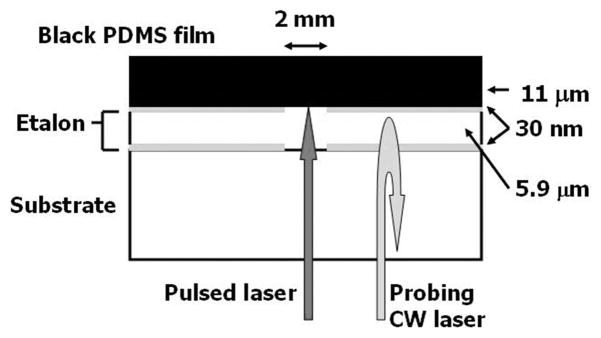
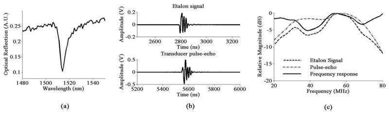
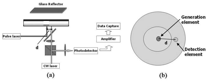
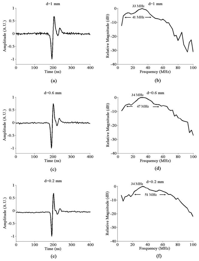
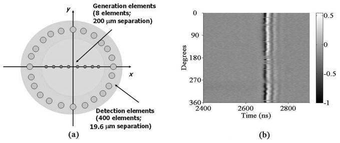
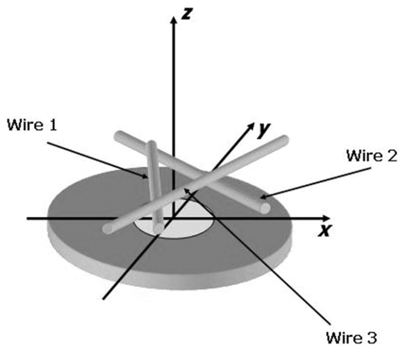
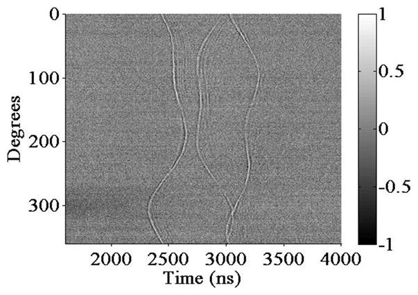
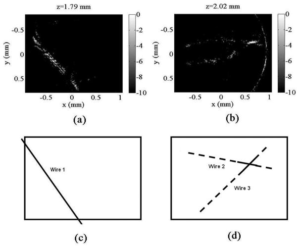
An integrated optoacoustic transducer combining etalon and black polydimethylsiloxane (PDMS) structures has been designed and developed. The device consists of an 11-μm-thick black PDMS film confined to a 2-mm-diameter circular region acting as an optoacoustic transmitter, surrounded by a 5.9-μm Fabry-Perot polymer etalon structure serving as an optoacoustic detector array. A pulsed laser is focused onto a 30-μm spot on the black PDMS film, defining the transmit element, while a CW laser probes a 20-μm spot on the etalon for ultrasound detection. Pulse-echo signals display center frequencies of above 30 MHz with bandwidths of at least 40 MHz. A theta-array is formed for 3-D ultrasound imaging by mechanically scanning the generation laser along a 1-D array and the detection laser around an annular array. Preliminary images with 3 metal wires as imaging targets are presented. Characterization of the device’s acoustical properties, as well as preliminary imaging results, suggest that all-optical ultrasound transducers are potential alternatives to piezoelectric techniques for high-frequency 2-D arrays enabling 3-D high-resolution ultrasound imaging.
Figures
References
-
- Turnbull DH, Starkoski BG, Harasiewicz KA, Semple JL, From L, Gupta AK, Sauder DN, Foster FS. A 40–100 MHz B-scan ultrasound backscatter microscope for skin imaging. Ultrasound Med Biol. 1995;21(1):79–88. - PubMed
-
- Passman C, Ermert H. A 100 MHz ultrasound imaging system for dermatologic and ophthalmologic diagnostics. IEEE Trans Ultrason Ferroelectr Freq Control. 1996;43(4):545–552.
-
- Foster FS, Pavlin CJ, Lockwood GR, Ryan LK, Harasiewicz KA, Berube L, Rauth AM. Principles and applications of ultrasonic backscatter microscopy. IEEE Trans Ultrason Ferroelectr Freq Control. 1993;40(5):608–616. - PubMed
-
- Coleman DJ, Silverman RH, Chabi A, Rondeau MJ, Shung KK, Cannata J, Lincoff H. High-resolution ultrasonic imaging of the posterior segment. Ophthalmology. 2004;111(7):1344–1351. - PubMed
-
- White RA, Donayre CE, Kopchock GE, Walot I, Mehinger CM, Wilson EP, de Virgilio C. Vascular imaging before, during and after endovascular repair. World J Surg. 1996;20(6):622–629. - PubMed
Publication types
MeSH terms
Substances
Grants and funding
LinkOut - more resources
Full Text Sources
Other Literature Sources
