. 2009 Mar 13;284(11):7177-89.
doi: 10.1074/jbc.M808895200. Epub 2009 Jan 5.

Curvature dynamics of alpha-synuclein familial Parkinson disease mutants: molecular simulations of the micelle- and bilayer-bound forms

Affiliations

Curvature dynamics of alpha-synuclein familial Parkinson disease mutants: molecular simulations of the micelle- and bilayer-bound forms

Jason D Perlmutter et al. J Biol Chem. .

Abstract

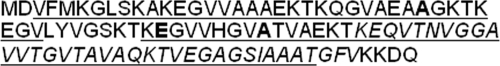
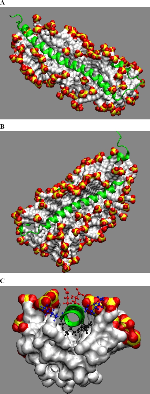
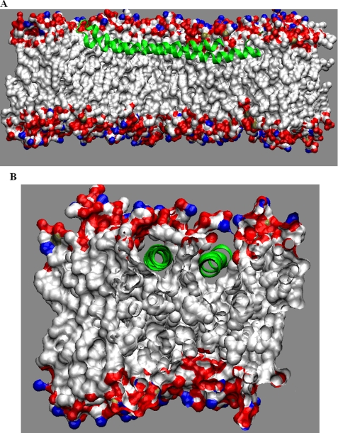
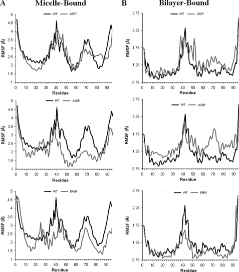
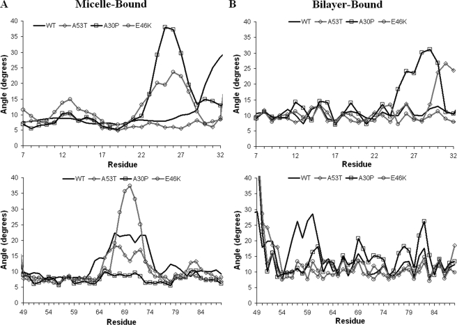
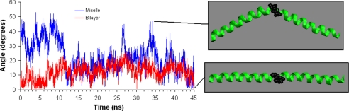
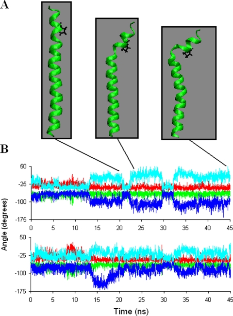
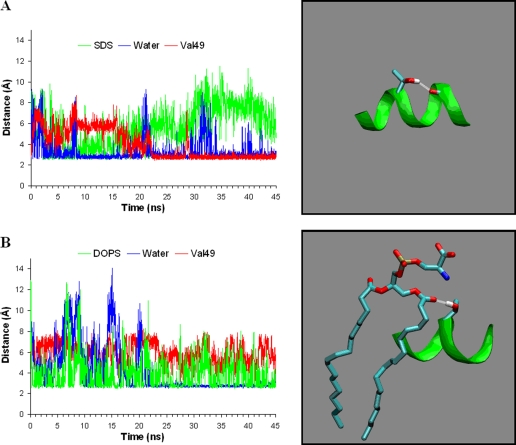
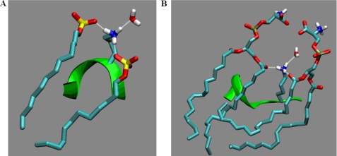
Alpha-synuclein remains a protein of interest due to its propensity to form fibrillar aggregates in neurodegenerative disease and its putative function in synaptic vesicle regulation. Herein, we present a series of atomistic molecular dynamics simulations of wild-type alpha-synuclein and three Parkinson disease familial mutants (A30P, A53T, and E46K) in two distinct environments. First, in order to match recent NMR experiments, we have simulated each protein bound to an SDS detergent micelle. Second, in order to connect more closely to the true biological environment, we have simulated the proteins bound to a 1,2-dioleoyl-sn-glycero-3-phosphoserine lipid bilayer. In the micelle-bound case, we find that the wild type and all of the variants of alpha-synuclein flatten the underlying micelle, decreasing its surface area. A30P is known to lessen alpha-synuclein/membrane affinity and, consistent with experiment, destabilizes the simulated secondary structure. In the case of A53T, our simulations reveal a range of stabilizing hydrogen bonds that form with the threonine. In both environments, the E46K mutation, which is known to increase bilayer affinity, leads to an additional hydrogen bond between the protein and either the detergent or lipid. Simulations indicate that alphaS and its variants are less dynamic in the bilayer than in the micelle. Furthermore, the simulations of the mutants suggest how changes in the structure and dynamics of alpha-synuclein may affect its biological role.


Figures

FIGURE 1.
FIGURE 1.
Amino acid sequence of membrane binding domain of αS. Only the first 99 amino acids are involved in lipid binding and were considered in these simulations. Underlined are the two helices in the lipid binding domain. The positions in the wild-type where the PD familial mutations occur are in boldface type. The highly hydrophobic NAC region consisting of residues 60-95 is in italic type.
FIGURE 2.
FIGURE 2.
The interaction of αS with an SDS micelle. A and B, snapshots of wild-type αS bound to the detergent micelle, after 45 ns of simulated dynamics. The protein embeds deeply into the core of the micelle, forming channels in both helix-C (A) and helix-N (B). C, a representative slice through the micelle, illustrating typical side chain orientations. Nonpolar side chains are directed toward the micelle center, basic side chains orient along the micelle surface, and acidic side chains orient toward the water. αS backbone is represented as a green ribbon, and side chains are represented with the following colors: nonpolar (black), basic (blue), and acidic (red). The SDS micelle is shown with the following color scheme: carbon (white), sulfur (yellow), and oxygen (red). Hydrogens, water, and ions have been removed for clarity of presentation.
FIGURE 3.
FIGURE 3.
The interaction of αS with a DOPS bilayer. A and B, snapshots of wild-type αS bound to the DOPS bilayer, after 45 ns of simulated dynamics. The protein embeds into the hydrophobic core of the bilayer, beneath the lipid headgroup.αS backbone is represented as a green ribbon, and the DOPS bilayer is shown with the following color scheme: carbon (white), oxygen (red), nitrogen (blue), and phosphorus (tan). Hydrogen, water, ions, and lipids have been removed for clarity of presentation.
FIGURE 4.
FIGURE 4.
Depth of αS in the DOPS bilayer. A, component electron density profiles (EDP) for DOPS bilayer and wild-type αS. B, Cα electron density, describing relative backbone depth of the protein helices.
FIGURE 5.
FIGURE 5.
Structural dynamics of the micelle- and bilayer-bound forms. Shown is the r.m.s.f. of αSCα calculated from the eight simulations, in each case averaged over the last 25 ns for the micelle-bound (A) and bilayer-bound (B) states.
FIGURE 6.
FIGURE 6.
Helical bending. The angle formed by the intersection of the residues four up- and downstream at each helical position describes the structural adaptation of the protein to the environment for the micelle-bound (A) and bilayer-bound (B) states.
FIGURE 7.
FIGURE 7.
Helical bending at Gly67/Gly68 is highly dynamic. The bending angle at Gly67/Gly68 was calculated at each time point in helix-C, as described under “Results,” for the micelle-bound (blue) and bilayer-bound (red) states. The micelle-bound state shows a large range of values, capturing nearly straight as well as highly bent structures, whereas the bilayer-bound state remains relatively straight. Representative snapshots from the micelle-bound state at the indicated time points show minimal and maximal bending conformations (the micelle, water, and ions have been omitted). The helix is represented as a green ribbon, and Gly67/Gly68 are shown in the space-filling representation as black.
FIGURE 8.
FIGURE 8.
The A30P mutation causes a decrease in helicity. A, snapshots illustrating different conformations of the A30P mutant from the micelle-bound simulation: straight helix, kinked helix, and unfolded (time points 21, 24, and 45 ns). Helix-N is represented as a green ribbon, and the proline is shown in black (micelle, water, and ions omitted). B, torsion angles for the A30P substitution mutant and wild-type protein for residues 26 and 28 from the micelle-bound simulations show reversible unfolding. The color scheme is as follows: WT phi (green), WT psi (red), A30P phi (dark blue), A30P psi (light blue).
FIGURE 9.
FIGURE 9.
Hydrogen bonding in the A53T mutant stabilizes the helicity. A, minimum distance between Thr53 side chain donor oxygen and water oxygens (blue), SDS oxygen hydrogen bond acceptors (green), and Val49 backbone carbonyl oxygen (red). Shown is a representative snapshot illustrating the hydrogen bond between the Thr53 side chain and Val49 backbone taken from the micelle-bound simulation. B, minimum distance between Thr53 side chain donor oxygen and water oxygens (blue), DOPS oxygen hydrogen bond acceptors (green), and Val49 backbone carbonyl oxygen (red). Shown is a representative snapshot illustrating the hydrogen bond between the Thr53 side chain and DOPS carbonyl from bilayer simulation. Micelle, bilayer, water, ions, and hydrogens have been omitted for clarity. Backbone is represented as a green ribbon; Thr53, Val49, and DOPS are represented with the following color scheme: threonine donor hydrogen (white), carbon (light blue), oxygen (red), nitrogen (blue), phosphorus (tan).
FIGURE 10.
FIGURE 10.
Hydrogen bonding in the E46K mutant. Representative snapshot illustrating the hydrogen bond between the Lys46 side chain and SDS detergent (A) or DOPS carbonyl (B) and water (other detergent molecules, lipids, water, ions, and aliphatic hydrogens omitted). Backbone is represented as a green ribbon; Lys46, SDS, DOPS, and water are represented with the following color scheme: hydrogen (white), carbon (light blue), sulfur (yellow), nitrogen (blue), and oxygen (red).

References

    1. Spillantini, M. G., Crowther, R. A., Jakes, R., Hasegawa, M., and Goedert, M. (1998) Proc. Natl. Acad. Sci. U. S. A. 95 6469-6473 - PMC - PubMed
    1. Kruger, R., Kuhn, W., Muller, T., Woitalla, D., Graeber, M., Kosel, S., Przuntek, H., Epplen, J. T., Schols, L., and Riess, O. (1998) Nat. Genet. 18 106-108 - PubMed
    1. Polymeropoulos, M. H., Lavedan, C., Leroy, E., Ide, S. E., Dehejia, A., Dutra, A., Pike, B., Root, H., Rubenstein, J., Boyer, R., Stenroos, E. S., Chandrasekharappa, S., Athanassiadou, A., Papapetropoulos, T., Johnson, W. G., Lazzarini, A. M., Duvoisin, R. C., Di Iorio, G., Golbe, L. I., and Nussbaum, R. L. (1997) Science 276 2045-2047 - PubMed
    1. Zarranz, J. J., Alegre, J., Gomez-Esteban, J. C., Lezcano, E., Ros, R., Ampuero, I., Vidal, L., Hoenicka, J., Rodriguez, O., Atares, B., Llorens, V., Gomez Tortosa, E., del Ser, T., Munoz, D. G., and de Yebenes, J. G. (2004) Ann. Neurol. 55 164-173 - PubMed
    1. Conway, K. A., Lee, S. J., Rochet, J. C., Ding, T. T., Williamson, R. E., and Lansbury, P. T., Jr. (2000) Proc. Natl. Acad. Sci. U. S. A. 97 571-576 - PMC - PubMed

LinkOut - more resources