. 2009 May;23(5):1347-57.
doi: 10.1096/fj.08-121574. Epub 2009 Jan 6.

Essential role of Rac1 and Rac3 GTPases in neuronal development

Affiliations

Essential role of Rac1 and Rac3 GTPases in neuronal development

Sara Corbetta et al. FASEB J. 2009 May.

Abstract

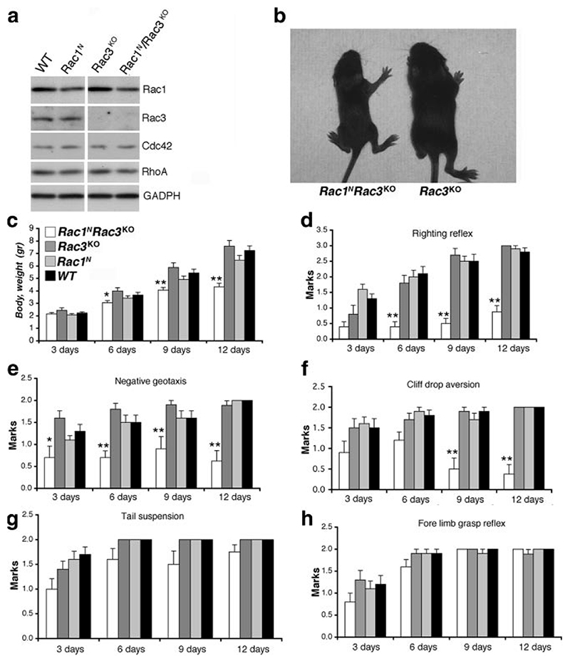
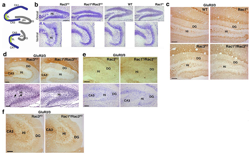
Rac GTPases are members of the Rho family regulating the actin cytoskeleton and implicated in neuronal development. Ubiquitous Rac1 and neuron-specific Rac3 GTPases are coexpressed in the developing mammalian brain. We used Cre-mediated conditional deletion of Rac1 in neurons combined with knockout of neuron-specific Rac3 to study the role of these GTPases in neural development. We found that lack of both genes causes motor behavioral defects, epilepsy, and premature death of mice. Deletion of either GTPase does not produce evident phenotypes. Double-knockout mice show specific defects in the development of the hippocampus. Selective impairment of the dorsal hilus of double-knockout animals is associated with alteration in the formation of the hippocampal circuitry. Axonal pathways to and from the dorsal hilus are affected because of the deficit of hilar mossy cells. Moreover, analysis of Rac function in hippocampal cultures shows that spine formation is strongly hampered only in neurons lacking both Rac proteins. These findings show for the first time that both Rac1 and Rac3 are important for the development of the nervous system, wherein they play complementary roles during late stages of neuronal and brain development.


Figures

Figure 1
Figure 1
Relative amounts of Rac3 and Rac1 in P7 mouse brain. Duplicate samples from P7 brain lysates from wild-type animals were immunoprecipitated (2 mg protein lysate/immunoprecipitation) with anti-Rac3 antibody and blotted with either anti-Rac1 recognizing both Rac1 and Rac3 (top filter) or anti-Rac3-specific antibodies (bottom filter). Lane 1: 200 μg of lysate; lanes 2 and 4: 200 μg of unbound fractions after immunoprecipitation with preimmune or immune anti-Rac3 Ab, respectively; lanes 3 and 5: immunoprecipitation from 2 mg of lysate with preimmune or immune anti-Rac3 antibody, respectively. Quantification on blots (see Materials and Methods) from two independent experiments shows that Rac3 and Rac1 represent ~7.6 ± 0.4 and 92.4 ± 0.44%, respectively, of the total Rac in P7 mouse brain lysates.
Figure 2
Figure 2. Activation of the SynI-Cre transgene.
X-Gal staining on P13 (a–c) and P8 (d) SynI-Cre/ROSA26 brain sections. In the hippocampus of P13 (b) and P8 mice (d), SynI-Cre expression is restricted mostly to the CA3 and dentate gyrus, including the hilus (c), and is absent in most cells of the CA1 region. Cb, cerebellum; Co, cortex; DG, dentate gyrus; Hi, hilus; Hp, hippocampus, Ob, olfactory bulb; Pn, pons; Th, thalamus. Scale bars = 500 μm (a); 200 μm (b, d); 100 μm (c).
Figure 3
Figure 3. Production of Rac1N and Rac1N/Rac3KO mice.
a) Scheme of the Rac1 flox allele before (left) and after (right) Cre-mediated recombination. b, c) Genotyping by PCR on DNA from tail (b) and spinal cord and kidney (c) of mice with the indicated genotypes. DNAs were screened with primer (Pr) 2 and Pr3 to produce a 0.33-kb fragment from the floxed allele (F) and a 0.27-kb fragment from the wild-type allele (wt). The 0.17-kb PCR fragment from the deleted allele (del) was obtained with Pr1 and Pr3 from spinal cord. b) A 0.45-kb PCR fragment (Cre) was obtained in the SynI-Cre-positive transgenic mice with Pr4 and Pr5. PrF1 and PrR3 generate a 0.37-kb fragment from the Rac3 mutant allele (Rac3KO). Sequences of all primers are reported in Materials and Methods. d) Northern blot analysis on total RNA (15 μg/lane) hybridized with probes specific for Rac1 (left panels) or Rac2 (right panels; spleen as a positive control for Rac2). Bottom panels: gels used for blotting, stained with ethidium bromide.
Figure 4
Figure 4. Characterization of double-knockout mice.
a) Immunoblotting for the indicated antigens on P13 brain lysates. Rac1 is decreased in brains of Rac1N and Rac1N/Rac3KO (P<0.035) mice. Levels of Cdc42 and RhoA appear unaltered in lysates from mutant mice. b) Rac1N/Rac3KO (left) and Rac3KO (right) P13 mice. c-h) Body weight (c), righting reflex (d), negative geotaxis (e), cliff drop aversion (f), tail suspension (g), and forelimb grasp reflex (h) were assessed as described in Material and Methods. Bars and error bars represent mean ± se scores; 10 animals/group.*P < 0.05, **P < 0.005 vs. control littermates; Student’s t test.
Figure 5
Figure 5. Reduced numbers of mossy cells in Rac1N/Rac3KO dorsal hilus at P13 and P7.
a) Scheme of the dorsal and ventral hippocampus. DG, dentate gyrus; dHi, dorsal hilus; vHi, ventral hilus; SL, stratum lucidum. b) Nissl staining on sections of dorsal (top) and ventral (bottom) dentate gyrus from P13 Rac3KO, Rac1N/Rac3KO, WT, and Rac1N mice, showing reduced thickness of the dorsal hilus in the hippocampus of Rac1N/Rac3KO mice. c) Immunohistochemistry on P13 brain sections immunostained for GluR2/3. GluR2/3-positive cells are strongly reduced in the dentate hilus of P13 Rac1N/Rac3KO mice compared with Rac3KO, WT, and Rac1N mice. d) Immunostaining for GluR2/3-positive cells (top) and hematoxylin and eosin staining (bottom) on P7 brain sections. Large cells (arrows) are strongly reduced in the dorsal hilus of P7 Rac1N/Rac3KO mice compared with P7 Rac3KO mice. e) Immunostaining for GluR2/3 (top) and Nissl staining (bottom) of sections from P4 Rac3KO and Rac1N/Rac3KO mice. No differences are detectable in the hilus of P4 double-knockout mice compared with Rac3KO mice. GluR2/3-positive mossy cells are not detectable at P4. f) Sections from the ventral hippocampus of P13 mice immunostained for GluR2/3: the reduction in mossy cells is not evident in the ventral hilus of Rac1N/Rac3KO mice compared with control Rac3KO mice. Scale bars = 200 μm (b); 100 μm (c–f).
Figure 6
Figure 6. Apoptosis is not affected by deletion of Rac1 and Rac3 in the dorsal hippocampus.
Immunostaining with anti-active caspase-3 antibody (green) and DAPI (blue). a, b) P4 (a),and P7 (b) dorsal hippocampi. Caspase-positive apoptotic cells are rarely seen in Rac3KO and double-knockout mice. c) Sections of the interdigit region from the anterior limb of E13.5 wild-type mice were used as positive controls for apoptosis. DG, dentate gyrus; Hi, hilus. Scale bars = 100 μm.
Figure 7
Figure 7. Mossy cell axonal projections to dentate granule cells are reduced in the dorsal hilus of Rac1N/Rac3KO mice.
a) Scheme of major connections within the hippocampus involving hilar mossy cells and dentate granule cells. Granule cells send their axons (mossy fibers) both to mossy cells in the hilus and to pyramidal neurons in the CA3. Mossy cells are bidirectionally linked to granule cells by a positive feedback loop that is strategically placed between the entorhinal cortex and the hippocampal CA3 region. b) Immunofluorescence on sections of P13 brains stained for calretinin (green) that stains immature granule cells near the hilus (Hi), and mossy cell axonal projections to the inner molecular layer (IML). The calretinin-positive projections of mossy cells to the IML of the dorsal dentate gyrus (arrows) are strongly reduced in Rac1N/Rac3KO mice compared with Rac3KO mice. GCL, granule cell layer. c) Quantification of the thickness of the inner molecular layer positive for calretinin and of the density of the calretinin signal in the inner molecular layer of the dorsal hippocampi of Rac1N/Rac3KO (dKO) and Rac3KO mice. Values represent mean ± se percentages with respect to control Rac3KO mice (n=8) after normalization (Rac3KO mice = 100%). d) Photomicrographs of ventral hippocampus of Rac3KO and Rac1N/Rac3KO mice stained with antibodies for calretinin (green). In the ventral hilus, mossy cells show strong immunoreactivity for calretinin. Calretinin-positive axonal projections and mossy cells are not affected in the ventral hippocampus of Rac1N/Rac3KO mice. Scale bars = 100 μm.
Figure 8
Figure 8. Alteration of circuitry in the dorsal hilus of P13 Rac1N/Rac3KO mice.
a) Distribution of synapsin I (green), tau (red), and nuclear DAPI (blue) in P13 hippocampi. Reduced thickness of the dorsal hilus (Hi) in Rac1N/Rac3KO mice correlates with reduced tau-positive axons and synapsin I-positive presynaptic terminals in this region. b) Enlargements of hilus from respective panels shown in a. c) Hippocampal sections from Rac3KO and Rac1N/Rac3KO P13 mice immunostained for synapsin (green) and MAP2 (red). A specific reduction of synapsin-positive presynaptic terminals and MAP2-positive dendrites is evident in the dorsal hilus (dHi) of Rac1N/Rac3KO mice compared with control Rac3KO mice (left panels). No differences between Rac3KO and Rac1N/Rac3KO mice are observed in the stratum lucidum (SL) of the dorsal CA3 subfield (central panels) or in the ventral hilus (vHi) (right panels). Scale bars = 100 μm (a); 25 μm (b); 50 μm (c).
Figure 9
Figure 9. Mossy fiber projections from granule cells to mossy cells are strongly reduced in Rac1N/Rac3KO mice.
a) Hippocampi from Rac3KO (top panels) and Rac1N/Rac3KO (bottom panels) P13 mice were stained with anti-ZnT-3 (green), a marker for granule cell mossy fibers from the dentate gyrus (DG), and with DAPI (blue). ZnT-3-positive axonal terminals are strongly reduced in the dorsal hilus (dHi) of Rac1N/Rac3KO mice compared with control Rac3KO mice (right panels), but not in the stratum lucidum (SL) of the dorsal CA3 subfield (left panels). Scale bars = 100 μm. b) Quantification of density of ZnT-3, MAP2, and synapsin I signals in the dorsal hilus of Rac1N/Rac3KO (dKO) and Rac3KO mice. Values represent mean ± se percentages with respect to control Rac3KO mice (n=8 for ZnT-3 and MAP2; n=7 for synapsin I) after normalization (Rac3KO mice = 100%).
Figure 10
Figure 10. Deletion of both Rac1 and Rac3 deeply affects spinogenesis.
a) Genomic DNA was extracted from the tail (T) of a Rac1F/+ SynI-Cre mouse, the spinal cord (SC) of a Rac1F/F SynI-Cre mouse, and 15 DIV hippocampal cultures obtained from two different Rac1F/+ SynI-Cre embryos (hc1 and hc2). DNAs were processed by PCR as detailed in Materials and Methods. PCR fragments are from floxed (F), wild-type (wt), and deleted (del) alleles. b) Hippocampal neurons from Rac1F/F mice were transfected at 4 DIV with EGFP and EGFP-Cre recombinase. Cotransfected cells fixed at 14 DIV were labeled with anti-GFP (green, cytoplasmic), anti-Cre (red, nuclear), and DAPI (blue). Same field is shown in both panels. Scale bar = 100 μm. c–f) Hippocampal neurons from Rac1F/F(c, d) or Rac1F/F/Rac3KO (e, f) mice were transfected at 4 DIV with GFP (c, e) or cotransfected with GFP-Cre and GFP (d, f). Cells were fixed at 14 DIV and stained with anti-GFP (green) and anti-PSD95 (red). Same fields are shown in the middle and bottom pictures of each panel. Scale bar = 5 μm. g) Quantification of the number of mature and immature spines on dendrites of hippocampal neurons, shown as average protrusion density (number/100 μm). Quantification was performed on ≥450 protrusions from 7-10 neurons per experiment per condition. Dendritic protrusions included mushroom and stubby spines, short (<4 μm) and long (>4 μm) filopodia, and lamellipodia. *P < 0.0005, **P < 0.00005 vs. respective control neurons; Student’s t test.

References

    1. Hall A. Rho GTPases and the actin cytoskeleton. Science. 1998;279:509–514. - PubMed
    1. Tada T, Sheng M. Molecular mechanisms of dendritic spine morphogenesis. Curr Opin Neurobiol. 2006;16:95–101. - PubMed
    1. De Curtis I. Functions of Rac GTPases during neuronal development. Dev Neurosci. 2008;30:47–58. - PubMed
    1. Corbetta S, Gualdoni S, Albertinazzi C, Paris S, Croci L, Consalez GG, de Curtis I. Generation and characterization of Rac3 knockout mice. Mol Cell Biol. 2005;25:5763–5776. doi: 10.1128/MCB.25.13.5763-5776.2005. - DOI - PMC - PubMed
    1. Didsbury J, Weber RF, Bokoch GM, Evans T, Snyderman R. Rac, a novel ras-related family of proteins that are botulinum toxin substrates. J Biol Chem. 1989;264:16378–16382. - PubMed

Publication types

LinkOut - more resources