. 2009 May;23(5):1415-30.
doi: 10.1096/fj.08-120568. Epub 2009 Jan 6.

Modification of surfactant protein D by reactive oxygen-nitrogen intermediates is accompanied by loss of aggregating activity, in vitro and in vivo

Affiliations

Modification of surfactant protein D by reactive oxygen-nitrogen intermediates is accompanied by loss of aggregating activity, in vitro and in vivo

Sadis Matalon et al. FASEB J. 2009 May.

Abstract

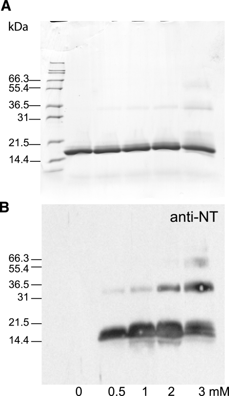
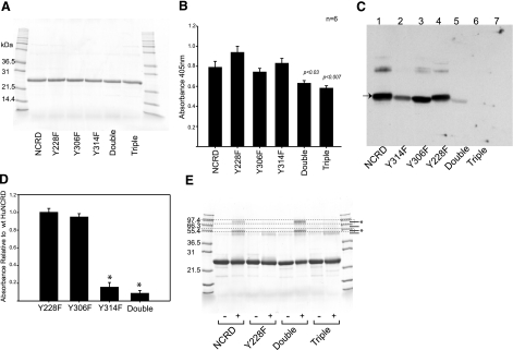
Surfactant protein D (SP-D) is an important effector of innate immunity. We have previously shown that SP-D accumulates at sites of acute bacterial infection and neutrophil infiltration, a setting associated with the release of reactive species such as peroxynitrite. Incubation of native SP-D or trimeric SP-D lectin domains (NCRDs) with peroxynitrite resulted in nitration and nondisulfide cross-linking. Modifications were blocked by peroxynitrite scavengers or pH inactivation of peroxynitrite, and mass spectroscopy confirmed nitration of conserved tyrosine residues within the C-terminal neck and lectin domains. Mutant NCRDs lacking one or more of the tyrosines allowed us to demonstrate preferential nitration of Tyr314 and the formation of Tyr228-dependent cross-links. Although there was no effect of peroxynitrite or tyrosine mutations on lectin activity, incubation of SP-D dodecamers or murine lavage with peroxynitrite decreased the SP-D-dependent aggregation of lipopolysaccharide-coated beads, supporting our hypothesis that defective aggregation results from abnormal cross-linking. We also observed nitration, cross-linking of SP-D, and a significant decrease in SP-D-dependent aggregating activity in the lavage of mice acutely exposed to nitrogen dioxide. Thus, modification of SP-D by reactive oxygen-nitrogen species could contribute to alterations in the structure and function of SP-D at sites of inflammation in vivo.


Figures

Figure 1.
Figure 1.
ONOO-dependent nitration and cross-linking of human SP-D dodecamers. A) Dose dependence of modification. Human SP-D dodecamers were exposed to the indicated concentration of ONOO (mM) prior to resolution of proteins by SDS-PAGE in the absence and presence of DTT. Proteins were immediately transferred to SDS sample buffer and resolved by SDS-PAGE (10% gel) in the absence and presence of DTT. Parallel gels were processed for protein staining with Coomassie blue (top panel) or transferred to nitrocellulose for immunoblotting with antinitrotyrosine (bottom panel). Note strong immunostaining of the ONOO exposed protein and the appearance of nitrated, high molecular weight species consistent with covalent cross-linking of the modified protein. In other experiments, addition of cysteine (5 mM) prior to adding ONOO, or preinactivation of the ONOOat neutral pH, prevented nitration and cross-linking (not shown). SP-D trimers and monomers are indicated with arrows; positions of Mark 12 protein standards are also shown. Findings are representative of at least 7 independent experiments. With longer exposures, modification and nitration could be readily detected at ONOO concentrations as low as 0.1 mM. B) pH inactivation of ONOO. Native human SP-D dodecamers (100 μg/ml) were exposed to assay buffer, 1 mM ONOO, or 1 mM pH-inactivated ONOO for 15 min at RT. Gel was processed for immunoblotting with anti-nitrotyrosine. Both the posttransfer protein-stained gel (top panel) and Western blot (bottom panel) are shown. Mark 12 standards are shown between reduced and nonreduced samples; approximate sizes are indicated at right (kDa). Positions of Mark 12 standards shown with the blot were estimated based on the migration of internal prestained standards. Positions of unreduced SP-D trimers (arrows, left) and reduced monomers (arrows, right) are indicated. Minor band below the unreduced trimer corresponds to a small amount of dimer in the purified protein. Note ONOO (+P)-dependent cross-linking and nitration that is not evident in the presence of pH-inactivated ONOO (+iP). Presence of abnormal higher molecular weight forms, both with and without sulfhydryl reduction, indicates the formation of nondisulfide covalent cross-links.
Figure 2.
Figure 2.
ONOO-dependent nitration and cross-linking of trimeric NCRDs. Tagless human NCRDs (50 μg/ml; total vol 20 μl) were exposed to ONOO at the indicated concentration. Proteins (10 μg/lane) were then resolved by SDS-PAGE (12% gel) in the presence of DTT prior to protein staining (top panel) or immunoblotting with anti-nitrotyrosine (anti-NT; bottom panel). Sample without ONOO (0 mM) contained same amount of NaOH as sample containing highest ONOOconcentration. Prior inactivation of ONOO at neutral pH prevented nitration and cross-linking (data not shown). Findings are representative of at least 4 independent experiments performed by 3 different individuals.
Figure 3.
Figure 3.
ONOO-dependent modification of SP-D by SIN-1. Human SP-D dodecamers (1 μg in 50 μl) were exposed to 1 mM SIN-1 or control buffer. Incubations were performed in a 5% CO2 atmosphere for 2 h at 37°C. Reaction products were resolved by SDS-PAGE with prior sulfhydryl reduction by DTT. Proteins were visualized by SYPRO staining (top panel) or by immunoblotting for nitrotyrosine (bottom panel). Positions of internal molecular weight standards are indicated at left. Position of reduced SP-D monomer (43 kDa, reduced) is indicated by arrow at right. Asterisks identify some of the most prominent cross-linked species. The 2 major species migrate near natural, unreduced, disulfide-cross-linked dimers and more slowly than unreduced, disulfide-cross-linked trimers. Note that control proteins sometimes contain small populations of nonreducible dimmers, as evident in the starting material.
Figure 4.
Figure 4.
Mass spectroscopic analysis of ONOO-modified dodecamers and NCRDs. Samples of purified human SP-D dodecamer or NCRDs were exposed to either ONOO (1 mM for 15 min) or pH-inactivated ONOO. Samples were digested with the endoproteinase Glu-C, which cuts after glutamic acid (E) or aspartic acid (D), allowing detection of all tyrosine (Y) residues within the neck and CRD domains. Resulting peptides were purified and analyzed by tandem mass spectral analyses as described in Materials and Methods. A) Mass chromatograms of control and NO2-modified fragments (Glu-C digests) of dodecamer. Bottom record indicates total ionic current generated from charged ions; x axis indicates elution times (s) through HPLC. Remaining records show m/z ratios of indicated control and nitrated fragments. Charge of each fragment is also shown (i.e., 2z=double protonated). Note shifts of m/z for nitrated fragments. Results are representative of 3 experiments. Calculations to estimate abundance of nitrated peaks are provided by MassLynx software. For ONOO-treated proteins, 6-14% of recovered peptides containing tyrosine residues were nitrated. B) Representative spectra. MS/MS spectra of the GKFTYPTGE peptide (containing Y306) obtained by Glu-C treatment of enterokinase-cleaved human NCRD, following exposure of the protein to inactive (top panel) or active ONOO (bottom panel). Both b and y fragments are shown. Tyrosine nitration is evident by the increase in m/z of y5 and b7 fragments by 45 m/z units. Glu-C control and nitrated fragments as well as m/z of various b and y fragments are shown. Results are representative of 3 experiments. C) Localization of modified sites within SP-D lectin domain. Sequences of the entire human SP-D neck and CRD are shown. Neck domain, as defined by exon structure, is underscored. Sites of nitration at 228, 306, and 314 (relative to mature protein) are indicated.
Figure 5.
Figure 5.
Tyrosine-deficient mutant NCRDs. Panel of tyrosine-deficient mutant NCRDs was generated by substituting phenylalanine (F) for tyrosine (Y) at positions 228, 306, and/or 314, as described in Materials and Methods. The 3 tyrosines are conserved in all known SP-Ds. There are no other tyrosines in the neck or lectin domain, and no tyrosine residues are found in the fusion tag. A) SDS-PAGE of mutant NCRDs. Equivalent amounts of purified mutant NCRD fusion protein (4 μg/lane) were reduced with DTT and resolved by SDS-PAGE on a 12% slab gel. Proteins were visualized by Coomassie blue staining. The 3 single-site mutants (Y228F, Y306F, and Y314F), a double mutant (Y306F+Y304F), and a triple mutant lacking tyrosine (Y228F+ Y306F+Y314F) are shown. B) Mannan binding assay. Carbohydrate binding activity was compared using a solid-phase mannan binding assay . Assays were performed in the absence and presence of maltose, and specific maltose-sensitive binding is shown; nonspecific binding was < 10%. Preliminary experiments demonstrated similar dose-dependent, saturable binding. Data are compiled from 6 independent experiments at a protein concentration of 20 μg/ml. Decrease in activity shown for double and triple mutants as compared to wild-type protein is small but statistically significant. C) Representative immunoblot showing nitration of mutants. Wild-type and mutant NCRD fusion proteins (8 μg) were exposed to ONOO (1 mM) in a total reaction volume of 40 μl. Proteins (4 μg) were resolved by SDS-PAGE, and nitrated species were visualized by immunoblotting with anti-nitrotyrosine. Lane 1, wild-type NCRD; lane 2, Y314F; lane 3, Y306F; lane 4, Y228; lane 5, Y306F + Y314F; lane 6, triple mutant; lane 7, NCRD with inactivated ONOO control. Arrow at left identifies the NCRD monomer. Differences in the distribution of larger cross-linked components are also evident; however, proportions of various forms cannot be reliably compared because of size-dependent differences in transfer efficiency. D) Summary of nitration data. Immunoblotting for nitrotyrosine was performed as for C. Nitration was quantified by densitometry; figure shows the mean ± se signal for the monomer band within the linear range of the film, normalized to the wild-type NCRD fusion protein. Data are derived from 5 independent experiments for Y314F, and 7 for other mutants. Signals for Y314F and double mutant were significantly decreased (∗). As illustrated in C, triple mutant showed no detectable signal, consistent with absence of tyrosine. E) ONOO-dependent cross-linking of mutants. Wild-type and mutant NCRD fusion proteins were exposed to ONOO as above, except the ONOO concentration was increased to 3 mM to increase the extent of cross-linking. Proteins (6.5 μg) were reduced with DTT, resolved by SDS-PAGE, and visualized by staining with Coomassie blue. Regions containing major dimeric (*) and larger cross-linked species (*) are identified in dashed boxes. Note that Y228F shows an obvious decrease in both of these cross-linked components, while Y306F + Y314F shows the same pattern as wild type. Note also that the triple mutant shows minor residual cross-linked species consistent with tyrosine-independent modifications. Findings are representative of 3 experiments.
Figure 6.
Figure 6.
Inhibition of agglutinating activity. A) Aggregation of LPS beads by SP-D and murine lavage. Aggregation of E. coli J5 LPS beads by rat SP-D dodecamers (left) or lavage (right) was examined in the absence (black bars) or presence (white bars) of competing maltose as described in Materials and Methods. Relative pixel density indicated on the y axis. Inset: representative light micrograph of beads incubated with lavage in the absence and presence of maltose. Maltose largely prevents aggregation of the beads. SP-D dose response is a single assay. Lavage shows the mean ± se for at least 3 experiments. *P < 0.01 vs. control. Density shown for 2.5 μg/ml RrSP-D was near maximal for this type of assay. B) Inhibition of SP-D-dependent aggregation by anti-SP-D. A similar experiment was performed except that aggregation was examined in the absence (black bars) or presence of rabbit antiserum to mouse SP-D (white bars). Aggregation of beads by SP-D or lavage was largely blocked by antibody to SP-D, and there was no inhibition by an irrelevant control rabbit antiserum (Ctrl Ig, anti-laminin, gray bars). Inset: linear correlation between aggregating activity of dilution of unconcentrated murine lavage supernatant (0.25-0.5, abscissa) and density of the 43 kDa SP-D monomer in aliquots of the corresponding sample as assessed by immunoblot (ordinate). C) Effects of ONOO on aggregation by lavage. Aggregation assays were performed as above except that SP-D or lavage was preincubated for 15 min with pH-inactivated ONOO or 1 mM active ONOO. Micrographs illustrate effects of ONOO and inactivated ONOO on bead ONOO aggregation, as visualized by scanning of representative wells. For this experiment, rat SP-D dodecamers (RrSP-D, 1.25 μg/ml) were compared with 3-fold concentrated murine lavage, which contains a comparable amount of SP-D as assessed by immunoblotting. Preliminary studies confirmed that pH remained near neutral. Findings are representative of 3 independent experiments. D) Effects of ONOO on lectin activity of purified and endogenous murine SP-D. Pull-down assay using LPS beads was performed as described in Materials and Methods. Bound SP-D was visualized by protein staining; specificity was confirmed using maltose as a competitor. Left panel (lanes 2-5): purified rat SP-D dodecamers (SP-D) were preincubated in the absence (−) or presence (+) of 1 mM ONOO or with pH-inactivated ONOO (iP) prior to incubation with beads in the absence or presence of 50 mM competing maltose. Right panel (lanes 7-10): unconcentrated murine lavage supernatant was similarly examined. Lanes 1 and 6 show protein standards. Findings are representative of 2 experiments.
Figure 7.
Figure 7.
Nitration and cross-linking of murine SP-D in vivo. A) Two-dimensional analysis of lavage. Mice were exposed to 10 ppm NO2, and aliquots of cell-free lavage were resolved by two-dimensional SDS-PAGE/IEF as described in Materials and Methods. Nitrated species were visualized by immunoblotting with anti-nitrotyrosine (anti-NT; left panel). Blot was then stripped and reprobed with anti-mouse SP-D (anti-SP-D; right panel). Components migrating in the expected position of SP-A (solid ellipse) and with the SP-D standard (dashed ellipse) are identified. Position of migration of reduced SP-D (43 kDa) is shown. Insert: in a separate experiment, superimposition of carefully registered, “pseudo-colored” images confirmed overlap (yellow) between SP-D isoforms (green) and nitrotyrosine (red). Higher molecular weight nitrated components were not specifically identified. No significant nitrated species were identified in aliquots of lavage from air-exposed controls (not shown). Appearance of the SP-D isoforms is typical and has been attributed to differences in sialylation (i.e., more heavily sialylated components are both slightly larger and more acidic). Comparable results were obtained in 3 experiments. B) Immunoprecipitation assay. Mice were exposed to 20 ppm NO2 or air. Aliquots of lavage supernatant were incubated with rabbit anti-mouse SP-D, and complexes were collected with Protein G beads. Complexes were solubilized in SDS sample buffer, and proteins were resolved by SDS-PAGE in the absence of sulfhydryl reduction prior to immunoblotting with mouse anti-NT (left panel). Following visualization of nitrated components, blots were stripped and reprobed with anti-SP-D (right panel). Lavage of NO2-treated mice contained a nitrated, immunoreactive species (bottom dashed box) that comigrated with nitrated recombinant rat SP-D (N-rSP-D). Higher molecular weight nitrated species were also identified (top dashed box). Loadings of nitrated rat SP-D standard were optimized to facilitate comparisons of electrophoretic mobility; higher loadings showed more obvious cross-linked components migrating near the position of aggregates formed in vivo. Note the presence of nitrated species (arrow, left) that does not react with anti-SP-D. The protein, which migrates more slowly than immunoglobulin heavy chains, was nonspecifically bound to the immune complex and/or beads. The increased intensity of this band in NO2-exposed animals was a reproducible finding.
Figure 8.
Figure 8.
Decreased SP-D-dependent agglutinating activity of lavage from NO2-exposed mice. Mice were exposed to 20 ppm NO2 or air, and unconcentrated lavage was analyzed for SP-D-dependent agglutinating activity as described in Fig. 6. Data are shown for 4 air-exposed mice and 3 NO2-exposed mice. Corresponding control dose response with purified rat SP-D is shown at left. Blood-tinged lavage samples excluded from the analysis. Difference between control and NO2-exposed mice was reproducible and significant (P<0.007). Inset: aliquots of unconcentrated lavage supernatant from air- and NO2-exposed mice (10 μl) and purified rat SP-D dodecamer (5 ng) were resolved by SDS-PAGE in the presence of dithiothreitol and examined by immunoblotting with anti-SP-D. Despite the marked loss of SP-D-dependent aggregating activity, the amount of immunoreactive SP-D monomer (arrow, 43 kDa) was not decreased in lavage of NO2-exposed mice.

References

    1. Kishore U, Greenhough T J, Waters P, Shrive A K, Ghai R, Kamran M F, Bernal A L, Reid K B, Madan T, Chakraborty T. Surfactant proteins SP-A and SP-D: structure, function and receptors. Mol Immunol. 2006;43:1293–1315. - PubMed
    1. Whitsett J A. Surfactant proteins in innate host defense of the lung. Biol Neonate. 2005;88:175–180. - PubMed
    1. Wright J R. Immunoregulatory functions of surfactant proteins. Nat Rev Immunol. 2005;5:58–68. - PubMed
    1. Madsen J, Kliem A, Tornoe I, Skjodt K, Koch C, Holmskov U. Localization of lung surfactant protein D (SP-D) on mucosal surfaces in human tissues. J Immunol. 2000;164:5866–5870. - PubMed
    1. Crouch E C. Surfactant protein D and pulmonary host defense. Respir Res. 2000;1:93–108. - PMC - PubMed

Publication types

MeSH terms

LinkOut - more resources