. 2009 Mar 1;81(4):669-77.
doi: 10.1093/cvr/cvn360. Epub 2009 Jan 6.

Expression of CYP1A1 and CYP1B1 in human endothelial cells: regulation by fluid shear stress

Affiliations

Expression of CYP1A1 and CYP1B1 in human endothelial cells: regulation by fluid shear stress

Daniel E Conway et al. Cardiovasc Res. .

Abstract

Aims: CYP1A1 and CYP1B1, members of the cytochrome P450 protein family, are regulated by fluid shear stress. This study describes the effects of duration, magnitude and pattern of shear stress on CYP1A1 and CYP1B1 expressions in human endothelial cells, towards the goal of understanding the role(s) of these genes in pro-atherogenic or anti-atherogenic endothelial cell functions.

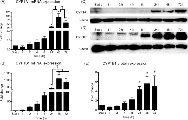
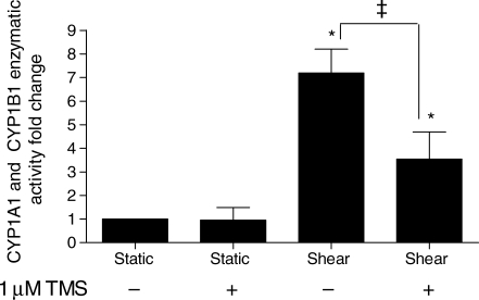
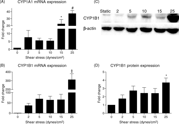
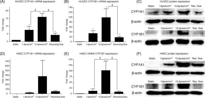
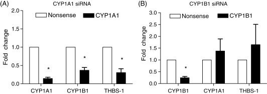
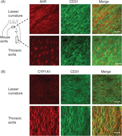
Methods and results: We investigated CYP1A1 and CYP1B1 expressions under different durations, levels, and patterns of shear stress. CYP1A1 and CYP1B1 mRNA, protein, and enzymatic activity were maximally up-regulated at > or =24 h of arterial levels of shear stress (15-25 dynes/cm2). Expression of both genes was significantly attenuated by reversing shear stress when compared with 15 dynes/cm2 steady shear stress. Small interfering RNA knockdown of CYP1A1 resulted in significantly reduced CYP1B1 and thrombospondin-1 expression, genes regulated by the aryl hydrocarbon receptor (AhR). Immunostaining of human coronary arteries showed constitutive CYP1A1 and CYP1B1 protein expressions in endothelial cells. Immunostaining of mouse aorta showed nuclear localization of AhR and increased expression of CYP1A1 in the descending thoracic aorta, whereas reduced nuclear localization of AhR and attenuated CYP1A1 expression were observed in the lesser curvature of the aortic arch.

Conclusion: CYP1A1 and CYP1B1 gene and protein expressions vary with time, magnitude, and pattern of shear stress. Increased CYP1A1 gene expression modulates AhR-regulated genes. Based on our in vitro reversing flow data and in vivo immunostained mouse aorta, we suggest that increased expression of both genes reflects an anti-atherogenic endothelial cell phenotype.


Figures

Figure 1
Figure 1
Time course of CYP1A1 and CYP1B1 mRNA and protein expression under shear stress. Human umbilical vein endothelial cells were exposed to 0 (static), 1, 2, 4, 8, 24, 48, or 72 h of 25 dynes/cm2 steady shear stress (n = 3–8). Both CYP1A1 (A) and CYP1B1 (B) mRNA were maximally induced by 24–72 h of shear stress. CYP1A1 protein (C) was usually detected only after 24 h of shear stress. CYP1B1 (D and E) protein was maximally induced after 24 h. Equal loading in (C) and (D) is indicated by reblotting the membranes with a β-actin antibody. Fold changes are relative to static values. Significance was assessed using analysis of variance followed by Tukey's post hoc test. *P < 0.05 vs. 0–8 h shear stress, P < 0.05 vs. indicated durations, #P < 0.05 vs. 0–8 h shear stress.
Figure 2
Figure 2
Effect of shear stress on CYP1A1 and CYP1B1 activity. Cells were exposed to 25 dynes/cm2 or maintained under static conditions for 48 h and subsequently incubated under static conditions with 100 µM luciferin-chloroethyl ether (CEE) for an additional 3 h (n = 8). The CYP1B1 inhibitor [1 µM 2,3′,4,5′-tetramethoxystilbene (TMS)] was added to the media simultaneously with luciferin-CEE in the indicated samples. Luminescence was normalized to total protein. Significance was assessed using analysis of variance, followed by Tukey's post hoc test; *P < 0.05 vs. static conditions and P < 0.05 between shear-stressed cells with and without TMS.
Figure 3
Figure 3
Effect of shear stress magnitude on CYP1A1 and CYP1B1 mRNA and protein expression. Human umbilical vein endothelial cells were subjected to 0 (static), 2, 5, 10, 15, or 25 dynes/cm2 shear stress for 24 h (n = 4–5). Both CYP1A1 (A) and CYP1B1 (B) mRNA expression and CYP1B1 protein (C and D) expression were maximal at 25 dynes/cm2 shear stress. Significance was assessed using analysis of variance, followed by Tukey's post hoc test. *P < 0.05 with respect to static conditions, #P < 0.05 vs. 0–10 dynes/cm2, P < 0.05 vs. 0–15 dynes/cm2.
Figure 4
Figure 4
Effect of shear stress pattern on CYP1A1 and CYP1B1 mRNA and protein expression. Human umbilical vein endothelial cells (HUVECs) (n = 5–9) or human aortic endothelial cells (HAECs) (n = 4) were subjected to one of three different flow regimes: low shear stress (1 dyne/cm2), steady arterial shear stress (15 dynes/cm2), or carotid sinus reversing shear stress for 24 h. CYP1A1 and CYP1B1 mRNA and protein expression in HUVECs (A–C) were highest under 15 dynes/cm2 and greatly reduced under reversing shear stress. CYP1A1 and CYP1B1 had similar trends of expression in HAECs (DF). Significance was assessed using analysis of variance, followed by Tukey's post hoc test; *P < 0.05 vs. static conditions and P < 0.05 between indicated conditions.
Figure 5
Figure 5
Effect of siRNA knockdown of CYP1A1 and CYP1B1 gene expressions. Human umbilical vein endothelial cells were treated with either CYP1A1 (A) or CYP1B1 (B) siRNA 48 h prior to 24 h of 25 dynes/cm2 shear stress (n = 4). mRNA levels of CYP1A1, CYP1B1, and thrombospondin-1 were compared with cells treated with a non-silencing nonsense control and subjected to identical shear conditions. The knockdown of CYP1A1 caused significant decreases in CYP1B1 and thrombospondin-1. The knockdown of CYP1B1 caused no significant gene changes other than the target knockdown of CYP1B1. Significance was measured using paired Student's t-test; *P < 0.05 between targeted knockdown and nonsense control.
Figure 6
Figure 6
Expression of aryl hydrocarbon receptor (AhR) and CYP1A1 protein in mouse aorta. Whole mouse aortas were stained for AhR (n = 3) or CYP1A1 (n = 7), and the lesser curvature and thoracic aorta were mounted en face. Endothelial cell integrity was confirmed with CD31 counterstaining. AhR (A) was expressed in the cytoplasm and nucleus in the region of lesser curvature, but was principally localized in the nucleus in the thoracic aorta. CYP1A1 (B) staining was strongest in the thoracic aorta, with attenuated staining in the region of lesser curvature. Bar is 20 µm.

Comment in

References

    1. Wootton DM, Ku DN. Fluid mechanics of vascular systems, diseases, and thrombosis. Annu Rev Biomed Eng. 1999;1:299–329. - PubMed
    1. Chien S. Effects of disturbed flow on endothelial cells. Ann Biomed Eng. 2008;36:554–562. - PMC - PubMed
    1. Davies PF. Endothelial transcriptome profiles in vivo in complex arterial flow fields. Ann Biomed Eng. 2008;36:563–570. - PubMed
    1. Michaelis UR, Fleming I. From endothelium-derived hyperpolarizing factor (EDHF) to angiogenesis: epoxyeicosatrienoic acids (EETs) and cell signaling. Pharmacol Ther. 2006;111:584–595. - PubMed
    1. Barouki R, Morel Y. Repression of cytochrome P450 1A1 gene expression by oxidative stress: mechanisms and biological implications. Biochem Pharmacol. 2001;61:511–516. - PubMed

Publication types

MeSH terms