. 2009 Feb 1;122(Pt 3):378-88.
doi: 10.1242/jcs.034124. Epub 2009 Jan 6.

Connexin mutation that causes dominant congenital cataracts inhibits gap junctions, but not hemichannels, in a dominant negative manner

Affiliations

Connexin mutation that causes dominant congenital cataracts inhibits gap junctions, but not hemichannels, in a dominant negative manner

Eric A Banks et al. J Cell Sci. .

Abstract

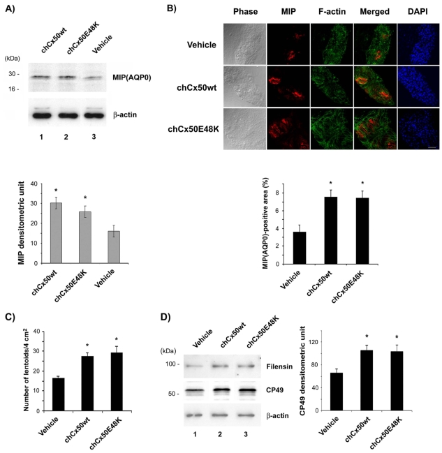
The connexin (Cx) 50, E48K, mutation is associated with a human dominant congenital cataract; however, the underlying molecular mechanism has not been characterized. The glutamate (E) residue at position 48 is highly conserved across animal species and types of connexins. When expressed in paired Xenopus oocytes, human (h) and chicken (ch) Cx50 E48K mutants showed no electrical coupling. In addition, this mutation acts in a dominant negative manner when paired hetero-typically or hetero-merically with wild-type Cx50, but has no such effect on Cx46, the other lens fiber connexin. A similar loss-of-function and dominant negative effect was observed using dye transfer assays in the same system. By using two different dye transfer methods, with two different tracer dyes, we found chCx50 E48K expressed in chicken lens embryonic fibroblast cells by retroviral infection similarly failed to induce dye coupling, and prevented wild-type chCx50 from forming functional gap junctions. In contrast to its effect on gap junctions, the E48K mutation has no effect on hemichannel activity when assayed using electrical conductance in oocytes, and mechanically induced dye uptake in cells. Cx50 is functionally involved in cell differentiation and lens development, and the E48K mutant promotes primary lens cell differentiation indistinguishable from wild-type chCx50, despite its lack of junctional channel function. Together the data show that mutations affecting gap junctions but not hemichannel function of Cx50 can lead to dominant congenital cataracts in humans. This clearly supports the model of intercellular coupling of fiber cells creating a microcirculation of nutrients and metabolites required for lens transparency.


Figures

Fig. 1.
Fig. 1.
E48K mutation of Cx50 is associated with human congenital cataracts. (A) A membrane topological diagram showing the location of E48K at the first extracellular domain of the chCx50 protein. (B,C) Sequence comparisons showing that E48 is a highly conserved amino acid residue of Cx50 across various animal species (B) and also between various human connexin isotypes (C).
Fig. 2.
Fig. 2.
chCx50 E48K mutant does not mediate electrical or dye coupling, and reveals a dominant negative behavior toward wild-type chCx50 in paired Xenopus oocytes. (A) Various pairings of oocytes expressing different combinations of chCx50, chCx46 and chCx50 E48K mutant were analyzed for net intercellular conductance. All oocytes were injected with antisense oligonucleotide directed against endogenous Xenopus Cx38. All experiments using chCx46 were performed in a similar manner to chCx50, except that in the case of chCx46 the calcium concentration of the bath media was kept at 4 mM Ca2+ in order to maintain healthy cells. Numbers of oocyte pairs tested are indicated at the top of each bar. **, P<0.005, indicates values significantly different to the negative control of chCx50 paired with oligo-only-injected oocytes. (B) Oocyte pairs expressing either wild-type or mutant chCx50 in different combinations, as indicated, were tested for electrical conductance before injection of one cell (donor) with Alexa 488. After 6 hours, images were taken (left panel) and the ratio of dye intensity [shown here in a pseudo-color scale from blue (lowest intensity) to red (highest intensity) in acceptor to donor determined (histogram on right, with mean conductances shown above each bar)]. Cells expressing mutant chCx50 alone (top right), or in combination with wild-type Cx50 and paired with a wild-type expressing cell (bottom right), showed no dye transfer above that seen in cells where only one cell in the pair expressed Cx50 (top left). Wild-type Cx50 pairs showed significant dye transfer over the same time frame (bottom left). Average acceptor:donor ratios from oocyte pairs (average junctional conductance shown above each bar; n=3 for each bar), show that only pairs expressing wild-type chCx50 in both cells show dye transfer significantly above control pairs (wild-type chCx50:oligo) (**, P<0.005).
Fig. 3.
Fig. 3.
Impaired gap junctional electrical conductance and dominant negative function of hCx50 E48K in paired Xenopus oocytes. Paired oocytes, both expressing wild-type Cx50 (hCx50wt:hCx50wt) show robust conductance, compared with paired oocytes where only one is injected with wild-type Cx50 (Oligo:hCx50wt) that show negligible coupling. Any oocyte pairing that includes Cx50E48K, whether homotypic (hCx50E48K:hCx50E48K), heterotypic (hCx50wt:hCx50E48K) or heteromeric (hCx50wt:hCx50wt+hCx50E48K) also failed to produce any intercellular conductance. The conductance is expressed as mean±s.e.m. The numbers of oocyte pairs tested are indicated at the top of each bar.
Fig. 4.
Fig. 4.
chCx50(E48K) mutant cannot mediate gap junction dye coupling in CEF cells. (A) Three days after infection of recombinant retroviruses, RCAS(A)-chCx50 and RCAS(A)-chCx50 E48K, the scrape loading dye transfer assay was performed using RD as a tracer dye and LY as transferring dyes. Extent of dye transfer was quantified by measuring the distance from the scrape line to the dye front for LY and comparing this to the dye front distance for RD. Data presented are mean±s.e.m. (n=5). Wild-type (chCx50wt) versus RCAS(A) (Vehicle) and mutant (chCx50E48K). (B) Six days after retroviral infection of retroviruses, RCAS(A) (Vehicle), RCAS(A)-chCx50 (chCx50wt) and RCAS(A)-chCx50 E48K (chCx50E48K) into CEF cells, the parachuting dye transfer assay was performed using Dil as a tracer dye and calcein as a transferring dye. The extent of dye transfer was quantified by measuring the area of calcein-fluorescence-stained cells (NIH image) (right panel). The data are presented as the mean±s.e.m. (n=3). chCx50wt versus Vehicle and chCx50E48K. (C) Lysates of cells expressing wild type (chCx50wt) (lane 1) and mutant (chCx50E48K) (lane 2) were immunblotted with affinity-purified anti-chCx50 antibody and stripped membrane replicas were probed with anti-β-actin antibody to demonstrate comparable expression levels of the two proteins. Bars, 10 μm. **P<0.005; ***P<0.001.
Fig. 5.
Fig. 5.
chCx50 E48K is a dominant negative mutant that inhibits intercellular coupling by wild-type chCx50 in CEF cells. CEF cells were infected with RCAS(A) (Vehicle), wild-type chCx50 (chCx50wt) and chCx46 (chCx46wt), mutant chCx50(E48K), wild-type chCx50 plus mutant (chCx50wt+chCx50E48K) and wild-type Cx46 plus mutant (chCx46wt+chCx50E48K). Six days after infection, CEF cells were examined for gap junction-mediated dye coupling. The extent of dye transfer was quantified by the scrape loading dye transfer assay using LY/RD (A) and parachuting dye transfer assay using calcein/Dil (B) as described in Fig. 4. chCx50 E48K mutant significantly diminished dye coupling mediated by wild-type chCx50 using both scrape loading and parachuting dye transfer. The data are presented as mean ±s.e.m. (n=3). chCx50wt versus chCx50wt+chCx50E48K, ***P<0.001.
Fig. 6.
Fig. 6.
chCx50 E48K has no effect on hemichannel function in Xenopus oocytes. Post-injection of cRNAs of wild type (chCx50wt) (A) or mutant (chCx50E48K) (B) in to Xenopus oocytes, the transmembrane hemichannel conductance was measured in the presence of 0.1 mM (left panels) or 4 mM Ca2+ (right panels). (C) Average hemichannel conductance of oocytes injected with antisense oligonucleotide (Oligo), wild type (chCx50wt) or mutant (chCx50E48K) in the presence of either 0.1 mM or 4 mM Ca2+ showed that both wild-type and mutant constructs induced Ca2+-sensitive membrane currents significantly different to oligo-only controls. The numbers of oocytes tested are indicated at the top of the bar. (D) Average hemichannel conductance of oocytes injected with antisense oligonucleotide (Oligo), wild-type chCx46 (chCx46wt), chCx50 (chCx50wt) or mutant (chCx50E48K) and other combinations in the presence of either 0.1 mM or 4 mM Ca2+ showed that both wild-type Cx46 and Cx50 and mutant constructs induced Ca2+-sensitive membrane currents significantly different from oligo-only controls. The numbers of oocytes tested are indicated at the top of the bar. (E) Average hemichannel conductance of wild-type and mutant hCx50 revealed similar results to that with the chick connexin. The numbers of oocytes tested are indicated at the top of the bar. **P<0.005.
Fig. 7.
Fig. 7.
The opening of chCx50 and chCx46-forming hemichannels induced by mechanical stimulation was not affected by E48K mutation. CEF cells were infected with RCAS(A) (Vehicle), wild-type Cx50 (chCx50wt), mutant chCx50(E48K) (chCx50E48K), different combination or non-infected control (Ctrl), and cultured at low cell density without (chCx50E48K) were fixed, labeled with DAPI (blue) or anti-FLAG (green) or anti-chCx50 (red) antibody. The primary antibodies were detected by fluorescein-conjugated anti-mouse IgG for anti-FLAG antibody and rhodamine-conjugated anti-rabbit IgG for anti-chCx50 antibody. The corresponding merged images (Merged) are shown on the right. Bar, 10 μm.
Fig. 8.
Fig. 8.
Exogenous chCx50 E48K expression is comparable to wild-type chCx50. (A) Lens primary cultures were infected with retroviruses containing RCAS(A) vector (Vehicle), cDNAs of wild-type chCx50 (chCx50wt) (lane 2) and mutant Cx50 (chCx50E48K) (lane 3). After 8 days of infection, crude membranes were prepared and analyzed by western blots probed with anti-chCx50 antibody. Stripped membrane replicas were re-probed with monoclonal antibody against β-actin. The intensity of the bands on western blots was quantified by densitometric measurement (right panel). Data are presented as mean ± s.e.m. (n=3). chCx50wt and chCx50E48K versus Vehicle, ***P<0.001. (B) At 8 days after infection, lens primary culture expressing exogenous wild-type (chCx50wt) and mutant (chCx50E48K) Cx50 were fixed, labeled with DAPI (blue) or anti-FLAG (green) or anti-chCx50 (red) antibody. The primary antibodies were detected by fluorescein-conjugated anti-mouse IgG for anti-FLAG antibody and rhodamine-conjugated anti-rabbit IgG for anti-chCx50 antibody. The corresponding merged images (Merged) are shown on the right. Bar, 10 μm.
Fig. 9.
Fig. 9.
chCx50 E48K mutant has similar capability to promote lens cell differentiation to wild-type chCx50. Lens primary cultures were infected with retroviruses containing RCAS(A) (Vehicle), RCAS(A)-chCx50 (chCx50wt) and RCAS(A)-chCx50(E48K) (chCx50E48K) for 8 days. (A) Crude membrane preparations from respective lens cell cultures: chCx50wt (lane 1), chCx50E48K (lane 2) and Vehicle (lane 3) were immunoblotted with anti-MIP(AQP0). The stripped membrane replicas were re-probed for β-actin. The intensity of the bands from western blot analysis was quantified by densitometry (lower panel). chCx50wt and chCx50E48K versus Vehicle, data presented as mean±s.e.m. (n=3). (B) Lens primary cells were immunolabeled with monoclonal MIP(AQP0) antibody and counterstained with Alex 488 phalloidin and DAPI. The primary antibody was detected by fluorescein-conjugated anti-mouse IgG, and the images were captured by fluorescence microscope. The intensity of the MIP(AQP0)-stained area was quantified (UTHSCSA ImageTool Software) and presented as a percentage in the x-axis (lower panel). chCx50wt and chCx50E48K versus Vehicle, data presented as mean±s.e.m. (n=3). (C) Lens cell differentiation was assessed by counting and quantifying numbers of lentoids. chCx50wt and chCx50E48K versus Vehicle, data presented as mean±s.e.m. (n=3). (D). Lysates from cells infected with retroviruses RCAS(A) (Vehicle) (lane 1), RCAS(A)-chCx50 (chCx50wt) (lane 2), and RCAS(A)-Cx45.6(E48K) (lane 3) were immunoblotted with anti-filensin, CP49 or β-actin antibodies. The CP49 protein bands from three separate western blot analyses were quantified by densitometry (right panel). chCx50wt and chCx50E48K versus Vehicle, data presented as mean±s.e.m. (n=3). Bar, 10 μm. * and **, P<0.05.

References

    1. Banks, E. A., Yu, X. S., Shi, Q. and Jiang, J. X. (2007). Promotion of lens epithelial-fiber differentiation by the C-terminus of connexin 45.6 a role independent of gap junction communication. J. Cell Sci. 120, 3602-3612. - PubMed
    1. Bao, L., Sachs, F. and Dahl, G. (2005). Connexins are mechanosensitive. Am. J. Physiol., Cell Physiol. 287 C1389-C1395. - PubMed
    1. Barrio, L. C., Suchyna, T., Bargiello, T., Xu, L. X., Roginski, R. S., Bennett, M. V. L. and Nicholson, B. J. (1991). Gap junctions formed by connexins 26 and 32 alone and in combination are differently affected by voltage. Proc. Natl. Acad. Sci. USA 88, 8410-8414. - PMC - PubMed
    1. Beahm, D. L. and Hall, J. E. (2002). Hemichannel and junctional properties of connexin 50. Biophys. J. 82, 2016-2031. - PMC - PubMed
    1. Berry, V., Mackay, D., Khaliq, S., Francis, P. J., Hameed, A., Anwar, K., Mehdi, S. Q., Newbold, R. J., Ionides, A., Shiels, A. et al. (1999). Connexin 50 mutation in a family with congenital “zonular nuclear” pulverulent cataract of Pakistani origin. Hum. Genet. 105, 168-170. - PubMed

Publication types

MeSH terms