Review
. 2009 Jan;89(1):279-308.
doi: 10.1152/physrev.00007.2008.

Cardiovascular actions of neurotrophins

Affiliations
Review

Cardiovascular actions of neurotrophins

Andrea Caporali et al. Physiol Rev. 2009 Jan.

Abstract

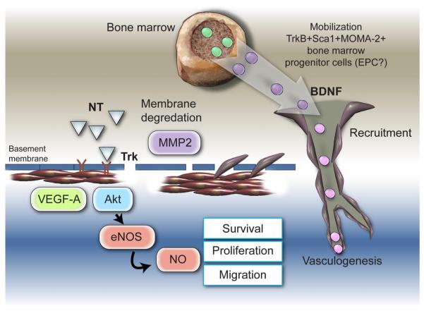
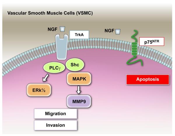
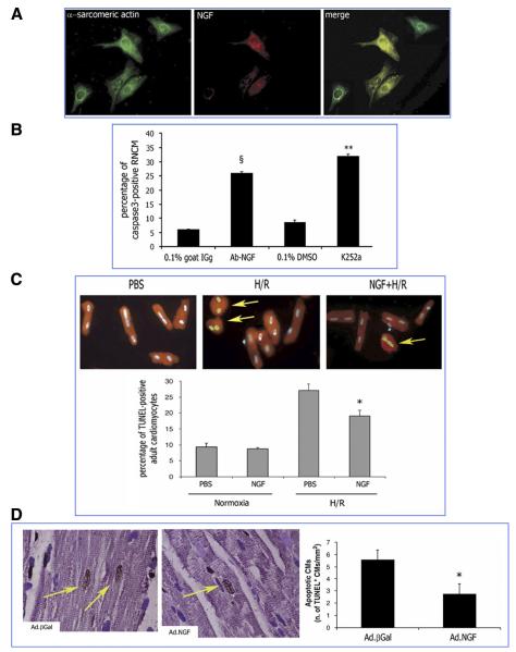
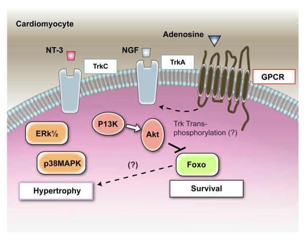
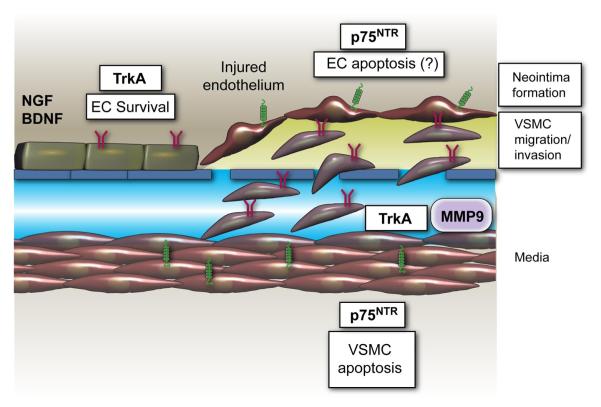
Neurotrophins were christened in consideration of their actions on the nervous system and, for a long time, they were the exclusive interest of neuroscientists. However, more recently, this family of proteins has been shown to possess essential cardiovascular functions. During cardiovascular development, neurotrophins and their receptors are essential factors in the formation of the heart and critical regulator of vascular development. Postnatally, neurotrophins control the survival of endothelial cells, vascular smooth muscle cells, and cardiomyocytes and regulate angiogenesis and vasculogenesis, by autocrine and paracrine mechanisms. Recent studies suggest the capacity of neurotrophins, via their tropomyosin-kinase receptors, to promote therapeutic neovascularization in animal models of hindlimb ischemia. Conversely, the neurotrophin low-affinity p75(NTR) receptor induces apoptosis of endothelial cells and vascular smooth muscle cells and impairs angiogenesis. Finally, nerve growth factor looks particularly promising in treating microvascular complications of diabetes or reducing cardiomyocyte apoptosis in the infarcted heart. These seminal discoveries have fuelled basic and translational research and thus opened a new field of investigation in cardiovascular medicine and therapeutics. Here, we review recent progress on the molecular signaling and roles played by neurotrophins in cardiovascular development, function, and pathology, and we discuss therapeutic potential of strategies based on neurotrophin manipulation.


Figures

FIG. 1
FIG. 1
Genetic deficiency of brain-derived neurotrophic factor (BDNF), neurotropin-3 (NT-3), or tropomyosin-related kinase receptor C (trKC) causes cardiac and vascular defects in the developing mammalian heart. I: BDNF−/− neonate mice exhibit ventricular wall hemorrhage. Histological analyses of BDNF+/+ (A, C, and E) or BDNF−/− (B, D, and F–H) littermates killed at P0. Hematoxylin and eosin-stained sections reveal hemorrhage in the epicardial third of both right and left ventricular walls of BDNF−/− neonates (B and D, arrowheads) and an atrial septal defect (B). Pulmonary hemorrhage is detectable in BDNF−/− (F) but not BDNF+/+ littermates (E). Hemorrhage was not detectable in other organs, such as kidneys (G) as well as skin and spinal cord (H). Ra and la, right and left atria; rv and lv, right and left ventricles; asd, atrial septal defect; s, skin; m, skeletal muscle; vb, vertebral body; sc, thoracic spinal cord. Scale bars, 150 μm (A and B), 50 μm (C and D), 100 μm (E–H). [From Donovan et al. (70), with permission from Development.] II: schematic representation of cardiac abnormalities in NT-3−/− neonate mice. Schematic representations of normal cardiac anatomy(A), the NT-3−/− mutant heart (B), the aorta (AO), ductus arteriousus (DA), right and left atrium (RA, LA), right and left ventricle (RV, LV), tricuspis (TV), and mitral (MV) valve are indicated. [From Donovan et al. (69), reprinted by permission from Macmillan Publishers Ltd.] III: table showing heart abnormalities in trkC−/− mice. [Adapted from Tessarollo et al. (258).]
FIG. 2
FIG. 2
The angiogenic potential of endothelial cells is impaired by p75NTR. A: human umbilical vein endothelial cells (HUVEC) were infected with different concentrations of an adenoviral vector carrying human the p75NTR gene (Ad.p75NTR) or with Ad.Null (control). After 48 h, cell lysates were collected and subjected to Western blotting with antibodies to p75NTR, the apoptosis marker cleaved caspase-3, and glyceraldehyde-3-phosphate dehydrogenase (GAPDH) (loading control). These analyses provide evidence that p75NTR expression induces EC apoptosis. B: apoptotic nuclei of transduced HUVEC were detected by the TUNEL assay. Fluorescent images are representative of apoptosis rate in Null-HUVEC and p75NTR-HUVEC. Original magnification: ×400; scale bar: 40 μm. Green fluorescence: TUNEL-positive nuclei; blue fluorescence: all the nuclei. Arrows point to TUNEL-positive nuclei. Bar graphs quantify apoptosis, which is expressed as percentage of TUNEL-positive nuclei to total nuclei. Data are presented as means ± SE. *P < 0.05 and **P < 0.001 vs. Ad.Null. C: HUVEC were transduced with p75NTR or Null and syncronized by serum starvation. Following the release from cycle arrest, the cell cycle progression was assessed by flow cytometric analysis, at the indicated time points. The percentage of cells in G1, S, and G2 phases is indicated in figures. These analyses showed defective cell cycle progression in p75NTR-HUVEC. D: migration toward the chemoattractant stroma derived factor-1 (SDF-1; 100 ng/ml) is reduced for p75NTR-HUVEC compared with Null-HUVEC. In bar graph, values are means ± SE. #P < 0.05 vs. Ad.Null combined with PBS, *P < 0. 05 vs. Ad.Null combined with SDF-1. Top panels show representative microscopic fields (original magnification: ×100; scale bar: 100 μm). E: the potential of HUVEC to form vessel-like structures on Matrigel is impaired by p75NTR transduction. Images (original magnification: ×100; scale bar: 100 μm) show the time course (up to 24 h from cell seeding) of cell organization on Matrigel. In bar graphs, the quantification of EC tube network formation at 24 h from seeding is expressed as the number of intersecting points of tubular structures for microscopic field (left) as well as the percent of microscopic field area covered by connected tubular structures (right). Values are means ± SE. **P < 0.01 vs. Ad.Null. [From Caporali et al. (35).]
FIG. 3
FIG. 3
Neurotrophin actions on the endothelial cell. Under basal conditions, the neurotrophin receptor trkA and trkB (and, in some tissues, also trkC) are expressed by EC. Trk receptor engagement by mature NTs initiates two major signaling pathways, Erk MAPK and IP3K/Akt. Nerve growth factor (NGF) treatment of EC causes a rapid phosphorylation of trkA, determining a parallel activation of ERK1/2 and a subsequent increase in EC proliferation and migration. Activation of the PI3K/Akt signaling pathway promotes EC survival. Moreover, via the PI3K/Akt, NGF stimulates EC invasion and cord formation by augmenting MMP-2. NGF, via trkA, supports EC survival in vitro and in vivo, an action which is, at least in part, mediated by increased VEGF-A. BDNF, via trkB, also supports EC survival. Under basal conditions, the p75NTR is scarcely expressed by EC. Following p75NTR induction or transduction, the receptor triggers EC apoptosis and inhibits EC cell cycling through mechanisms that may be dependent or not from ligand (mature NT and pro-NGF) activation.
FIG. 4
FIG. 4
NGF promotes angiogenesis and arteriogenesis in ischemic hindlimbs. A: time course of NGF content in adductor muscles following induction of hindlimb ischemia. Values are means ± SE (n = 4 for each time point). *P < 0.05 vs time 0. B: endogenous NGF promotes angiogenesis in ischemic limb muscles. Twenty-one days from ischemia induction, control IgG-treated mice (n = 6) showed higher capillary density in ischemic adductors (I) than in contralateral normoperfused muscles (C). The capillary response to ischemia was prevented in mice given a NGF-neutralizing antibody (anti-NGF Ab, n = 8). Values are means ± SE. *P < 0.05 vs. contralaterals. §P < 0.05 vs IgG. C and D: supplementation of NGF to ischemic limb muscles promotes angiogenesis and increases arteriole numbers. Local daily injections of NGF (full columns) or vehicle (V, dotted columns) in ischemic muscles were repeated over 5 or 14 days, starting from the day of ischemia induction. Capillary (C) and arteriole (D) density was evaluated 14 days thereafter. Microvascular density of untouched adductors (open column) is shown as a reference. Neovascularization was potentiated at arteriole and capillary level by 14-day NGF treatment. Five days of treatment exerted an effect on arteriole growth only. Values are means ± SE. *P < 0.05 vs. controls. §P < 0.05 vs. V; +P < 0.05 vs. 5-day treatment. E and F: supplementation of NGF reduces apoptosis of endothelial cells and skeletal myocytes in ischemic adductor muscles. NGF or vehicle was intramuscularly injected every day starting on the day of femoral artery excision. Muscles were harvested 5 days later. Apoptosis found at the level of capillary endothelial cells (ECs) and skeletal myocytes was expressed as the number of TUNEL-positive cells per 1,000 capillaries (A) or myofiber (B), respectively. Values are means ± SE (n = 4 mice per group). §P < 0.05 vs. V. G: supplementation of NGF to ischemic limb muscles improves postischemic blood flow recovery. Line graph shows the effect of NGF supplementation for 14 days on foot postischemic BF recovery, expressed as ischemic to contralateral BF ratio. NGF (full symbols, n = 8) improved perfusion recovery compared with vehicle (open symbols, n = 10). Values are means ± SE. *P < 0.05 vs. time 0. §P < 0.05 vs. vehicle. H: NGF induces angiogenesis via VEGF-A, Akt, and nitric oxide (NO). Daily injection of 20 μg NGF for 7 days stimulates capillary growth in normoperfused muscles (closed column) compared with vehicle-treated muscles (V, open column). The angiogenic effect of NGF was blocked by a VEGF-neutralizing antibody (Ab-VEGF), the NO synthase inhibitor l-nitroarginine methyl ester (l-NAME), or an adenovirus carrying dominant-negative Akt (DNAkt). Values are means ± SE (at least n = 6 per group). §P < 0.05 vs. vehicle; +P < 0.05 vs NGF alone. [From Emanueli et al. (78).]
FIG. 5
FIG. 5
Schematic representation of mechanisms by which NTs stimulate blood vessel growth. NGF promotes angiogenesis through trkA by increasing the expression level of VEGF-A, and through activation of the Akt intracellular pathways leading to NO production and upregulation of matrix metalloproteinase (MMP)-2 expression. Some of the angiogenesis-related actions of NGF have been later proven to be shared by BDNF. Moreover, BDNF induces the mobilization of trkB+Sca1+MOMA-2+ bone marrow progenitor cells, suggesting a promotional role of BDNF on postnatal vasculogenesis, although this hypothesis needs to be validated.
FIG. 6
FIG. 6
Neurotrophin actions on vascular smooth muscle cells. Human and rodent VSMC express NTs, p75NTR, and trk receptors in vivo and in culture. Activaction of trkA by NGF, via PI3K and Akt, promotes chemotaxis of human aortic VSMC, without influencing VSMC proliferation. Moreover, the NGF/trkA receptor signal, via Shc/MAPK pathway, induces MMP-9 expression in primary rat aortic VSMC, which, in vivo, may favor the invasion of VSMC through the extracellular matrix and endothelial basal membrane. Stimulation with NGF of p75NTR-expressing VSMC promotes apoptotic death.
FIG. 7
FIG. 7
Prosurvival activity of NGF on cardiac myocytes. A: rat neonatal cardiomyocytes (RNMCs) express NGF and trkA. Immunofluorescence analysis is shown of the cardiac marker α-sarcomeric actin (green fluorescence) and NGF (red fluorescence) and merged images (costaining results in yellow fluorescence). B: NGF is an endogenous prosurvival factor for RNCMs. RNCMs were incubated for 48 h in serum-free medium with a goat-raised anti-NGF neutralizing antibody (Ab-NGF) or the trkA inhibitor K252a. Controls consisted of nonimmune goat serum (0.1% in PBS) or 0.1% DMSO, respectively. Apoptosis was detected by cleaved caspase-3 immunostaining (green fluorescence). Nuclei were counterstained by DAPI (blue fluorescence). The fluorescent images (×400) are representative of the experiment. Bar graph shows the percentage of cleaved caspase 3-positive RNCMs. Data are presented as means ± SE (n = 3). §P < 0.05 vs. 0.1% goat IgG; **P < 0.01 vs. 0.1% DMSO. C: NGF inhibits apoptosis in rat adult cardiomyocytes. A, isolated rat adult cardiomyocytes were maintained under normoxia or submitted to 6 h of hypoxia followed by 18 h reoxygenation and cotreated with NGF (50 ng/ml) or its vehicle PBS. Apoptotic nuclei were identified by TUNEL staining (green fluorescence). α-Sarcomeric actin stains cardiomyocytes (red fluorescence). The pictures were captured at ×400 magnification. Arrows point to TUNEL-positive cardiomyocytes. Bar graphs quantify apoptosis, which is expressed as percentage of TUNEL-positive cardiomyocyte. Data are presented as means ± SE (n = 3). *P < 0.05 vs. PBS plus H/R (B) D: local NGF gene transfer prevents apoptosis of cardiomyocytes in the rat infarcted heart. Myocardial infarction was induced in adult Wistar rat. Ad.NGF or Ad.βGal (each at 108 p.f.u.) was injected in the peri-infarct myocardium. After 7 days, the heart was arrested in diastole and perfusion/fixed. Apoptosis of cardiomyocytes (CMs) was revealed by double staining for TUNEL (TUNEL-positive nuclei are stained in dark brown) and the cardiac marker α-sarcomeric actin (in purple). Nuclei were counterstained with hematoxylin. In the pictures captured (optical microscopy, ×1,000) from Ad.NGF and Ad.βGal specimens, TUNEL-positive apoptotic cardiomyocytes are pointed by arrows. Graph quantifies apoptosis of cardiomyocytes per mm2 of peri-infarct myocardium section. Values are means ± SE (n = 7) *P < 0.05 vs. Ad.βGal. [From Caporali et al. (36).]
FIG. 8
FIG. 8
Neurotrophin actions and signalling in cardiomyocytes. NGF is an autocrine prosurvival factor for cardiomyocytes, and an increased level of this neurotrophin protects cardiomyocytes from apoptosis. The prosurvival signal of NGF in cardiac myocytes is mediated by trkA and downsream Akt phosphorylation. In response to Akt activation by NGF, Forkhead transcription factors Foxo are phosphorylated and excluded from the nucleus, thus resulting in increased cardiomyocyte survival. In primary-cultured rat neonatal cardiomyocytes, NT-3, via trkC, activates p38 MAPK and Erk1/2, thus resulting in increased cell size. Interestingly, in neural cells, the possibility that trk receptors can be transactivated in response to G protein-coupled receptor (GPCR) signaling has been proven. The GPCR adenosine receptors are expressed by cardiomyocytes, where they trigger a range of responses, including activation PI3K. We speculate that transactivation of trk receptors by adenosine receptors may happen in cardiomyocytes.
FIG. 9
FIG. 9
Neurotrophin actions could be involved in neointima formation. Neurotrophins, which, via trk receptors and p75NTR, regulate survival of both EC and VSMC may have a dual role in neointima formation. In fact, the trk-mediated survival of EC may contribute to avoid intravascular adhesion of monocytes and to maintain VSMC in a contractile rather than proliferative status. In contrast, p75NTR triggers apoptosis of both EC (negative event) and VSMC (possibily a positive event, as it may inhibit VSMC growth). Moreover, NGF is a potent chemotactic agent for human aortic VSMC and, by inducing MMP-9 expression, it may also favor VSMC invasion through the basal membrane and internal elastic lamina. Arterial balloon injury upregulates the expression levels of NGF, BDNF, trkA, and trkB. Increased expression levels of NTs and trk receptors persist during neointima formation.
FIG. 10
FIG. 10
p75NTR is implicated in diabetes-induced impairment in reparative neovascularization of ischemic limb muscles. A: diabetes upregulates the content of endogenous p75NTR protein in capillary EC of ischemic limb muscles. Staining of ischemic muscular sections is shown from diabetic and normoglycemic mice with an antibody for mouse p75NTR (green fluorescence) and with the endothelial marker isolectin-B4 (red fluorescence). Capillary EC which are positive for p75NTR express yellow fluorescence (from merging of the red and green fluorescence) and are pointed by arrows. Representative sections show higher density of p75NTR-expressing capillary EC in the ischemic muscles of diabetics in comparison with normoglycemic mice. (Original magnification: ×400; scale bar: 500 μm.). B–F: inhibition of p75NTR signaling in diabetic limb muscles restores proper neovascularization and BF recovery following limb ischemia. Unilateral limb ischemia was induced in diabetic and normoglycemic mice before an adenovirus carrying a dominant negative mutant of p75NTR (Ad.DN-p75NTR) or Ad.GFP (control) was delivered to the ischemic adductor. B, left: time course of postischemic BF recovery (calculated by laser color Doppler flowmetry) in diabetic and normoglycemic mice treated with Ad.DN-p75NTR (◆, diabetic; ◇, normoglycemic) or Ad.GFP (●, diabetic; ○, normoglycemic). Right: representative laser Doppler images taken at 14 days after induction of ischemia are shown. Squares include the ischemic feet. C: recovery of BF (measured by Oxford Optronic) is expressed as the ratio between the BF to ischemic muscle and the BF to the contralateral muscle at 14 days after ischemia induction. Values are means ± SE. *P < 0.05 vs. normoglycemic mice with Ad.GFP. §P < 0.05 vs. diabetic with Ad.GFP. D: capillary density in the ischemic adductor at 14 days postischemia. Values are means ± SE. **P < 0.01 vs. normoglycemic mice with Ad.GFP. §§P < 0.05 vs. diabetic with Ad.GFP. Apoptosis (revealed by TUNEL assay) (E) and proliferation (revealed by immunhistochemistry for the proliferation antigen MCM-2) (F) of capillary EC in diabetic limb muscles at 14 days postischemia. Values are means ± SE. *P < 0.05 vs. Ad.GFP. [From Caporali et al. (35).]

References

    1. Ackah E, Yu J, Zoellner S, Iwakiri Y, Skurk C, Shibata R, Ouchi N, Easton RM, Galasso G, Birnbaum MJ, Walsh K, Sessa WC. Akt1/protein kinase Balpha is critical for ischemic and VEGF-mediated angiogenesis. J Clin Invest. 2005;115:2119–2127. - PMC - PubMed
    1. Adams RH, Alitalo K. Molecular regulation of angiogenesis and lymphangiogenesis. Nat Rev Mol Cell Biol. 2007;8:464–478. - PubMed
    1. Anand P. Neurotrophic factors and their receptors in human sensory neuropathies. Prog Brain Res. 2004;146:477–492. - PubMed
    1. Anand P, Foley P, Navsaria HA, Sinicropi D, Williams-Chestnut RE, Leigh IM. Nerve growth factor levels in cultured human skin cells: effect of gestation and viral transformation. Neurosci Lett. 1995;184:157–160. - PubMed
    1. Anand P, Terenghi G, Warner G, Kopelman P, Williams-Chestnut RE, Sinicropi DV. The role of endogenous nerve growth factor in human diabetic neuropathy. Nat Med. 1996;2:703–707. - PubMed

Publication types

MeSH terms