Comparative Study
. 2009 Mar;296(3):E503-12.
doi: 10.1152/ajpendo.90517.2008. Epub 2009 Jan 6.

Estrogen replacement enhances EDHF-mediated vasodilation of mesenteric and uterine resistance arteries: role of endothelial cell Ca2+

Affiliations
Comparative Study

Estrogen replacement enhances EDHF-mediated vasodilation of mesenteric and uterine resistance arteries: role of endothelial cell Ca2+

Natalie Z Burger et al. Am J Physiol Endocrinol Metab. 2009 Mar.

Abstract

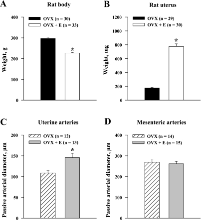
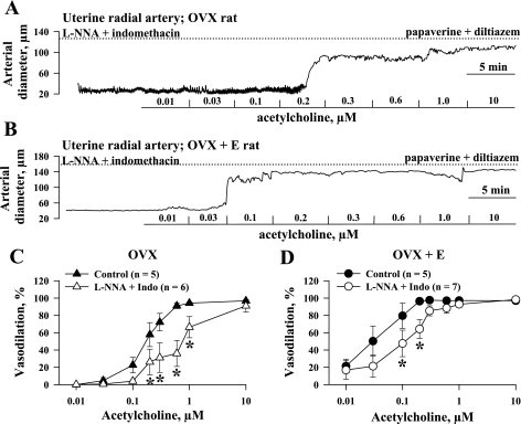
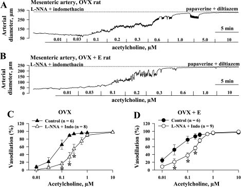
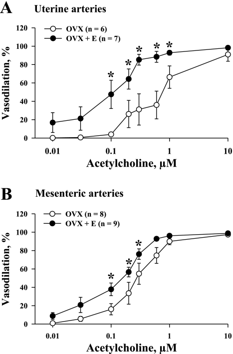
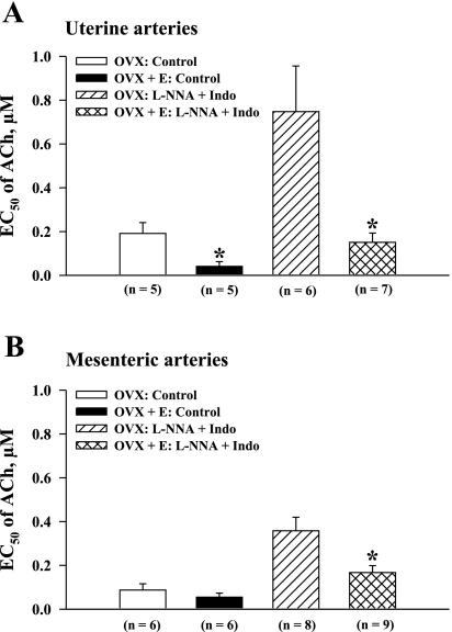
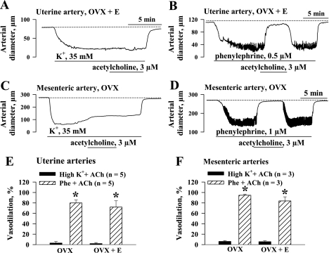
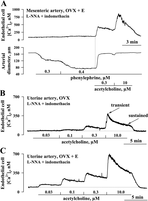
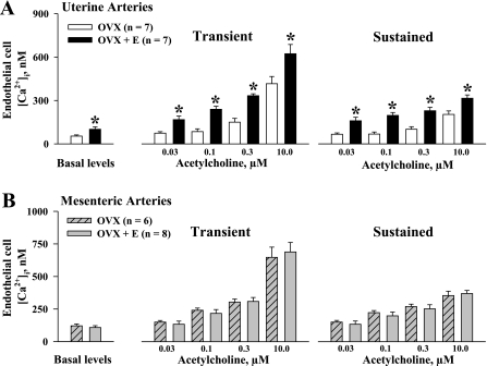
Endothelium-derived hyperpolarizing factor (EDHF) plays an important role in the regulation of vascular microcirculatory tone. This study explores the role of estrogen in controlling EDHF-mediated vasodilation of uterine resistance arteries of the rat and also analyzes the contribution of endothelial cell (EC) Ca(2+) signaling to this process. A parallel study was also performed with mesenteric arteries to provide comparison with a nonreproductive vasculature. Mature female rats underwent ovariectomy, with one half receiving 17beta-estradiol replacement (OVX+E) and the other half serving as estrogen-deficient controls (OVX). Uterine or mesenteric resistance arteries were harvested, cannulated, and pressurized. Nitric oxide and prostacyclin production were inhibited with 200 microM N(G)-nitro-l-arginine and 10 microM indomethacin, respectively. ACh effectively dilated the arteries preconstricted with phenylephrine but failed to induce dilation of vessels preconstricted with high-K(+) solution. ACh EC(50) values were decreased by estrogen replacement by five- and twofold in uterine and mesenteric arteries, respectively. As evidenced by fura-2-based measurements of EC cytoplasmic Ca(2+) concentration ([Ca(2+)](i)), estrogen replacement was associated with increased basal and ACh-stimulated EC [Ca(2+)](i) rise in uterine, but not mesenteric, vessels. These data demonstrate that EDHF contributes to endothelium-dependent vasodilation of uterine and mesenteric resistance arteries and that estrogen controls EDHF-related mechanism(s) more efficiently in reproductive vs. nonreproductive vessels. Enhanced endothelial Ca(2+) signaling may be an important underlying mechanism in estrogenic modulation of EDHF-mediated vasodilation in small resistance uterine arteries.


Figures

Fig. 1.
Fig. 1.
Effects of long-term exposure of ovariectomized (OVX) rats to estrogen (OVX+E) on body weight (A), uterine weight (B), and passive diameters of uterine (C) and mesenteric (D) arteries. Passive diameters were determined in arteries pressurized at 50 mmHg in the presence of 10 μM diltiazem and 100 μM papaverine. n = no. of tested arteries. *Significantly different at P < 0.5 (unpaired Student's t-test).
Fig. 2.
Fig. 2.
Estrogen replacement potentiates nitric oxide (NO)- and prostacyclin-resistant vasodilation of uterine arteries in response to ACh. A and B: representative changes in arterial diameter of uterine arteries from ovariectomized, estrogen-deficient (OVX, A) and ovariectomized, estrogen-replaced (OVX+E, B) rats induced by a cumulative application of ACh in concentrations from 0.01 to 10 μM. All arteries were pretreated for 20 min with 200 μM NG-nitro-l-arginine (l-NNA) and 10 μM indomethacin and preconstricted with phenylephrine before testing with ACh. Solid lines indicate the time of administration of ACh at different concentrations. Dotted lines show maximally dilated diameters of the arteries measured in a relaxing solution containing 10 μM diltiazem and 100 μM papaverine. C and D: summary graphs demonstrating effect of NO and prostacyclin inhibition on ACh-induced vasodilation of uterine arteries from OVX (C) and OVX+E (D) rats. n = no. of tested arteries. *Significant difference between groups at P < 0.05 (2-way repeated-measures ANOVA).
Fig. 3.
Fig. 3.
Effects of estrogen supplementation on ACh-induced dilation of mesenteric arteries. A and B: representative changes in diameter of mesenteric arteries from OVX (A) and OVX+E (B) rats induced by a cumulative application of ACh. Arteries were pretreated for 20 min with 200 μM l-NNA and 10 μM indomethacin and preconstricted with phenylephrine before testing with ACh. Solid lines indicate the time of administration of ACh at different concentrations. Dotted lines show maximally dilated diameters of arteries measured in a relaxing solution containing 10 μM diltiazem and 100 μM papaverine. C and D: summary graphs showing effect of NO and prostacyclin inhibition on ACh-induced vasodilation of mesenteric arteries from OVX (C) and OVX+E (D) rats. n = no. of tested arteries. *Significant difference between groups at P < 0.5 (2-way repeated-measures ANOVA).
Fig. 4.
Fig. 4.
Chronic administration of estrogen to ovariectomized rats enhances endothelium-derived hyperpolarizing factor (EDHF)-mediated vasodilation of mesenteric and uterine arteries. Vasodilator responses of uterine (A) and mesenteric (B) arteries from OVX and OVX+E rats are shown as a function of ACh concentration. Vasodilation is expressed as % of maximal dilator response to papaverine and diltiazem. n = no. of tested arteries. All experiments were performed in the presence of 200 μM l-NNA and 10 μM indomethacin. *Significant difference between groups at P < 0.05 (2-way repeated-measures ANOVA).
Fig. 5.
Fig. 5.
Estrogen replacement increases the vasodilator sensitivity of uterine and mesenteric arteries to ACh. Summary graphs demonstrate an increase in the concentration of ACh producing half-maximal dilation (EC50) of uterine (A) and mesenteric (B) arteries from estrogen-deficient (OVX) vs. estrogen-replaced (OVX+E) rats before and after blockade of NO and prostacyclin production. n = no. of tested arteries. *Significantly different at P < 0.05 (unpaired Student's t-test).
Fig. 6.
Fig. 6.
Abolition of EDHF-mediated arterial responses by treatment with high-K+ solution. A: application of 3 μM ACh results in no vasodilation of a uterine artery preconstricted with 35 mM K+. B: ACh-induced dilation of a uterine artery preconstricted with phenylephrine. l-NNA (200 μM) and indomethacin (10 μM) were present throughout whole experiment. Dotted line shows maximal level of dilation in response to application of 10 μM diltiazem and 100 μM papaverine. C and D: responses of mesenteric arteries to the same concentration of ACh (3 μM) preconstricted with high-K+ solution (C) or phenylephrine (D) in the presence of 200 μM l-NNA and 10 μM indomethacin. E and F: effects of ACh in uterine (E) and mesenteric (F) arteries preconstricted with high-K+ solution or phenylephrine. Vasodilation is expressed as % of maximal response obtained in a relaxing solution containing diltiazem and papaverine. n = no. of tested arteries. *Significant difference at P < 0.05 (unpaired Student's t-test).
Fig. 7.
Fig. 7.
A: temporal correlations between ACh-induced elevations in cytoplasmic concentration of Ca2+ in endothelial cells (EC [Ca2+]i) and associated vasodilation. Representative changes in EC [Ca2+]i (top) and diameter of a mesenteric artery (bottom) from an OVX+E rat to phenylephrine and ACh are shown. Artery was preconstricted with phenylephrine to 50% of initial diameter in the presence of 200 μM l-NNA and 10 μM indomethacin. ACh induced a rise in EC [Ca2+]i followed by immediate vasodilation. Dotted line indicates the starting point of the EC [Ca2+]i rise, while solid lines show the time the artery was exposed to phenylephrine and ACh. B and C: representative changes in EC [Ca2+]i in response to increasing concentrations of ACh in pressurized uterine arteries from an estrogen-deficient (OVX, B) and an estrogen-replaced (OVX+E, C) rat. Arteries were pretreated with 200 μM l-NNA and 10 μM indomethacin. Solid lines indicate time of exposure of arteries to ACh, while dotted lines show basal levels of EC [Ca2+]i. Transient and sustained (5 min after ACh application) components of the responses are shown as well. n = no. of tested arteries.
Fig. 8.
Fig. 8.
Estrogen replacement results in significant enhancement of ACh-induced elevation in EC [Ca2+]i in uterine but not in mesenteric arteries. A: summary graphs show a significant increase in basal levels of EC [Ca2+]i, and transient and sustained [Ca2+]i responses to application of ACh in uterine arteries of OVX vs. OVX+E rats. B: summary graphs demonstrate lack of differences in EC [Ca2+]i responses to ACh in mesenteric arteries of OVX vs. OVX+E rats. All experiments were conducted in the presence of l-NNA and indomethacin. n = no. of tested arteries. *Significantly different at P < 0.05 (2-way repeated-measures ANOVA).

References

    1. Abdalla FM, Marostica E, Picarelli ZP, Abreu LC, Avellar MC, Porto CS. Effect of estrogen on muscarinic acetylcholine receptor expression in rat myometrium. Mol Cell Endocrinol 213: 139–148, 2004. - PubMed
    1. Adeagbo AS, Triggle CR. Varying extracellular [K+]: a functional approach to separating EDHF- and EDNO-related mechanisms in perfused rat mesenteric arterial bed. J Cardiovasc Pharmacol 21: 423–429, 1993. - PubMed
    1. Anari MR, Bakhtiar R, Zhu B, Huskey S, Franklin RB, Evans DC. Derivatization of ethinylestradiol with dansyl chloride to enhance electrospray ionization: application in trace analysis of ethinylestradiol in rhesus monkey plasma. Anal Chem 74: 4136–4144, 2002. - PubMed
    1. Bell C Oestrogen-induced sensitization of the uterine artery of the guinea-pig to acetylcholine. Br J Pharmacol 49: 595–601, 1973. - PMC - PubMed
    1. Bosch MA, Kelly MJ, Ronnekleiv OK. Distribution, neuronal colocalization, and 17beta-E2 modulation of small conductance calcium-activated K+ channel (SK3) mRNA in the guinea pig brain. Endocrinology 143: 1097–1107, 2002. - PubMed

Publication types

MeSH terms