

Special article: gait measures indicate underlying focal gray matter atrophy in the brain of older adults
- PMID: 19126852
- PMCID: PMC2648808
- DOI: 10.1093/gerona/63.12.1380
Special article: gait measures indicate underlying focal gray matter atrophy in the brain of older adults
Abstract
Objective: Our objective was to identify the spatial distribution of focal atrophy within mobility-related brain regions in relationship with quantitative gait characteristics.
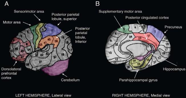
Methods: Gray matter volume was obtained from 220 older adults (78.0 years old, 63% women, 77% white) for brain regions of five domains: motor (motor, sensorimotor and supplementary areas, basal ganglia, cerebellum), visuospatial attention (inferior and superior posterior parietal lobules), cognitive processing speed/executive control function (dorsolateral prefrontal cortex), memory (hippocampus), and motor imagery (parahippocampus, posterior cingulated cortex) domains. Spatial (step width, step length) and temporal (double support time) gait characteristics were measured using the GaitMat II instrumented walking surface. Multivariable linear regression models were adjusted for demographics, total brain volume, and peripheral (body mass index, ankle-arm index, arthritis, vibratory sensation) and central (markers of diffuse brain structural abnormalities and of brain function) risk factors for gait impairment.
Results: Shorter steps and longer double support times were associated with smaller sensorimotor regions and also with smaller frontoparietal regions within the motor, visuospatial, and cognitive processing speed domains. The associations between wider step and smaller pallidum and inferior parietal lobule were less robust. None of the gait measures were associated with the cerebellum or with regions of the memory or motor imagery domains.
Conclusions: Spatial and temporal characteristics of gait are associated with distinct brain networks in older adults. Addressing focal neuronal losses in these networks may represent an important strategy to prevent mobility disability.
Figures
References
-
- Guttmann CR, Benson R, Warfield SK, et al. White matter abnormalities in mobility-impaired older persons. Neurology. 2000;54:1277–1283. - PubMed
-
- Camicioli R, Moore MM, Sexton G, Howieson DB, Kaye JA. Age-related brain changes associated with motor function in healthy older people. J Am Geriatr Soc. 1999;47:330–334. - PubMed
-
- Longstreth WT, Jr, Arnold AM, Beauchamp NJ, Jr, et al. Incidence, manifestations, and predictors of worsening white matter on serial cranial magnetic resonance imaging in the elderly: the Cardiovascular Health Study. Stroke. 2005;36:56–61. - PubMed
-
- Tell GS, Lefkowitz DS, Diehr P, Elster AD. Relationship between balance and abnormalities in cerebral magnetic resonance imaging in older adults. Arch Neurol. 1998;55:73–79. - PubMed
Publication types
MeSH terms
Grants and funding
- U01 HL080295/HL/NHLBI NIH HHS/United States
- R03 AG025076/AG/NIA NIH HHS/United States
- N01-HC-85081/HC/NHLBI NIH HHS/United States
- N01-HC-85086/HC/NHLBI NIH HHS/United States
- N01-HC-85082/HC/NHLBI NIH HHS/United States
- 1K23 AG028966-01/AG/NIA NIH HHS/United States
- N01 HC055222/HL/NHLBI NIH HHS/United States
- N01-HC-55222/HC/NHLBI NIH HHS/United States
- N01-HC-85079/HC/NHLBI NIH HHS/United States
- N01 HC075150/HL/NHLBI NIH HHS/United States
- N01 HC045133/HC/NHLBI NIH HHS/United States
- N01 HC035129/HC/NHLBI NIH HHS/United States
- 1 P30 AG024827/AG/NIA NIH HHS/United States
- P30 AG024827/AG/NIA NIH HHS/United States
- 1 R01 AG029232-01/AG/NIA NIH HHS/United States
- N01-HC-85085/HC/NHLBI NIH HHS/United States
- N01 HC015103/HC/NHLBI NIH HHS/United States
- N01 HC085086/HL/NHLBI NIH HHS/United States
- K23 AG028966/AG/NIA NIH HHS/United States
- N01 HC085079/HL/NHLBI NIH HHS/United States
- N01-HC-85083/HC/NHLBI NIH HHS/United States
- N01-HC-75150/HC/NHLBI NIH HHS/United States
- N01-HC-85080/HC/NHLBI NIH HHS/United States
- R01 AG029232/AG/NIA NIH HHS/United States
- N01-HC-85084/HC/NHLBI NIH HHS/United States
