Comparative Study
. 2009 May;16(5):479-88.
doi: 10.1177/1933719108329818. Epub 2009 Jan 6.

Placenta-derived chymotrypsin-like protease (CLP) disturbs endothelial junctional structure in preeclampsia

Affiliations
Comparative Study

Placenta-derived chymotrypsin-like protease (CLP) disturbs endothelial junctional structure in preeclampsia

Yang Gu et al. Reprod Sci. 2009 May.

Abstract

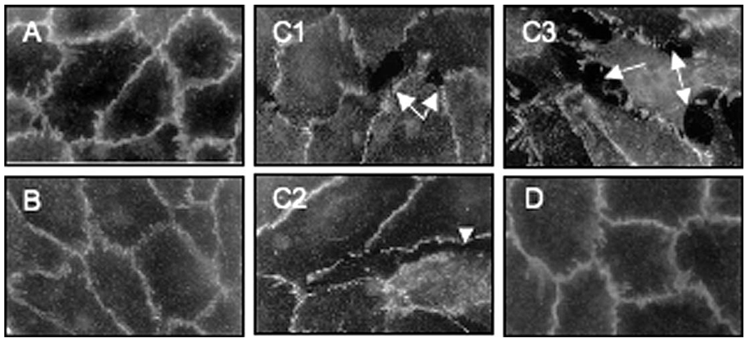
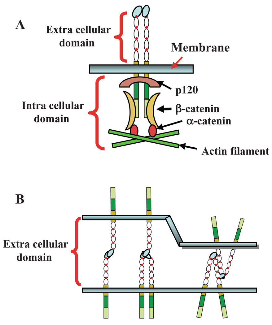
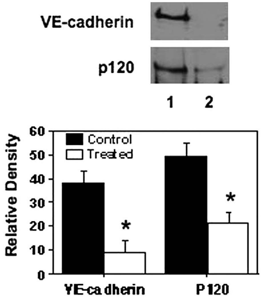
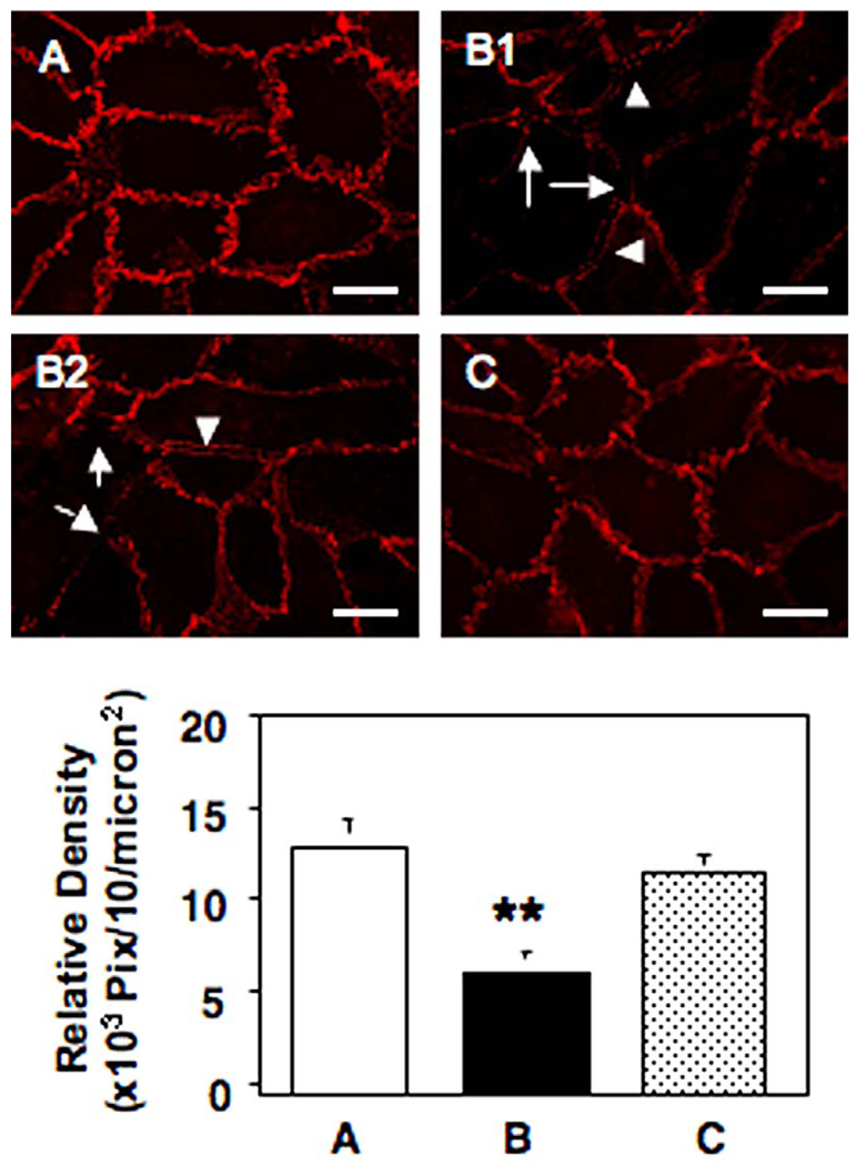
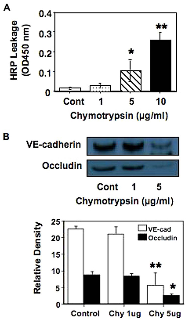
Placenta-derived chymotrypsin-like protease may contribute to endothelial activation in preeclampsia. In this study, we determined if placenta-derived chymotrypsin-like protease could disturb endothelial junctional integrity to promote endothelial permeability in preeclampsia. Confluent endothelial cells were cocultured with placental trophoblasts or treated with preeclampsia placenta-conditioned medium. Endothelial junction protein vascular endothelial cadherin expression and distribution were examined by fluorescent staining of endothelial cells with or without depletion of chymotrypsin. The association of endothelial cell junction protein complex VE-cadherin/beta-catenin/p120 was examined by a combined immuno-precipitation and immuno-blotting assay. Our results showed that endothelial cells cocultured with preeclampsia trophoblasts or exposed to preeclampsia placental conditioned medium exhibited a discontinuous distribution and reduced expression of vascular endothelial cadherin at cell contact regions. Vascular endothelial cadherin and p120 were expressed in control endothelial cells, but reduced or lost in endothelial cells exposed to preeclampsia placental conditioned medium, suggesting that the junctional protein complex of VE-cadherin/beta-catenin/p120 was disrupted in endothelial cells exposed to preeclampsia placental conditioned medium. We also observed that removal of trophoblasts from the coculture system and depletion of the protease from the preeclampsia placental conditioned medium could restore the dysregulated endothelial junction protein expression and distribution. Chymotrypsin also induced a dose dependent increase in endothelial monolayer permeability. We conclude that chymotrypsin-like protease released by the placenta is at least one important mediator responsible for disrupting endothelial cell integrity and inducing endothelial permeability in preeclampsia.


Figures

Figure 1
Figure 1
Vascular endothelial cadherin (VE-cadherin) expression and distribution. A, control endothelial cells (ECs); B, ECs cocultured with normal TCs; C, ECs cocultured with preeclampsia (PE) TCs; and D, EC recovery after cocultured with PE-TCs. Vascular endothelial cadherin was continuously expressed and distributed at region of cell contact in control cells and in cells cocultured with normal TCs (A and B). Multimorphology changes were observed in cells cocultured with preeclamptic TCs (C1–3): elongation, pore formation (arrow), and separation of VE-cadherin expression between adjacent cells (arrowhead). Disorganized VE-cadherin expression and distribution was restored after PE-TCs were removed from the coculture (D).
Figure 2
Figure 2
Schematic drawing of endothelial Vascular endothelial cadherin (VE-cadherin) domain organization and models for VE-cadherin based adhesion between adjacent endothelial cells. A, Endothelial VE-cadherin domains and intracellular protein complex. The cytoplasmic tail of VE-cadherin directly binds to β-catenin and p120 forming an intracellular junction complex. B, Vascular endothelial cadherin intercellular binding models. The dual homophilic binding model of VE-cadherin molecules between cells shows increased strength of cell-to-cell adhesion and binding force between adjacent cells.
Figure 3
Figure 3
Disassembly of vascular endothelial cadherin (VE-cadherin) and intracellular protein complex in endothelial cells (ECs) by factors released from preeclampsia (PE) placentas. Lane 1: control cells; Lane 2: cells treated with PE placental conditioned medium (CM). Total cellular protein was precipitated with antibody against β-catenin. The precipitated protein was run on SDS-PAGE and immunoblotted with antibodies against VE-cadherin and p120, respectively. Vascular endothelial cadherin and p120 were expressed in control cells, but not in cells treated with PE CM. These data suggest that dissociation occurred between the interaction of VE-cadherin and intracellular protein p120 and β-catenin in cells exposed to PE placental factors. The blot was representative from 3 independent experiments. The bar graph shows relative density of VE-cadherin and p120 expression, * P < .05, respectively.
Figure 4
Figure 4
Effects of placental chymotrypsin-like protease (CLP) on endothelial vascular endothelial cadherin (VE-cadherin) expression and distribution examined by fluorescent staining. A, control cells; B1 & B2: cells treated with preeclampsia (PE) placental conditioned medium (CM); and C, cells treated with PE placental CM after depletion of chymotrypsin. Reduced VE-cadherin expression and separation of VEcadherin expression between adjacent cells were observed in cells treated with PE placental CM, but not in cells treated with PE placental CM after depletion of chymotrypsin. Arrow: pore formation; and arrowhead: separation of VE-cadherin expression between adjacent cells. The bar graph shows a quantitative measure for VE-cadherin expression at endothelial junctions for control cells (A), cells treated with PE placental CM (B): cells treated with PE placental CM after depletion of chymotrypsin (C). Data are means from 5 independent experiments each in triplicate.
Figure 5
Figure 5
Chymotrypsin protease induced altered endothelial barrier dysfunction. A: Endothelial permeability measured by horseradish peroxidase (HRP) leakage in ECs treated with chymotrypsin, which induced a dose-dependent increase in HRP leakage, * P < .05, ** P < .01 compared to control cells, respectively. Data are means ± S.E. from 5 independent experiments each in duplicate. B, VE-cadherin and occludin expression in ECs treated with different concentrations of chymotrypsin. The blot was representative from 3 independent experiments. The bar graph shows relative density of VE-cadherin and occludin expression, chymotrypsin treated vs. control: ** P < .01 and * P < .05, respectively

Similar articles

Cited by

References

    1. Campbell DM, Campbell AJ. Evans blue disappearance rate in normal and preeclamptic pregnancy. Clin Exp Hypertens. 1983;2:163–169. - PubMed
    1. Brown MA, Zammit VC, Lowe SA. Capillary permeability and extracellular fluid volumes in pregnancy-induced hypertension. Clin Sci. 1989;77:599–604. - PubMed
    1. Svedas E, Islam KB, Nisell H, Kublickiene KR. Vascular endothelial growth factor induced functional and morphologic signs of endothelial dysfunction in isolated arteries from normal pregnant women. Am J Obstet Gynecol. 2003;188:168–176. - PubMed
    1. Wang Y, Gu Y, Granger DN, Roberts JM, Alexander JS. Endothelial junctional protein redistribution and increased monolayer permeability in HUVECs isolated during preeclampsia. Am J Obstet Gynecol. 2002;186:214–220. - PubMed
    1. Svedas E, Nisell H, Vanwijk MJ, Nikas Y, Kublickiene KR. Endothelial dysfunction in uterine circulation in preeclampsia: Can estrogens improve it? Am J Obstet Gynecol. 2002;187:1608–1616. - PubMed

Publication types

MeSH terms