. 2008;30(6):410-8.
doi: 10.1159/000191212. Epub 2009 Jan 7.

Oxidative imbalance in nonstimulated X-adrenoleukodystrophy-derived lymphoblasts

Affiliations

Oxidative imbalance in nonstimulated X-adrenoleukodystrophy-derived lymphoblasts

Takuhiro Uto et al. Dev Neurosci. 2008.

Abstract

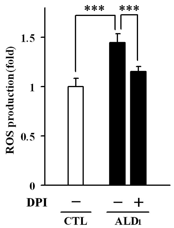
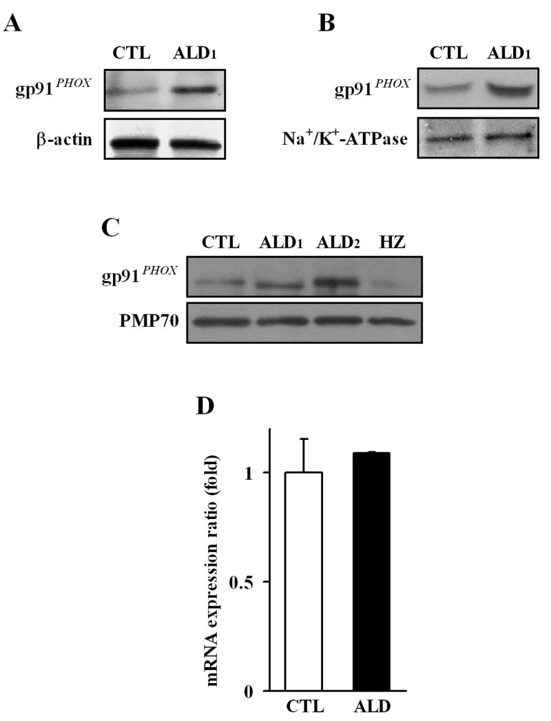
X-Adrenoleukodystrophy (X-ALD) is a peroxisomal disorder characterized by accumulation of very-long-chain (VLC) fatty acids, which induces inflammatory disease and alterations in cellular redox, both of which are reported to play a role in the pathogenesis of the severe form of the disease (childhood cerebral ALD). Here, we report on the status of oxidative stress (NADPH oxidase activity) and inflammatory mediators in an X-ALD lymphoblast cell line under nonstimulated conditions. X-ALD lymphoblasts contain nearly 7 times higher levels of the C(26:0) fatty acid compared to controls; these levels were downregulated by treatment with sodium phenylacetate (NaPA), lovastatin or the combination of both drugs. In addition, free-radicals synthesis was elevated in X-ALD lymphoblasts, and protein levels of the NADPH oxidase gp91(PHOX) membrane subunit were significantly upregulated, but no changes were observed in the p47(PHOX) and p67(PHOX) cytoplasmic subunits. Unexpectedly, there was no increase in gp91(PHOX) mRNA levels in X-ALD lymphoblasts. Furthermore, X-ALD lymphoblasts produced higher levels of nitric oxide (NO) and cytokines (tumor necrosis factor-alpha and interleukin 1 beta), and treatment with NaPA or lovastatin decreased the synthesis of NO. Our data indicate that X-ALD lymphoblasts are significantly affected by the accumulation of VLC fatty acids, which induces changes in the cell membrane properties/functions that may, in turn, play a role in the development/progression of the pathogenesis of X-ALD disease.


Figures

Fig. 1
Fig. 1
Molecular and biochemical alterations in an X-ALD lymphoblast cell lines and pharmacological restoration of very long chain fatty acids levels. Human B-lymphoblasts derived from a healthy control (CTL: GM03798), cerebral X-ALD patients (ALD1: GM04673; ALD2: GM13496), and heterozygote (HZ: GM 03798) were cultured and used to analyze protein levels of the peroxisomal integral membrane proteins transporters ALDP (ABCD1) and PMP70 (ABCD3) (A); cellular levels of very long chain fatty acids (C26:0)(B, C), and the effect of lovastatin and NaPA on C26:0 levels (D). Protein levels of the peroxisomal transporters were analyzed by Western blot in membranes fractions obtained by carbonate treatment (membrane preparation containing integral membrane proteins), as indicated in Methods section. Lipid analysis was performed by gas chromatography of the total fatty acids methyl ester of a organic extract of whole cell and C26:0 values are expressed as percent of total fatty acids (B), and the ratio of C26:0 to C22:0 (C). Western blots are representative of three different experiments. Data represent the average ± SD of n = 3 experiments done in duplicated. (B, C) ***: p < 0.0001; (D) ***: p < 0.0001 vs. untreated X-ALD lymphoblasts.
Fig. 2
Fig. 2
NADPH oxidase-dependent free radical production in control and X-ALD lymphoblasts. The fluorescent dye H2DCFDA was utilized to measure the synthesis of free radical in cultured control (white bar) and X-ALD (black bars) lymphoblasts under non-stimulated conditions. NADPH oxidase contribution to free radical synthesis in X-ALD was determined by pre-incubation of lymphoblasts with the enzyme’s inhibitor DPI, as indicated in Methods section. The data represent the average ± SD of n = 6 experiments done in triplicate. ***: p < 0.0001
Fig. 3
Fig. 3
Protein level and expression of the NADPH oxidase membrane subunit gp91PHOX in different cell fractions from control and X-ALD lymphoblasts. Protein levels of the membrane anchored subunit gp91PHOX of NADPH oxidase was determined in total homogenate (A), and total membrane fractions (B), using specific antibodies. β-Actin and Na+/K+-ATPase (plasma membrane protein) were used as indicators of protein loading for homogenate (A) and membrane fractions (B) respectively. The levels of gp91PHOX subunit anchored in the cell membrane was analyzed by Western blot on the carbonate membrane fractions obtained from control (CTL: GM03798), cerebral X-ALD patients (ALD1: GM04673; ALD2: GM13496), and heterozygote (HZ: GM 03798)(C). PMP70 was screened as indicators of protein loading for the carbonate membranes preparations (integral membrane proteins). Western blots are representative of three different experiments. The gp91PHOX gene expression was determined by qRT-PCR analysis, and normalized to the expression of GAPDH (D), as indicated in Methods section. The data represent the average ± SD of n = 3 experiments done in triplicate.
Fig. 4
Fig. 4
Protein levels and expression of the NADPH oxidase cytoplasmic subunits p47PHOX and p67PHOX in control and X-ALD cultured lymphoblasts. Protein levels of the cytoplasmic subunits of NADPH oxidase (p47PHOX and p67PHOX) were determined in total cell homogenate (A), and membrane and cytosolic cell fractions (B), using Western blot analysis as indicated in Methods. β-Actin and Na+/K+-ATPase (plasma membrane protein) were used as indicator of protein loading for cell homogenate (A), and plasma membrane fractions (B) respectively. Levels of p47PHOX and p67PHOX gene expression were determined by quantitative qRT-PCR analysis and represented after normalization to the expression of GAPDH (C, D). Western blots are representative of three different experiments. Data represent the average ± SD of n = 3 experiments done in triplicate.
Fig. 5
Fig. 5
Nitric oxide production and cytokine synthesis in control and X-ALD cultured lymphoblasts and pharmacological regulation of nitric oxide synthesis. Nitric oxide (NO) levels (A) and cytokines synthesis (TNF-α (B) and IL-1β (C)) were determined in cell-free supernatants from cultured control (GM03798; white bars), and a cerebral X-ALD (GM04673; black bars) lymphoblasts as indicated in Methods sections. The pharmacologic effect of drugs (Lovastatin (1μM), NaPA (1 mM), or combination of both drugs) on the production of NO by Control and X-ALD lymphoblasts were analyzed after a week of treatment. The data represent the average ± SD of n = 3 experiments done in triplicate. (A-C) **: p < 0.003; ***: p < 0.0001; (D) *: p < 0.05; ***: p < 0.0001.

References

    1. Wanders RJ, Waterham HR. Biochemistry of mammalian peroxisomes revisited. Annu Rev Biochem. 2006;75:295–332. - PubMed
    1. Mosser J, Douar AM, Sarde CO, Kioschis P, Feil R, Moser H, Poustka AM, et al. Putative X-linked adrenoleukodystrophy gene shares unexpected homology with ABC transporters. Nature. 1993;361:726–730. - PubMed
    1. Contreras M, Mosser J, Mandel JL, Aubourg P, Singh I. The protein coded by the X-adrenoleukodystrophy gene is a peroxisomal integral membrane protein. FEBS Lett. 1994;344:211–215. - PubMed
    1. Contreras M, Sengupta TK, Sheikh F, Aubourg P, Singh I. Topology of ATP-binding domain of adrenoleukodystrophy gene product in peroxisomes. Arch Biochem Biophys. 1996;334:369–379. - PubMed
    1. Lazo O, Contreras M, Hashmi M, Stanley W, Irazu C, Singh I. Peroxisomal lignoceroyl-CoA ligase deficiency in childhood adrenoleukodystrophy and adrenomyeloneuropathy. Proc Natl Acad Sci U S A. 1988;85:7647–7651. - PMC - PubMed

Publication types

MeSH terms