. 2009;149(2):119-26.
doi: 10.1159/000189194. Epub 2009 Jan 6.

Pimecrolimus reduces eosinophil activation associated with calcium mobilization

Affiliations

Pimecrolimus reduces eosinophil activation associated with calcium mobilization

Douglas A Plager et al. Int Arch Allergy Immunol. 2009.

Abstract

Background: Pimecrolimus is a calcineurin inhibitor that inhibits T cell and mast cell activation and effectively treats atopic dermatitis. However, its effects on eosinophils, a cell type implicated in allergic disease pathology, are unknown. Therefore, we examined the effects of pimecrolimus on eosinophil superoxide anion production, degranulation and survival.

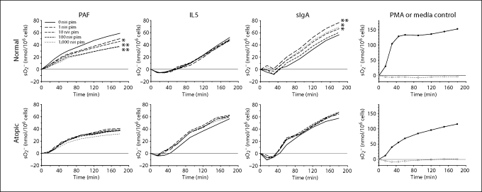
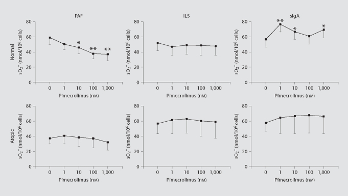
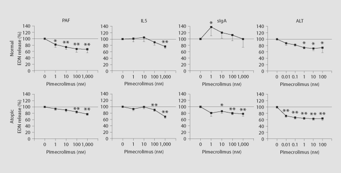
Methods: Purified eosinophils from normal or atopic donors were incubated with serial dilutions of pimecrolimus (microM to nM) and then stimulated with platelet activating factor (PAF), interleukin 5 (IL5), secretory immunoglobulin A (sIgA) or Alternaria alternata (Alt) fungus extract. Eosinophil activation was monitored by cytochrome c reduction resulting from superoxide anion production and by a 2-site immunoassay for eosinophil-derived neurotoxin (EDN) in cellular supernatants, as a marker of degranulation. Eosinophil survival was measured by propidium iodide exclusion using flow cytometry after 4 days in culture.

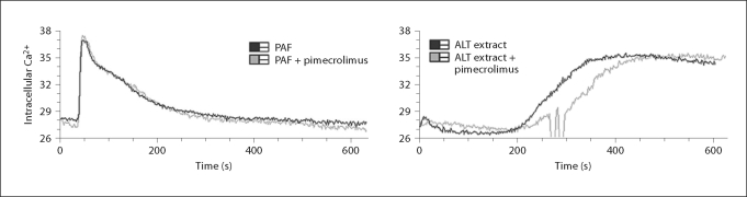
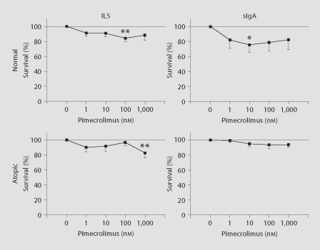
Results: Normal and atopic eosinophil superoxide anion production induced by PAF, and associated with increased intracellular calcium, was inhibited up to 37% with 1 microM pimecrolimus. However, superoxide anion production induced by IL5 and sIgA was not consistently inhibited. EDN release, which ultimately depends on calcium, was inhibited about 30% with PAF, IL5 and sIgA stimulation for normal and atopic donor eosinophils. Furthermore, calcium-dependent Alt-induced EDN release was inhibited up to 49% with nanomolar pimecrolimus. Finally, increased eosinophil survival promoted by IL5 and sIgA was not influenced by pimecrolimus.

Conclusion: Pimecrolimus moderately inhibits eosinophil superoxide anion production and EDN release associated with calcium mobilization, which may contribute to its efficacy in treating atopic dermatitis.


Figures

Fig. 1
Fig. 1
Effect of pimecrolimus on eosinophil superoxide production. Mean superoxide anion (sO2) production by purified eosinophils from normal (n = 13) or atopic donors (n = 9 or 10) is shown following stimulation with PAF, IL5 or immobilized sIgA after pre-incubating eosinophils with no pimecrolimus (i.e. 0 nM pim; solid line) or with pimecrolimus at 1 nM (long dashes), 10 nM (medium dashes), 100 nM (short dashes) or 1,000 nM (dotted). Positive (PMA, closed circles) and negative (medium alone, open circles) control responses are shown (n = 11 for normals and n = 4 for atopics). Significant changes in superoxide anion production relative to stimuli alone (i.e. 0 nM pimecrolimus) at the 180-min time point are indicated by asterisks. ∗ p < 0.05; ∗∗ p < 0.01.
Fig. 2
Fig. 2
Pimecrolimus dose effect on endpoint eosinophil superoxide production. Mean superoxide anion (sO2) production after incubating for 180 min in the absence (0 nM) and presence of increasing concentrations of pimecrolimus (1 to 1,000 nM) are shown. Values are from figure 1 following stimulation of normal or atopic donor eosinophils with PAF, IL5 or immobilized sIgA. Significant changes in superoxide anion production relative to stimuli alone (i.e. 0 nM pimecrolimus) are indicated by asterisks. ∗ p < 0.05; ∗∗ p < 0.01. Standard errors of the means are indicated by unidirectional error bars.
Fig. 3
Fig. 3
Effect of pimecrolimus on eosinophil EDN release. Cellular supernatant collected at the end of superoxide anion (sO2) production experiments (fig. 1) were assayed for EDN content (n = 11 for normals, n = 4 for atopics). Data are presented as percent EDN release relative to that induced by stimuli (PAF, IL5 or immobilized sIgA) alone. In separate experiments (ALT extract complicates measurement of superoxide anion production), EDN release from purified eosinophils (n = 3 for normals, n = 5 for atopics) induced by ALT fungal extract was measured. In all instances, the mean percent EDN release in the presence of medium alone (i.e. with no stimulant) was within the range of 15 to 28%. Significant changes in percent EDN release relative to stimuli alone (i.e. 0 nM pimecrolimus) are indicated by asterisks. ∗ p < 0.05; ∗∗ p < 0.01. Standard errors of the means are indicated by unidirectional error bars.
Fig. 4
Fig. 4
Intracellular calcium flux induced by PAF and ALT in the absence and presence of pimecrolimus. Purified eosinophils loaded with a fluorescent calcium indicator (indo-1/AM) were stimulated with 1 μM PAF or 75 μg/ml ALT fungal extract (black lines). In a second round of experiments, eosinophils were pretreated with 1 nM pimecrolimus for 1 h on ice before PAF or ALT stimulation (gray lines). Eosinophils were all from a single individual, and the sharp fluctuations in the ALT extract + pimecrolimus trace are artificial.
Fig. 5
Fig. 5
Effect of pimecrolimus on eosinophil survival. Purified eosinophils were preincubated for 30 min without or with varying concentrations of pimecrolimus [n = 11 (IL5) or 7 (sIgA) for normals, n = 7 for atopics]. IL5 (75 pg/ml) or soluble sIgA (10 μg/ml) was then added and cell survival was measured 4 days later. Data are presented as percent survival relative to that induced by stimuli (IL5 or sIgA) alone. In all instances, the mean percent survival in the presence of medium alone (i.e. with no stimulant) was within the range of 17 to 25%. Significant changes in percent survival relative to stimuli alone (i.e. 0 nM pimecrolimus) are indicated by asterisks. ∗ p < 0.05; ∗∗ p < 0.01. Standard errors of the means are indicated by unidirectional error bars.

References

    1. Leung DY, Bieber T. Atopic dermatitis. Lancet. 2003;361:151–160. - PubMed
    1. Laughter D, Istvan JA, Tofte SJ, Hanifin JM. The prevalence of atopic dermatitis in Oregon schoolchildren. J Am Acad Dermatol. 2000;43:649–655. - PubMed
    1. Worldwide variation in prevalence of symptoms of asthma, allergic rhinoconjunctivitis, and atopic eczema: ISAAC. The International Study of Asthma and Allergies in Childhood (ISAAC) Steering Committee. Lancet. 1998;351:1225–1232. - PubMed
    1. Diepgen TL. Is the prevalence of atopic dermatitis increasing? In: Williams HC, editor. Atopic Dermatitis: The Epidemiology, Causes and Prevention of Atopic Eczema. Cambridge: Cambridge University Press; 2000. pp. 96–109.
    1. Linna O, Kokkonen J, Lahtela P, Tammela O. Ten-year prognosis for generalized infantile eczema. Acta Paediatr. 1992;81:1013–1016. - PubMed

Publication types

MeSH terms