. 2009 May;65(5):524-9.
doi: 10.1203/PDR.0b013e3181998baf.

Thalamic damage in periventricular leukomalacia: novel pathologic observations relevant to cognitive deficits in survivors of prematurity

Affiliations

Thalamic damage in periventricular leukomalacia: novel pathologic observations relevant to cognitive deficits in survivors of prematurity

Poonam Ligam et al. Pediatr Res. 2009 May.

Abstract

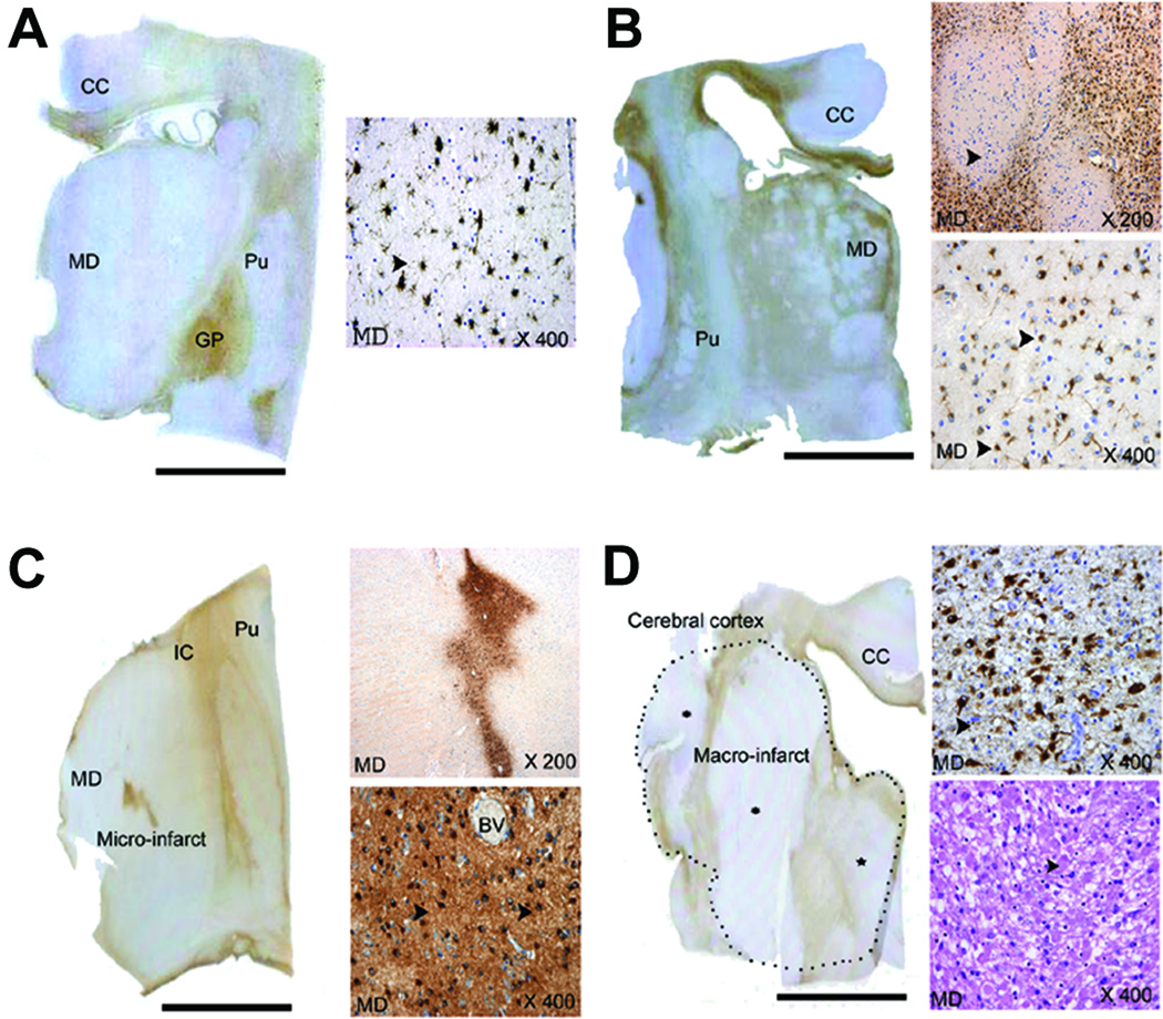
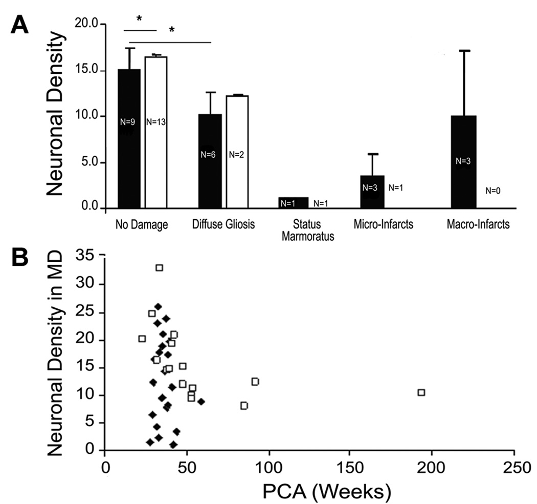
Despite major advances in the long-term survival of premature infants, cognitive deficits occur in 30-50% of very preterm (<32 gestational weeks) survivors. Impaired working memory and attention despite average global intelligence are central to the academic difficulties of the survivors. Periventricular leukomalacia (PVL), characterized by periventricular necrosis and diffuse gliosis in the cerebral white matter, is the major brain pathology in preterm infants. We tested the novel hypothesis that pathology in thalamic nuclei critical for working memory and attention, i.e. mediodorsal nucleus and reticular nucleus, respectively, occurs in PVL. In 22 PVL cases (gestational age 32.5 +/- 4.8 wk) and 16 non-PVL controls (36.7 +/- 5.2 wk) who died within infancy, the incidence of thalamic pathology was significantly higher in PVL cases (59%; 13/22) compared with controls (19%; 3/16) (p = 0.01), with substantial involvement of the mediodorsal, and reticular nuclei in PVL. The prevention of thalamic damage may be required for the eradication of defects in survivors with PVL.


Figures

Figure 1
Figure 1
We observed four patterns of injury in the thalamus (Patterns 1–4) compared to no histopathology (Pattern 0)(not shown). The four patterns are: a. Pattern 1—Diffuse gliosis throughout the thalamus (left-hand whole mount) with arrows indicating reactive astrocytes immunostained with GFAP in the right-hand insert of mediodorsal nucleus at x400; of note, gliosis is striking in the globus pallidus in the left-hand whole mount. b. Pattern 2—Status marmoratus with linear scarring at low magnification (whole mount of tissue section) and islands of profound neuronal loss and gliosis (highlighted with GFAP immunostaining) interspersed with better preserved islands (arrow head, upper right-hand insert) detected microscopically in the mediodorsal and other nuclei at x200; reactive astrocytes immunostained with GFAP are demonstrated in the lower right insert at x400. Gliosis in the internal capsule, corpus callosum, and deep white matter are associated with the thalamic damage in the left-hand whole mount. c. Pattern 3—Micro-infarcts detected with GFAP immunostaining at low and high magnification and characterized by severe reactive gliosis (and neuronal loss) embedded in an otherwise intact parenchyma in the left-hand whole mount and in right hand inserts at x200 and x400; the sections are immunostained with GFAP; and d. Pattern 4—Macro-infarcts: here the dotted outline in the left-hand whole mount defines the outer boundary of the macro-infarct, and includes the dorsal thalamus, internal capsule, and insular cortex, i.e., regions in the distribution of the posterior and middle cerebral artery; the infarct in the mediodorsal nucleus in the right-hand inserts is characterized by reactive astrocytes (x400, upper right-hand insert, GFAP immunostaining), macrophagocytic infiltration, and axonal spheroids (lower right-hand insert, hematoxylin-and-eosin). Abbreviations: BV, blood vessels; CC, corpus callosum; GP, globus pallidus; IC, internal capsule; MD, mediodorsal nucleus; Pu, putamen. Scale bar=1 centimeter.
Figure 2
Figure 2
A. Neuronal density in the four patterns of injury (Patterns 1–4) in PVL and control cases compared to the neuronal density in PVL and control cases without histopathologic changes (Pattern 0) in the mediodorsal nucleus. The neuronal density in Pattern 0 and Pattern 1 is presented as the mean adjusted for postconceptional age. There was significantly reduced neuronal density in the PVL cases compared to controls (p<0.05) in Pattern 0 in the mediodorsal nucleus, but not in the lateral posterior nucleus (data not shown). There was also a significant difference in neuronal density in the mediodorsal nucleus between Pattern 0 and Pattern 1 in PVL cases (p=0.001) but not controls (p=0.37). Neuronal density was markedly reduced in status marmoratus, micro-infarcts within infarct itself), and macro-infarcts. PVL, ■; Control, □. B. In PVL cases without status marmoratus or micro- or macro-infarcts, the neuronal density tended to decrease with increasing postconceptional age in both PVL and control cases in the mediodorsal nucleus, with a marginal difference in the mean density adjusted for postconceptional age in the PVL cases (11.9±1.6 neurons/mm2) compared to controls (16.7 neurons/mm2) (p=0.07). PVL, ◆; Control, □
Figure 3
Figure 3
The relationship of reactive astrocyte (GFAP-positive) density to MDA-immunopositive neuronal density. As the density of reactive astrocyte increases, the density of MDA-positive neurons decreases, suggesting that MDA expression is an early marker of neuronal injury (see text). PVL,◆; Control, □

References

    1. Marlow N, Wolke D, Bracewell M, Samara M. Neurologic and developmental disability at six years after extremely preterm birth. N Engl J Med. 2005;352:9–19. - PubMed
    1. Bhutta AT, Cleves MA, Casey PH, Cradock MM, Anand KJ. Cognitive and behavioural outcomes of school-aged children who were born preterm: a meta-analysis. JAMA. 2002;288:728–737. - PubMed
    1. Woodward LJ, Edgin JO, Thompson D, Inder TE. Object working memory deficits predicted by early brain injury and development in the preterm infant. Brain. 2005;128:2578–2587. - PubMed
    1. Bayless S, Stevenson J. Executive functions in school-age children born very prematurely. Early Hum Dev. 2007;83:247–254. - PubMed
    1. Böhm B, Smedler A-C, Forssberg H. impulse control, working memory and other executive functions in preterm children when starting school. Acta Paediatr. 2004;93:1363–1371. - PubMed

Publication types

MeSH terms