

Spinal extradural angiolipoma: report of two cases and review of the literature
- PMID: 19127373
- PMCID: PMC2899409
- DOI: 10.1007/s00586-008-0858-8
Spinal extradural angiolipoma: report of two cases and review of the literature
Abstract
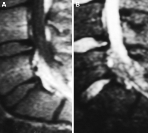
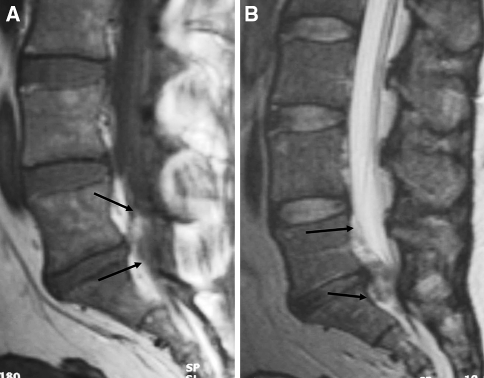
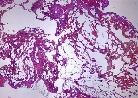
Spinal angiolipomas are benign uncommon neoplasm composed of mature lipocytes admixed with abnormal blood vessels. They account for only 0.04-1.2% of all spinal tumors. We report two cases of lumbar extradural angiolipoma and review previously reported cases. We found 118 cases of spinal epidural angiolipoma (70 females and 48 males; age range 1.5-85 years, mean 44.03) spanning from 1890 to 2006. Prior to diagnosis 40.6% of the patients had weakness of the lower limbs. The interval between the initial symptoms and tumor diagnosis ranged from 1 day to 17 years (mean 20.2 months). Except for four cases diagnosed at autopsy, 109 patients underwent surgery and gross-total resection was performed in 79 cases (72.4%). Spinal angiolipomas are tumors containing angiomatous and lipomatous tissue, predominantly located in the mid-thoracic region. All angiolipomas show iso- or hyperintensity on T1-weighted images and hyperintensity on T2-weighted images and most lesions enhance with gadolinium administration. The treatment for spinal extradural angiolipomas is total surgical resection and no adjuvant therapy should be administered.
Figures
References
-
- Akhaddar A, Gazzaz M, Derraz S, Rifi L, Amarti A, Aghzadi A, El Ouahabi A, El Khamlichi A. Spinal epidural angiolipomas: a rare cause of spinal cord compression. A report of 8 cases and review of the literature. Neurochirurgie. 2000;46:523–533. - PubMed
-
- Amlashi SF, Morandi X, Chabert E, Riffaud L, Haegelen C, Rolland Y. Spinal epidural angiolipoma. J Neuroradiol. 2001;28:253–256. - PubMed
Publication types
MeSH terms
LinkOut - more resources
Full Text Sources
