. 2009 Feb;49(2):618-26.
doi: 10.1002/hep.22656.

Hepatic function is preserved in the absence of mature microRNAs

Affiliations

Hepatic function is preserved in the absence of mature microRNAs

Nicholas J Hand et al. Hepatology. 2009 Feb.

Abstract

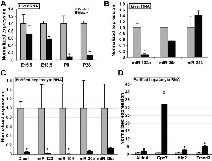
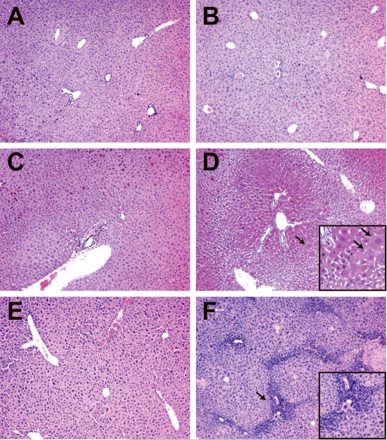
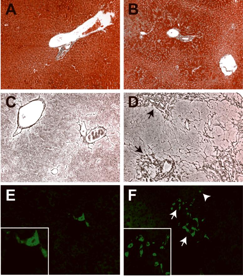
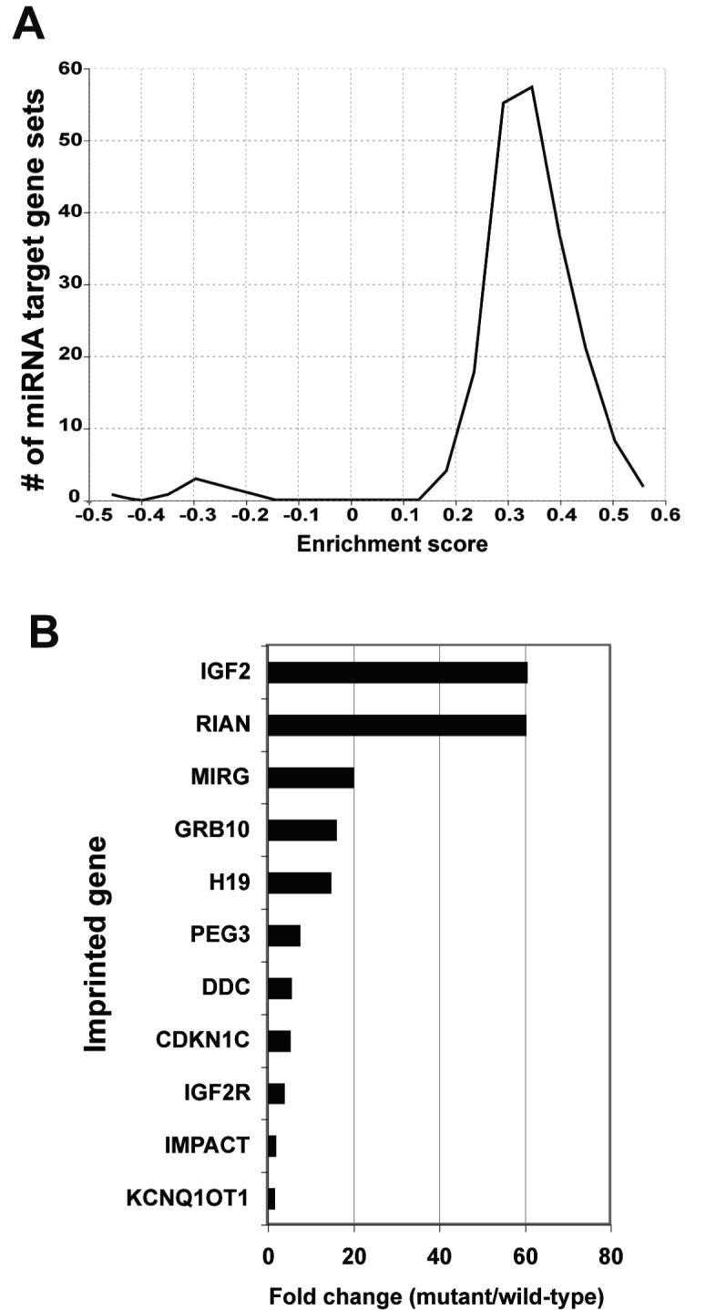
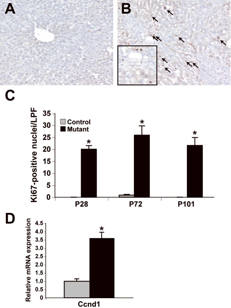
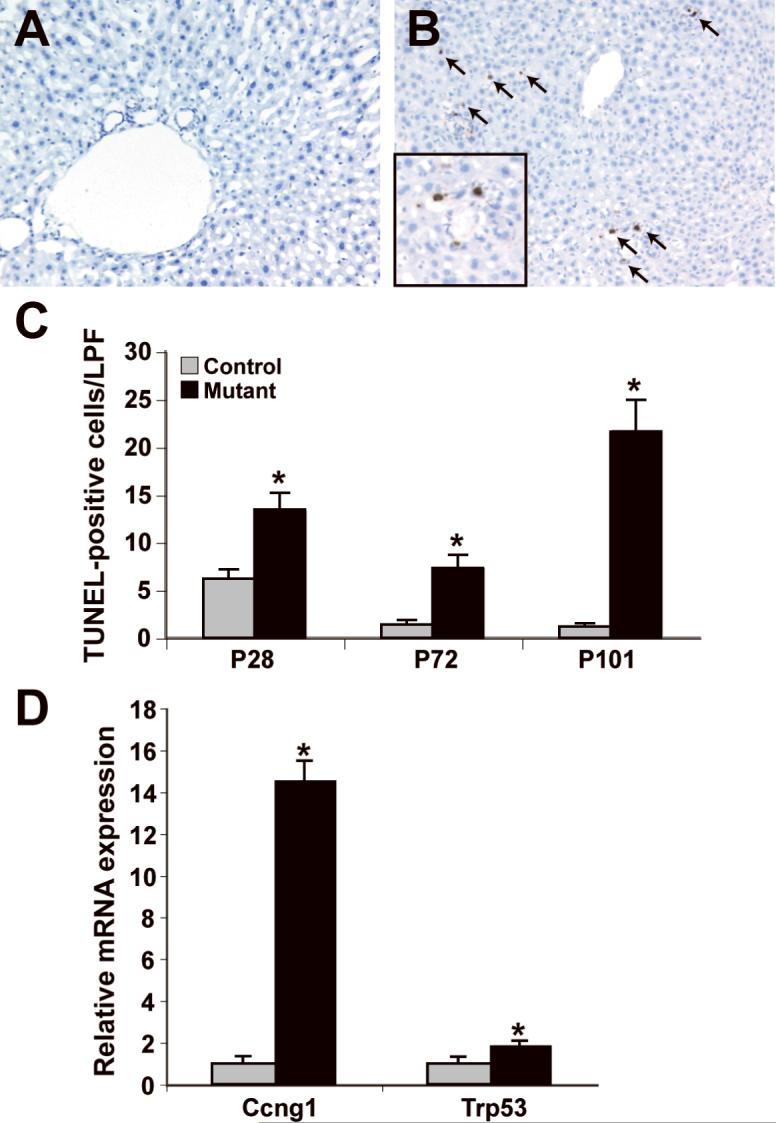
MicroRNAs (miRNAs) are small noncoding RNA molecules that regulate gene expression through partial or complete complementarity with target messenger RNAs. The function of miRNAs in normal liver physiology is largely unknown. We address the role of Dicer1 in the differentiated liver. We derived mice lacking Dicer1 function in hepatocytes and assessed the loss of mature miRNA via quantitative polymerase chain reaction. Gene expression microarray analysis was performed on liver RNA from mutant and control mice. Liver sections from mutant and control mice were examined and liver function tests were performed. Mice lacking Dicer1 function in hepatocytes appeared and behaved normally. Despite the loss of mature miRNAs, hepatic function was maintained, as reflected by normal blood glucose, albumin, cholesterol, and bilirubin. However, mutant mice between 2 and 4 months of age exhibited progressive hepatocyte damage with elevated serum alanine aminotransferase and aspartate aminotransferase. Liver mass was increased in mutant mice, as were cellular markers of both proliferation and apoptosis. Microarray analysis indicated large-scale changes in gene expression, with increased expression of many miRNA targets, particularly imprinted genes.

Conclusions: Loss of miRNA processing in the liver at late gestation has a remarkably mild phenotype, suggesting that miRNAs do not play an essential role in hepatic function. However, miRNA deficiency results in hepatocyte apoptosis, hepatocyte regeneration, and portal inflammation. Finally, microarray analysis of gene expression in the mutant liver supports a previously hypothesized role for Dicer1 in the repression of imprinted genes.


Figures

Figure 1
Figure 1. Functional depletion of Dicer1 in liver
A: Wild-type Dicer1 transcript levels in total liver RNA from mutant and control animals were assayed by qRT-PCR and normalized to Hprt transcript levels. Control levels are scaled to 1. By P0, wild-type Dicer1 transcript levels in mutant liver are 9% of those in control liver (p<0.05), and remain at 12% at P28 (p<0.0002). B: MiRNA transcript levels in total liver RNA from mutant and control animals were assayed by qRT-PCR, and normalized to U6 RNA transcript levels. Levels of miR-122a are reduced by 93% in liver lacking functional Dicer1 (p<0.01). (C) Expression of Dicer1 and selected miRNA was assayed in total RNA isolated from P40 purified hepatocytes and normalized either to Hprt (Dicer1) or U6 RNA (miRNA). (D) Transcript levels of selected experimentally validated targets of miR-122a regulation in P40 purified hepatocytes, normalized to Hprt. C, D (control, n=3; mutant, n=5). Mutant, AlfpCre+; Dicer1flox/flox. Control, AlfpCre-; Dicer1flox/flox.
Figure 2
Figure 2. Dicer1-deficient liver develops progressive hepatitis
Hematoxylin and eosin stained liver sections. A, C, E: Controls (AlfpCre-; Dicer1flox/flox). B, D, F: Mutant, AlfpCre+; Dicer1flox/flox. A, B: P28. No significant differences were observed between control and mutant tissue. C, D: P72. Hepatocytes close to portal tracts appear to be enlarged (arrows) relative to those close to central veins, resulting in a pseudonodular appearance. E, F: P101. Mutant tissue exhibits extensive infiltration of inflammatory cells, particularly in peri-portal regions (arrows).
Figure 3
Figure 3. Dicer1 mutant livers exhibit hepatocyte loss and increased CK19 expression
A, C, E: Controls. B, D, F: Mutants. A, B: Trichrome staining of P72 liver sections reveals that despite the pseudonodular appearance of the mutant tissue, no fibrosis is evident at this age. C, D: Reticulin staining of P101 mutant liver sections to highlight the sinusoidal architecture reveals extensive compaction in peri-portal regions (arrows), indicating loss of hepatocytes. E, F: CK19 immunofluorescent antibody staining of P101 liver sections reveals a ductular reaction (arrows), with increased numbers of CK19-positive cells, both in portal areas and occasionally within the lobule (arrowhead). The intensity of CK19 in the mutant sections was increased relative to controls.
Figure 4
Figure 4. Microarray analysis of gene expression in Dicer1 mutant liver
A: Global histogram of gene set enrichment scores for the 192 sets of predicted miRNA target genes. The data is remarkable for the asymmetry between the large number of sets having increased expression and the absence of any sets having decreased expression in Dicer-deficient liver. B: The expression of several imprinted genes is increased in the Dicer1-deficient liver, as determined by microarray analysis (n=5).
Figure 5
Figure 5. Dicer1 mutant livers display increased cellular proliferation
A, B: Ki67 immunohistochemistry on control (A) and mutant (B) liver sections reveals numerous proliferating cells (arrows) in mutant liver (P101 shown). C: Quantification of the results of the Ki67 immunohistochemistry (n=3; *p<0.05). D: Transcript levels of Ccnd1 normalized to Hprt. in P40 purified hepatocytes (control, n=3; mutant, n=5). LPF, low powered field.
Figure 6
Figure 6. Dicer1 mutant livers display increased apoptosis
A, B: TUNEL assay of control (A) and mutant (B) liver sections reveals numerous apoptotic bodies (arrows) per low powered field in mutant liver sections (P101 shown). C: Quantification of the results of the TUNEL assay (n=3; *p<0.05). D: Ccng1 and Trp53 expression in P40 purified hepatocytes, normalized to Hprt (control, n=3; mutant, n=5). LPF, low powered field.

References

    1. Ambros V. microRNAs: tiny regulators with great potential. Cell. 2001;107:823–826. - PubMed
    1. Ambros V. The functions of animal microRNAs. Nature. 2004;431:350–355. - PubMed
    1. Bernstein E, Caudy AA, Hammond SM, Hannon GJ. Role for a bidentate ribonuclease in the initiation step of RNA interference. Nature. 2001;409:363–366. - PubMed
    1. Hutvagner G, McLachlan J, Pasquinelli AE, Balint E, Tuschl T, Zamore PD. A cellular function for the RNA-interference enzyme Dicer in the maturation of the let-7 small temporal RNA. Science. 2001;293:834–838. - PubMed
    1. Ketting RF, Fischer SE, Bernstein E, Sijen T, Hannon GJ, Plasterk RH. Dicer functions in RNA interference and in synthesis of small RNA involved in developmental timing. C. elegans. Genes Dev. 2001;15:2654–2659. - PMC - PubMed

Publication types

MeSH terms