

doi: 10.1002/prot.22338.
Crystal structure of the Fic (Filamentation induced by cAMP) family protein SO4266 (gi|24375750) from Shewanella oneidensis MR-1 at 1.6 A resolution
Affiliations
- PMID: 19127588
- PMCID: PMC2674511
- DOI: 10.1002/prot.22338
Item in Clipboard
Crystal structure of the Fic (Filamentation induced by cAMP) family protein SO4266 (gi|24375750) from Shewanella oneidensis MR-1 at 1.6 A resolution
Proteins.
2009 Apr.
No abstract available
Figures
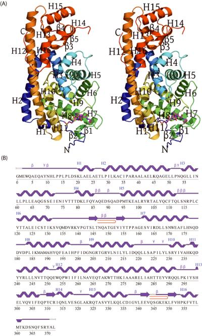
A) Stereo representation of the crystal structure of the chain A monomer of the SO4266 protein. The peptide modeled as `MEWQ' corresponds to residues 1-4 of the crystallographic symmetry-related B chain and is shown in ball-and-stick. B) Diagram showing the secondary structure elements of SO4266 superimposed on the primary amino acid sequence. The helices, β-strands, γ-turns, and β-turns are indicated. The β-hairpins are indicated as red loops. Residues absent from the monomer A (G0, M1, Q344, S345, A371 and L372) are indicated by lack of a secondary structure trace.
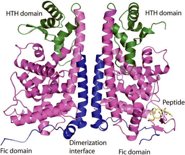
The monomers of SO4266 assume a closed fist-like shape, with a back-to-back association of the monomers forming a “transcription factor-like” dimer. The core of the Fic domain is made up of residues 100-290 (magenta), with a long, mostly α-helical insert region at the N-terminus (residues 1-100; blue) that is involved in dimerization. Residues 290-370 of the C-terminus form a winged helix-turn-helix (wHTH) DNA-binding domain (green) similar to that seen in several transcriptional regulators (e.g. PDB id 2d1h). The peptide `MEWQ' bound to chain A that corresponds to residues 1-4 of the crystallographic symmetry-related B chain is shown (yellow).
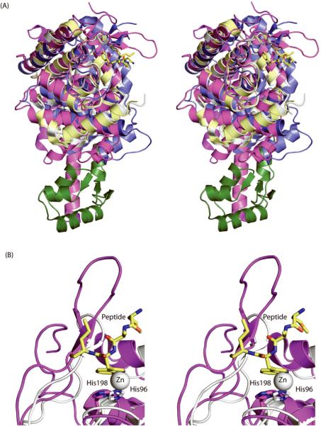
A) Superimposition of the SO4266 monomers (magenta and green) with the Fic family proteins from Neisseria meningitidis (194 residues, 2g03, yellow), Helicobacter pylori (201 residues, 2f6s, grey) and Bacteroides thetaiotaomicron vpi-5482 (262 residues, 3cuc, blue), reveal the similarity in their structures despite the absence of the C-terminal domain of SO4266 (green) in the other Fic proteins. They align on SO4266 with RMSDs of 3.1 Å, 3.2 Å and 2.8 Å, Z-scores of 10.5, 10.4 and 18.5 (DaliLite), and sequence identities of 12%, 14% and 16%, respectively. The peptide is shown (yellow ball-and-stick). B) The structure of the H. pylori Fic protein (grey) contains a zinc ion (grey) interacting with His 96 of the conserved HPFXXGNG motif. In the structure of SO4266 (magenta), the tryptophan residue of the peptide (yellow ball-and-stick) partially overlaps with the location of the Zn2+ ion and it is unlikely that the protein can bind metal and the peptide at the same time (grey).
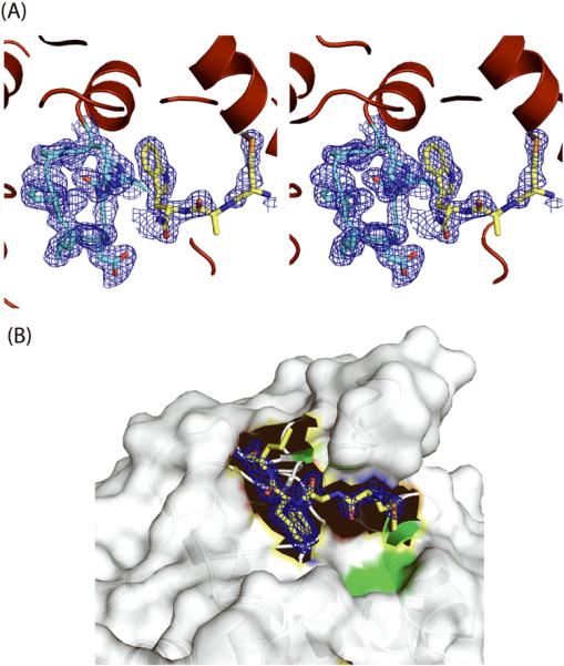
A) Density modified electron density map from experimental phases at 1.0 σ with the N-terminal B1-B4 peptide sequence `MEWQ' (yellow) modeled near the HPFXXGNG motif (cyan) in SO4266, with the tryptophan facing and within interaction distance to His198. B) A surface rendering of the most prominent cleft in SO4266, with the B1-B4 peptide shown (`MEWQ', yellow). The rest of the peptide is close to residues 143-148 of chain A, which forms part of a β-hairpin between helices H6 and H7. This region of the protein forms a lid over the binding cleft. In addition to this lid, the binding cleft is situated between helices H8, H9, and H11. The green region corresponds to Thr248, Leu244, Tyr241, Leu145, Tyr155, and Leu245 that are proximal to the other extra electron density that was modeled as a PGE molecule from the crystallization condition.
Similar articles
-
The three-dimensional crystal structure of the PrpF protein of Shewanella oneidensis complexed with trans-aconitate: insights into its biological function.Protein Sci. 2007 Jul;16(7):1274-84. doi: 10.1110/ps.072801907. Epub 2007 Jun 13. Protein Sci. 2007. PMID: 17567742 Free PMC article.
-
Structures of the first representatives of Pfam family PF06938 (DUF1285) reveal a new fold with repeated structural motifs and possible involvement in signal transduction.Acta Crystallogr Sect F Struct Biol Cryst Commun. 2010 Oct 1;66(Pt 10):1218-25. doi: 10.1107/S1744309109050416. Epub 2010 Mar 5. Acta Crystallogr Sect F Struct Biol Cryst Commun. 2010. PMID: 20944214 Free PMC article.
-
Structural dissection of Shewanella oneidensis old yellow enzyme 4 bound to a Meisenheimer complex and (nitro)phenolic ligands.FEBS Lett. 2017 Oct;591(20):3391-3401. doi: 10.1002/1873-3468.12833. Epub 2017 Sep 15. FEBS Lett. 2017. PMID: 28869767
-
Adenylylation control by intra- or intermolecular active-site obstruction in Fic proteins.Nature. 2012 Jan 22;482(7383):107-10. doi: 10.1038/nature10729. Nature. 2012. PMID: 22266942
-
Structural insights into substrate recognition in proton-dependent oligopeptide transporters.EMBO Rep. 2013 Sep;14(9):804-10. doi: 10.1038/embor.2013.107. Epub 2013 Jul 19. EMBO Rep. 2013. PMID: 23867627 Free PMC article.
Cited by
-
Protein expression, characterization, crystallization and preliminary X-ray crystallographic analysis of a Fic protein from Clostridium difficile.Acta Crystallogr F Struct Biol Commun. 2014 Jun;70(Pt 6):827-31. doi: 10.1107/S2053230X1400987X. Epub 2014 May 25. Acta Crystallogr F Struct Biol Commun. 2014. PMID: 24915103 Free PMC article.
-
Acquisition through horizontal gene transfer of plasmid pSMA198 by Streptococcus macedonicus ACA-DC 198 points towards the dairy origin of the species.PLoS One. 2015 Jan 13;10(1):e0116337. doi: 10.1371/journal.pone.0116337. eCollection 2015. PLoS One. 2015. PMID: 25584532 Free PMC article.
-
Comparative analysis of Histophilus somni immunoglobulin-binding protein A (IbpA) with other fic domain-containing enzymes reveals differences in substrate and nucleotide specificities.J Biol Chem. 2011 Sep 16;286(37):32834-42. doi: 10.1074/jbc.M111.227603. Epub 2011 Jul 27. J Biol Chem. 2011. PMID: 21795713 Free PMC article.
-
The DNA-binding induced (de)AMPylation activity of a Coxiella burnetii Fic enzyme targets Histone H3.Commun Biol. 2023 Nov 6;6(1):1124. doi: 10.1038/s42003-023-05494-7. Commun Biol. 2023. PMID: 37932372 Free PMC article.
-
Structure and function of Fic proteins.Nat Rev Microbiol. 2015 Oct;13(10):631-40. doi: 10.1038/nrmicro3520. Epub 2015 Aug 24. Nat Rev Microbiol. 2015. PMID: 26299785 Review.
References
-
- Komano T, Utsumi R, Kawamukai M. Functional analysis of the fic gene involved in regulation of cell division. Res Microbiol. 1991;142:269–277. - PubMed
-
- Li W, Godzik A. Cd-hit: a fast program for clustering and comparing large sets of protein or nucleotide sequences. Bioinformatics. 2006;22:1658–1659. - PubMed
-
- Clark HF, Gurney AL, Abaya E, Baker K, Baldwin D, Brush J, Chen J, Chow B, Chui C, Crowley C, Currell B, Deuel B, Dowd P, Eaton D, Foster J, Grimaldi C, Gu Q, Hass PE, Heldens S, Huang A, Kim HS, Klimowski L, Jin Y, Johnson S, Lee J, Lewis L, Liao D, Mark M, Robbie E, Sanchez C, Schoenfeld J, Seshagiri S, Simmons L, Singh J, Smith V, Stinson J, Vagts A, Vandlen R, Watanabe C, Wieand D, Woods K, Xie MH, Yansura D, Yi S, Yu G, Yuan J, Zhang M, Zhang Z, Goddard A, Wood WI, Godowski P, Gray A. The secreted protein discovery initiative (SPDI), a large-scale effort to identify novel human secreted and transmembrane proteins: a bioinformatics assessment. Genome Res. 2003;13:2265–2270. - PMC - PubMed
Publication types
MeSH terms
Substances
Grants and funding
LinkOut - more resources
Full Text Sources
Molecular Biology Databases
