. 2009 Apr 15;419(2):437-45.
doi: 10.1042/BJ20082003.

Peroxiredoxin 5 confers protection against oxidative stress and apoptosis and also promotes longevity in Drosophila

Affiliations

Peroxiredoxin 5 confers protection against oxidative stress and apoptosis and also promotes longevity in Drosophila

Svetlana N Radyuk et al. Biochem J. .

Abstract

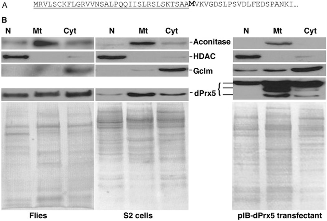
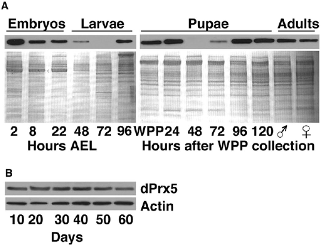
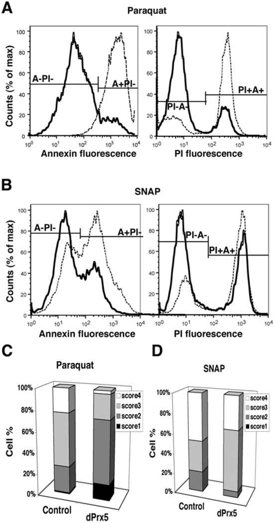
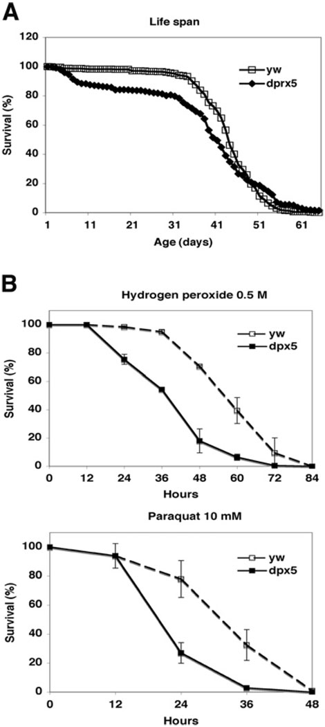
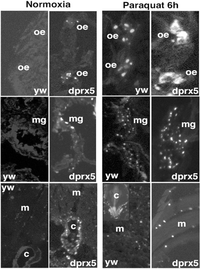
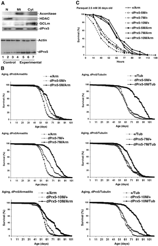
Peroxiredoxin 5 is a distinct isoform of the peroxiredoxin gene family. The antioxidative and anti-apoptotic functions of peroxiredoxin 5 have been extensively demonstrated in cell culture experiments. In the present paper, we provide the first functional analysis of peroxiredoxin 5 in a multicellular organism, Drosophila melanogaster. Similar to its mammalian, yeast or human counterparts, dPrx5 (Drosophila peroxiredoxin 5) is expressed in several cellular compartments, including the cytosol, nucleus and the mitochondrion. Global overexpression of dPrx5 in flies increased resistance to oxidative stress and extended their life span by up to 30% under normal conditions. The dprx5(-/-) null flies were comparatively more susceptible to oxidative stress, had higher incidence of apoptosis, and a shortened life span. TUNEL (terminal deoxynucleotidyl transferase-mediated dUTP nick-end labelling) analysis revealed that the dprx5(-/-) null mutant had discernible tissue-specific apoptotic patterns, similar to those observed in control flies exposed to paraquat. In addition, apoptosis was particularly notable in oenocytes. During development the dPrx5 levels co-varied with ecdysone pulses, suggesting inter-relationship between ecdystreroids and dPrx5 expression. The importance of dPrx5 for development was further underscored by the embryonic lethal phenotype of progeny derived from the dprx5(-/-) null mutant. Results from the present study suggest that the antioxidant and anti-apoptotic activities of dPrx5 play a critical role in development and aging of the fly.


Figures

Figure 1
Figure 1. Analysis of subcellular localization of Drosophila dPrx5 protein
(A) The N-terminal part of the deduced amino acid sequence of dPrx5 protein. The predicted mitochondrial targeting sequence is underlined and the putative second start site is bold. (B) Immunoblot analysis of protein lysates extracted from nuclear (N), mitochondrial (Mt) and cytosolic (Cyt) fractions prepared from flies, S2 cells and S2 cells overexpressing dPrx5 tagged with V5/His6 epitopes (pIB-dPrx5 transfectant). Signals were obtained with anti-dPrx5 antibodies and with anti-aconitase (mitochondrial protein), anti-HDAC (nuclear protein) and anti-GCLm (cytosolic protein) antibodies to control for a specificity of subcellular fractions. Staining with Coomassie Blue (lower panels) was performed to control for a loading.
Figure 2
Figure 2. Immunoblot analysis of dPrx5 expression during development (A) and aging (B)
(A) Protein was extracted from embryos, larvae and pupae, which were allowed to develop for different periods of time after egg laying (AEL) or after white prepupae (WPP) collection. Male and female symbols indicate freshly hatched animals (0–3 h). (B) Adults were collected at different ages. Immunoblots were performed using anti-dPrx5 antibodies and re-probed with anti-actin antibodies to control for a loading. Control for a loading in samples isolated during development was performed with Coomassie Blue staining, as actin levels vary more significantly at these stages.
Figure 3
Figure 3. Analysis of the 5′-flanking region of dPrx5 gene
The transcription start site is indicated by an arrow. ATG translation start codons are indicated by bold capital letters. Predicted transcription factor binding sites are underlined. The prediction was performed using TFsearch and AliBaba2 softwares (www.gene-regulation.com). alp/alph, alpha (α); AP-2, activator protein-2; BRCZ, BRC zinc finger; C/EBP, CCAAT/enhancer-binding protein; COUP, chicken ovalbumin upstream promoter; ENKTF, enkephalin transcription factor; GCN4, general control non-derepressible 4; GLO, GLOBOSA; HNF, hepatocyte NF; HOXA, homeobox A4; PR, progesterone receptor; RAR-a1, retinoicacid receptor α1; SRF, serum-response factor; T3R-a1, tri-iodothyronine receptor α1; TBP, TATA-box-binding protein.
Figure 4
Figure 4. dPrx5 overexpression protects S2 cells from oxidants
(A and B) Flow cytometry analysis of cell death after exposure to paraquat and SNAP. Control cells (transfected with empty vector) and cells overexpressing dPrx5 were exposed to 10 mM paraquat or 1 mM SNAP for 8 h followed by incubation with anti-Annexin-V-FITC antibodies and PI. Cells undergoing apoptosis (Annexin-V-FITC-positive, PI negative; A + PI−) are seen in the right peaks; non-apoptotic viable cells (Annexin-V-FITC- and PI-negative; A−PI−) are seen in the peaks on the left. PI-positive cells (right peaks) are dead cells or cells in the late stage of apoptosis (Annexin-V-FITC- and PI-positive; PI + A+). Continuous lines represent cells overexpressing dPrx5 and broken lines represent control cells transfected with empty vector. (C and D) Evaluation of DNA damage in control cells and cells expressing dPrx5 by single cell Comet assay. Cells were treated with 10 mM paraquat or 1 mM SNAP for 24 h. Frequency distribution of nuclei in four different classes of increasing total DNA damage is shown (see the Experimental section). The results are mean percentage values of three separate experiments. Differences in scores 1–4 for dPrx5 overexpressor compared with control exposed to paraquat (C) were statistically significant (P < 0.05).In samples treated with SNAP, statistically significant differences were determined for cells at the score 2 and 3 (D).
Figure 5
Figure 5. Survival of dprx5−/− mutant under normal (A) and oxidative stress (B) conditions
(A) Pooled data of three independent experiments are shown for each line. In all experiments, 200–250 flies for each fly strain were used. The median age was 43 ± 16 and 40 ± 12 days and mean life span was 65.3 and 57.4 days for y w and dprx5 strains correspondingly. There were no significant differences in maximum age (10% survival) between y w strain and dprx5−/− mutant, which in both cases was 53–54 days. (B) Approx. 100–150 flies of each group (dprx5 mutant and y w control) were fed 1% sucrose solution containing 10 mM paraquat or 0.5 M H2O2. Results are means ± S.D. of two independent experiments.
Figure 6
Figure 6. TUNEL analysis of cell death in y w control and dprx5 mutant
Representative images show DNA fragmentation in cryosections made from 10-day-old flies reared under normal conditions (left panels) or fed 10 mM paraquat for 6 h (right panels). Shown are selected images of the abdomen and thoraces regions, where differences in DNA fragmentation between dprx5−/− mutant and control were detected. oe, oenocytes; mg, midgut; c, cardia and m, muscles.
Figure 7
Figure 7. The effects of dPrx5 overexpression on aging and resistance to oxidative stress
(A) Immunoblot analysis of dPrx5 overexpression. Subcellular fractions (upper panel) were prepared as described in Figure 1. Lower panel, samples were prepared from whole flies. Lanes 1, 2, 3 and 4, driver (Tub-GAL4/+) and three different transgene (UAS-dPrx5/+) controls; lanes 5, 6, and 7, three different experimental (UAS-dPrx5/Tub-GAL4) lines. (B) Life spans of flies overexpressing dPrx5 globally at low levels (with Arm-GAL4 driver) and at high levels (with Tub-GAL4 driver). Pooled data of two independent experiments are shown for each line. The median ages were extended in dPrx5/Arm experimental flies by 24.7 ± 3.7% compared with driver and by 13.4 ± 3.9% compared with transgenic controls. With Tub-GAL4 driver, median age was extended by 29.2 ± 7.7% compared with driver and by 30.0 ± 4.6% compared with transgenic controls. (C) Resistance of 30-day-old flies to 2.5 mM paraquat. Approx. 100–150 flies of each group were fed 1% sucrose solution containing 5 mM paraquat. Similar results were obtained in two independent experiments. The median survival time in experimental flies was increased by 52.6% to 60.0% compared with driver control and by 41.5% to 52.3% compared with transgene controls.

Similar articles

Cited by

References

    1. Wang X, Phelan SA, Forsman-Semb K, Taylor EF, Petros C, Brown A, Lerner CP, Paigen B. Mice with targeted mutation of peroxiredoxin 6 develop normally but are susceptible to oxidative stress. J. Biol. Chem. 2003;278:25179–25190. - PubMed
    1. Wang Y, Feinstein SI, Manevich Y, Ho YS, Fisher AB. Peroxiredoxin 6 gene-targeted mice show increased lung injury with paraquat-induced oxidative stress. Antioxid. Redox Signaling. 2006;8:229–237. - PubMed
    1. Nagy N, Malik G, Fisher AB, Das DK. Targeted disruption of peroxiredoxin 6 gene renders the heart vulnerable to ischemia-reperfusion injury. Am. J. Physiol. 2006;291:H2636–H2640. - PubMed
    1. Li L, Shoji W, Oshima H, Obinata M, Fukumoto M, Kanno N. Crucial role of peroxiredoxin III in placental antioxidant defense of mice. FEBS Lett. 2008;582:2431–2434. - PubMed
    1. Egler RA, Fernandes E, Rothermund K, Sereika S, de Souza-Pinto N, Jaruga P, Dizdaroglu M, Prochownik EV. Regulation of reactive oxygen species, DNA damage, and c-Myc function by peroxiredoxin 1. Oncogene. 2005;24:8038–8050. - PubMed

Publication types