

Analysis of islet inflammation in human type 1 diabetes
- PMID: 19128359
- PMCID: PMC2675247
- DOI: 10.1111/j.1365-2249.2008.03860.x
Analysis of islet inflammation in human type 1 diabetes
Abstract
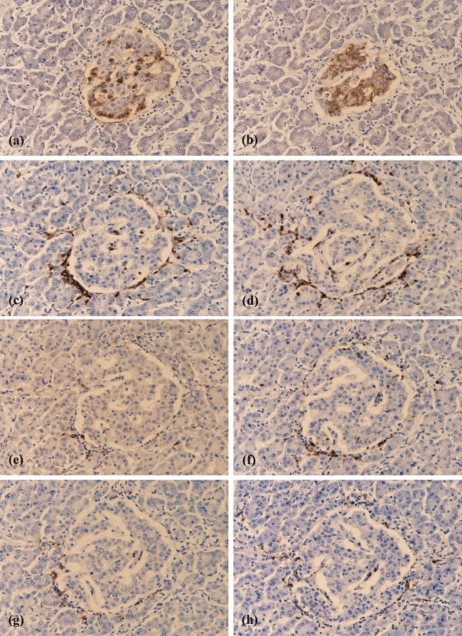
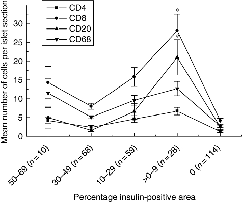
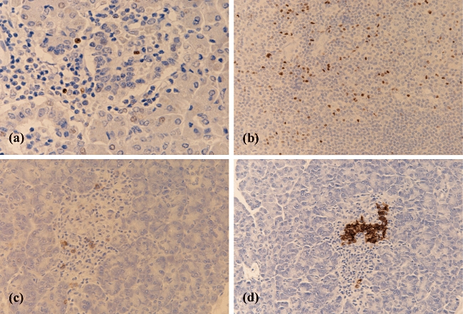
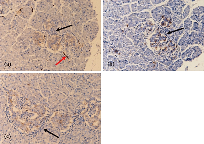
The immunopathology of type 1 diabetes (T1D) has proved difficult to study in man because of the limited availability of appropriate samples, but we now report a detailed study charting the evolution of insulitis in human T1D. Pancreas samples removed post-mortem from 29 patients (mean age 11.7 years) with recent-onset T1D were analysed by immunohistochemistry. The cell types constituting the inflammatory infiltrate within islets (insulitis) were determined in parallel with islet insulin content. CD8(+) cytotoxic T cells were the most abundant population during insulitis. Macrophages (CD68(+)) were also present during both early and later insulitis, although in fewer numbers. CD20(+) cells were present in only small numbers in early insulitis but were recruited to islets as beta cell death progressed. CD138(+) plasma cells were infrequent at all stages of insulitis. CD4(+) cells were present in the islet infiltrate in all patients but were less abundant than CD8(+) or CD68(+) cells. Forkhead box protein P3(+) regulatory T cells were detected in the islets of only a single patient. Natural killer cells were detected rarely, even in heavily inflamed islets. The results suggest a defined sequence of immune cell recruitment in human T1D. They imply that both CD8(+) cytotoxic cells and macrophages may contribute to beta cell death during early insulitis. CD20(+) cells are recruited in greatest numbers during late insulitis, suggesting an increasing role for these cells as insulitis develops. Natural killer cells and forkhead box protein P3(+) T cells do not appear to be required for beta cell death.
Figures
Comment in
-
Post-mortem analysis of islet pathology in type 1 diabetes illuminates the life and death of the beta cell.Clin Exp Immunol. 2009 Feb;155(2):125-7. doi: 10.1111/j.1365-2249.2008.03864.x. Clin Exp Immunol. 2009. PMID: 19128357 Free PMC article. No abstract available.
References
-
- Yang Y, Santamaria P. Lessons on autoimmune diabetes from animal models. Clin Sci (Lond) 2006;110:627–39. - PubMed
-
- Lally FJ, Ratcliff H, Bone AJ. Apoptosis and disease progression in the spontaneously diabetic BB/S rat. Diabetologia. 2001;44:320–24. - PubMed
-
- Jorns A, Gunther A, Hedrich HJ, Wedekind D, Tiedge M, Lenzen S. Immune cell infiltration, cytokine expression, and beta-cell apoptosis during the development of type 1 diabetes in the spontaneously diabetic LEW.1AR1/Ztm-iddm rat. Diabetes. 2005;54:2041–52. - PubMed
-
- Itoh N, Hanafusa T, Miyazaki A, et al. Mononuclear cell infiltration and its relation to the expression of major histocompatibility complex antigens and adhesion molecules in pancreas biopsy specimens from newly diagnosed insulin-dependent diabetes mellitus patients. J Clin Invest. 1993;92:2313–22. - PMC - PubMed
-
- Foulis AK. The pathogenesis of beta cell destruction in type I (insulin-dependent) diabetes mellitus. J Pathol. 1987;152:141–8. - PubMed
Publication types
MeSH terms
Substances
LinkOut - more resources
Full Text Sources
Other Literature Sources
Medical
Research Materials
