. 2009 Jan 7:9:1.
doi: 10.1186/1472-6750-9-1.

Reporter gene-expressing bone marrow-derived stromal cells are immune-tolerated following implantation in the central nervous system of syngeneic immunocompetent mice

Affiliations

Reporter gene-expressing bone marrow-derived stromal cells are immune-tolerated following implantation in the central nervous system of syngeneic immunocompetent mice

Irene Bergwerf et al. BMC Biotechnol. .

Abstract

Background: Cell transplantation is likely to become an important therapeutic tool for the treatment of various traumatic and ischemic injuries to the central nervous system (CNS). However, in many pre-clinical cell therapy studies, reporter gene-assisted imaging of cellular implants in the CNS and potential reporter gene and/or cell-based immunogenicity, still remain challenging research topics.

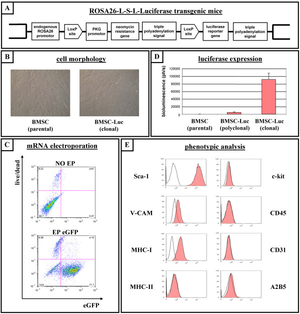
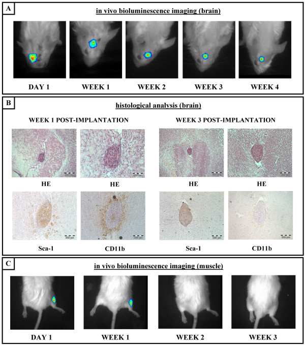
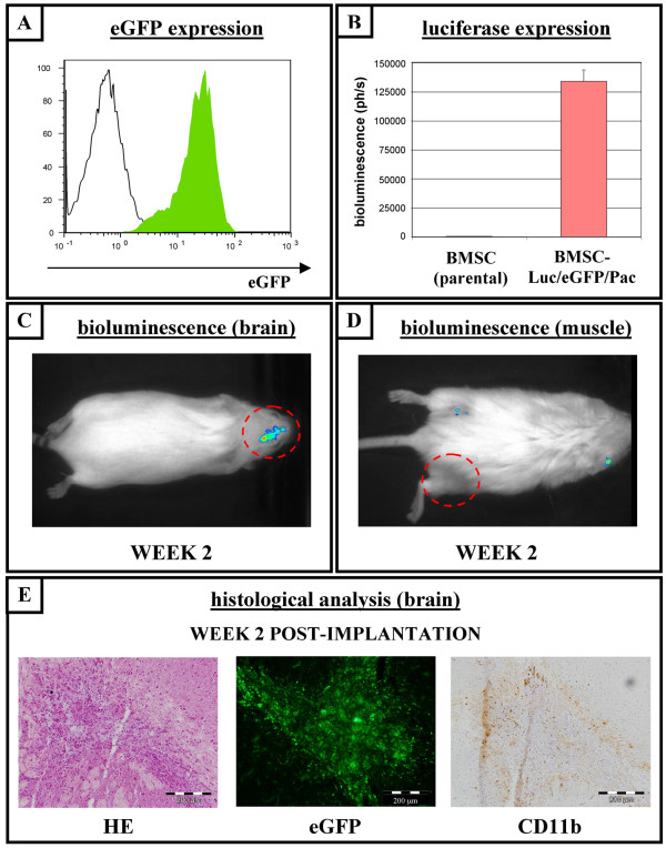
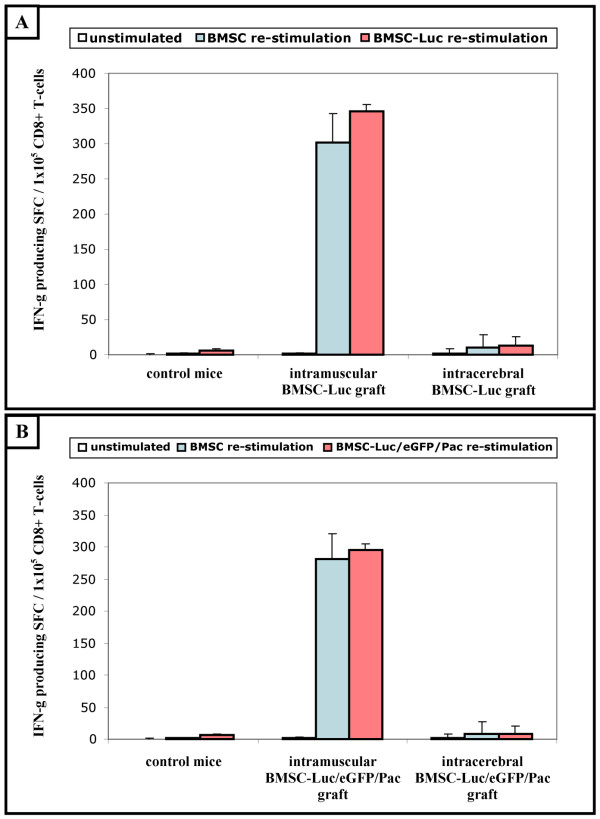
Results: In this study, we performed cell implantation experiments in the CNS of immunocompetent mice using autologous (syngeneic) luciferase-expressing bone marrow-derived stromal cells (BMSC-Luc) cultured from ROSA26-L-S-L-Luciferase transgenic mice, and BMSC-Luc genetically modified using a lentivirus encoding the enhanced green fluorescence protein (eGFP) and the puromycin resistance gene (Pac) (BMSC-Luc/eGFP/Pac). Both reporter gene-modified BMSC populations displayed high engraftment capacity in the CNS of immunocompetent mice, despite potential immunogenicity of introduced reporter proteins, as demonstrated by real-time bioluminescence imaging (BLI) and histological analysis at different time-points post-implantation. In contrast, both BMSC-Luc and BMSC-Luc/eGFP/Pac did not survive upon intramuscular cell implantation, as demonstrated by real-time BLI at different time-points post-implantation. In addition, ELISPOT analysis demonstrated the induction of IFN-gamma-producing CD8+ T-cells upon intramuscular cell implantation, but not upon intracerebral cell implantation, indicating that BMSC-Luc and BMSC-Luc/eGFP/Pac are immune-tolerated in the CNS. However, in our experimental transplantation model, results also indicated that reporter gene-specific immune-reactive T-cell responses were not the main contributors to the immunological rejection of BMSC-Luc or BMSC-Luc/eGFP/Pac upon intramuscular cell implantation.

Conclusion: We here demonstrate that reporter gene-modified BMSC derived from ROSA26-L-S-L-Luciferase transgenic mice are immune-tolerated upon implantation in the CNS of syngeneic immunocompetent mice, providing a research model for studying survival and localisation of autologous BMSC implants in the CNS by real-time BLI and/or histological analysis in the absence of immunosuppressive therapy.


Figures

Figure 1
Figure 1
Culture and characterisation of a clonal luciferase-expressing bone marrow-derived stromal cell line from ROSA26-L-S-L-Luciferase transgenic mice. (A) Molecular organisation of the ROSA26 locus in ROSA26-L-S-L-Luciferase transgenic mice. (B) Representative pictures of cultured bone marrow-derived stromal cells (BMSC) taken under phase contrast microscopy. Left: unmodified parental BMSC derived from ROSA26-L-S-L-Luciferase transgenic mice (BMSC parental). Right: clonal luciferase-expressing BMSC derived from ROSA26-L-S-L-Luciferase transgenic mice (BMSC-Luc clonal). (C) Parental BMSC derived from ROSA26-L-S-L-Luciferase transgenic mice were non-electroporated (NO EP, upper dot plot) or electroporated with eGFP mRNA (EP eGFP, lower dot plot), and were analyzed by flow cytometry for eGFP fluorescence (x-axis) versus viability (GelRed-staining, y-axis) after 24 hours of culture. The percentage indicated in the lower left quadrant is the number of viable eGFP-negative cells. The percentage indicated in the lower right quadrant is the number of viable eGFP-positive cells. The percentages indicated in the upper left and right quadrant are numbers of non-viable cells. Representative dot plots are shown. (D) In vitro luminescence assay on parental BMSC (BMSC parental), on Cre-recombined polyclonal luciferase-expressing BMSC (BMSC-Luc polyclonal), and on Cre-recombined clonal luciferase-expressing BMSC (BMSC-Luc clonal), all derived from ROSA26-L-S-L-Luciferase transgenic mice. (E) Representative flow cytometric analysis showing expression pattern of membrane proteins on clonal BMSC-Luc derived from ROSA26-L-S-L-Luciferase transgenic mice (i.e. expression of Sca-1, V-CAM and MHC-I, but no expression of MHC-II, c-kit, CD45, CD31 and A2B5). Open histograms: control. Filled histograms: specific antibody staining.
Figure 2
Figure 2
Survival of luciferase-expressing BMSC derived from ROSA26-L-S-L-Luciferase transgenic mice following implantation in the central nervous system of syngeneic immunocompetent mice. (A) Representative time course for in vivo bioluminescence imaging of clonal BMSC-Luc derived from ROSA26-L-S-L-Luciferase transgenic mice following implantation in the central nervous system of syngeneic immunocompetent mice. (B) Representative histological analysis of clonal BMSC-Luc grafts in the central nervous system of syngeneic immunocompetent mice. Week 1 post-implantation: Upper pictures, haematoxylin-eosin (HE) staining indicating localisation and general appearance of the implantation site. Lower left picture, Sca-1 staining indicating the BMSC origin of the observed cell graft. Lower right picture, CD11b staining indicating the presence of activated microglia surrounding the observed cell graft. Week 3 post-implantation: Upper pictures, HE staining indicating localisation and general appearance of the implantation site. Lower left picture, Sca-1 staining indicating the BMSC origin of the observed cell graft. Lower right picture, CD11b staining indicating the absence of activated microglia surrounding the observed cell graft. All slides were examined using a conventional bright field microscope and digital pictures were taken under magnification as indicated by the scale bars. (C) Representative time course for in vivo bioluminescence imaging of clonal BMSC-Luc derived from ROSA26-L-S-L-Luciferase transgenic mice following intramuscular implantation in syngeneic immunocompetent mice.
Figure 3
Figure 3
Survival of BMSC genetically modified with multiple reporter genes following implantation in the central nervous system of syngeneic immunocompetent mice. (A) Histogram overlay showing a representative flow cytometric analysis of eGFP expression by BMSC expressing the luciferase-, eGFP- and puromycin resistance genes (BMSC-Luc/eGFP/Pac, filled histogram). Parental BMSC were used as negative control (open histogram). (B) In vitro luminescence assay on parental BMSC and on clonal BMSC-Luc/eGFP/Pac. (C) In vivo real time bioluminescence imaging (BLI) of clonal BMSC-Luc/eGFP/Pac grafts in the central nervous system (CNS) of syngeneic immunocompetent mice at week 2 post-implantation. (D) In vivo real time BLI of clonal BMSC-Luc/eGFP/Pac intramuscular grafts in syngeneic immunocompetent mice at week 2 post-injection. (E) Representative histological analysis of clonal BMSC-Luc/eGFP/Pac implants in the CNS of syngeneic immunocompetent mice at week 2 post-implantation. Left picture: haematoxylin-eosin (HE) staining showing general appearance of the cell implantation site. Middle picture: direct eGFP-fluorescence indicating the BMSC-Luc/eGFP/Pac origin of the observed cell implant. Right picture: CD11b staining indicating a limited number of microglia surrounding the observed cell graft. All slides were examined using a conventional bright field microscope and digital pictures were taken under magnification as indicated by the scale bars.
Figure 4
Figure 4
Induction of BMSC-specific CD8+ T-cell responses following intramuscular, but not intracerebral, cell implantation in syngeneic immunocompetent mice. (A) Spleen CD8+ T-cells (1 × 10⁵ cells/well) from non-transplanted ROSA26-L-S-L-Luciferase mice (control mice, n = 4) and ROSA26-L-S-L-Luciferase mice with BMSC-Luc implants, either intramuscularly (intramuscular BMSC-Luc graft, n = 5) or intracerebrally (intracerebral BMSC-Luc graft, n = 6), were cultured in quadruplet in an IFN-γ ELISPOT assay alone (unstimulated), with addition of parental BMSC (BMSC re-stimulation) or with addition of BMSC-Luc (BMSC-Luc re-stimulation) (1 × 10⁴ cells/well, ratio 10:1). Data are expressed as the mean number of IFN-γ spot forming cells (SFC)/1 × 105 CD8+ T-cells for each experimental group. (B) Spleen CD8+ T-cells (1 × 10⁵ cells/well) from non-transplanted ROSA26-L-S-L-Luciferase mice (control mice, n = 4) and ROSA26-L-S-L-Luciferase mice with BMSC-Luc/eGFP/Pac implants, either intramuscularly (intramuscular BMSC-Luc/eGFP/Pac graft, n = 5) or intracerebrally (intracerebral BMSC-Luc/eGFP/Pac graft, n = 6), were cultured in quadruplet in an IFN-γ ELISPOT assay alone (unstimulated), with addition of parental BMSC (BMSC re-stimulation) or with addition of BMSC-Luc/eGFP/Pac (BMSC-Luc/eGFP/Pac re-stimulation) (1 × 10⁴ cells/well, ratio 10:1). Data are expressed as the mean number of IFN-γ spot forming cells (SFC)/1 × 105 CD8+ T-cells for each experimental group.

Similar articles

Cited by

References

    1. Arvidson A, Collin T, Kirik D, Kokaia Z, Lindvall O. Neuronal replacement from endogenous precursors in the adult brain after stroke. Nat Med. 2002;8:963–970. doi: 10.1038/nm747. - DOI - PubMed
    1. Kokaia Z, Lindvall O. Neurogenesis after ischaemic brain insults. Curr Opin Neurobiol. 2003;13:127–132. doi: 10.1016/S0959-4388(03)00017-5. - DOI - PubMed
    1. Webber DJ, Bradbury EJ, McMahon SB, Minger SL. Transplanted neural progenitor cells survive and differentiate but achieve limited functional recovery in the lesioned adult rat spinal cord. Regen Med. 2007;2:929–945. doi: 10.2217/17460751.2.6.929. - DOI - PubMed
    1. Li J, Sun CR, Zhang H, Tsang KS, Li JH, Zhang SD, An YH. Induction of functional recovery by co-transplantation of neural stem cells and Schwann cells in a rat spinal cord contusion injury model. Biomed Environ Sci. 2007;20:242–249. - PubMed
    1. Ormerod BK, Palmer TD, Caldwell MA. Neurodegeneration and cell replacement. Philos Trans R Soc Lond B Biol Sci. 2008;363:153–170. doi: 10.1098/rstb.2006.2018. - DOI - PMC - PubMed

Publication types