. 2009 Mar;37(4):1193-201.
doi: 10.1093/nar/gkn1050. Epub 2009 Jan 7.

The G140S mutation in HIV integrases from raltegravir-resistant patients rescues catalytic defect due to the resistance Q148H mutation

Affiliations

The G140S mutation in HIV integrases from raltegravir-resistant patients rescues catalytic defect due to the resistance Q148H mutation

Olivier Delelis et al. Nucleic Acids Res. 2009 Mar.

Abstract

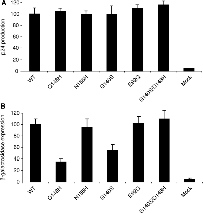
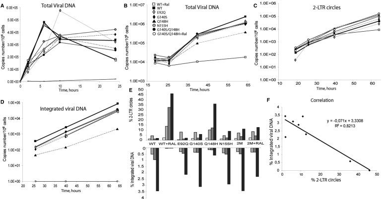
Raltegravir (MK-0518) is the first integrase (IN) inhibitor to be approved by the US FDA and is currently used in clinical treatment of viruses resistant to other antiretroviral compounds. Virological failure of Raltegravir treatment is associated with mutations in the IN gene following two main distinct genetic pathways involving either the N155 or Q148 residue. Importantly, in most cases, an additional mutation at the position G140 is associated with the Q148 pathway. Here, we investigated the viral DNA kinetics for mutants identified in Raltegravir-resistant patients. We found that (i) integration is impaired for Q148H when compared with the wild-type, G140S and G140S/Q148H mutants; and (ii) the N155H and G140S mutations confer lower levels of resistance than the Q148H mutation. We also characterized the corresponding recombinant INs properties. Enzymatic performances closely parallel ex vivo studies. The Q148H mutation 'freezes' IN into a catalytically inactive state. By contrast, the conformational transition converting the inactive form into an active form is rescued by the G140S/Q148H double mutation. In conclusion, the Q148H mutation is responsible for resistance to Raltegravir whereas the G140S mutation increases viral fitness in the G140S/Q148H context. Altogether, these results account for the predominance of G140S/Q148H mutants in clinical trials using Raltegravir.


Figures

Figure 1.
Figure 1.
p24 and infectivity of IN mutant viruses. (A) Quantification of p24 protein, 48 h after transfection of 5 µg of each virus DNA. (B) Viral infectivity for WT and mutants. Viral infectivity was determined in a single-cycle replication assay using HeLa p4 indicator cells and 3 ng of p24 antigen for each virus. Cells were exposed to virus during 48 h. Early steps of infections were assessed by measuring β-galactosidase activity in cell extracts by the CPRG method. For panels A and B, the results are expressed as percentages of the value obtained for the WT. The data shown are the means of three independent experiments.
Figure 2.
Figure 2.
Resistance of IN mutants to RAL. (A) HeLa p4 cells were infected, in triplicate, with 3 ng of each virus, in the presence of various RAL concentrations. β-Galactosidase production was quantified by the CPRG assay. Data from a representative experiment (performed three times) is shown. The IC50 was determined as the concentration of RAL inhibiting β-galactosidase production by 50% with respect to untreated infected cells. (B) MTT assay. The MTT assay was performed 48 and 72 h after infection for all viruses. For the WT and G140S/Q148H mutant, the assay was performed with and without 500 nM RAL. The data shown are the means of three independent experiments.
Figure 3.
Figure 3.
Kinetics of viral DNA synthesis. CEM cells were infected with 40 ng of p24 antigen and levels of intracellular HIV-1 DNA species were monitored by qPCR. (A) Dynamics of total viral DNA during the first 24 h of infection. WT+RAL (open square), WT (filled square), E92Q (gray diamond), G140S (filled diamond), Q148H (filled triangle), N155H (gray circle), G140S/Q148H (filled circle), G140S/Q148H+Ral (open circle), WT + AZT (straight line). (B-D) Quantification of viral DNA over a 3-day period. (B) Total Viral DNA (C) 2-LTR circles (D) integrated HIV-1 DNA. The scales in panels A to D are logarithmic. Depicted results were obtained from a representative experiment. (E) The percentage of 2-LTR circles and integrated viral DNA were determined by calculating the ratio of 2-LTR circles copy number and integrated DNA levels, respectively, over total viral DNA. (F) Correlation between the percentages of 2-LTR circles and integrated viral DNA. The data shown are the means of three independent experiments.
Figure 4.
Figure 4.
p24 production. Viral particles released in the supernatant were determined 48 and 72 h after infection by quantification of the p24 protein (see Materials and methods section). The data shown are the means of four independent experiments.
Figure 5.
Figure 5.
Comparative study of the DNA-binding and catalytic properties of wild-type and RAL-resistant INs. (A) 3′-Processing activity—after 3 h of incubation at 37°C—as a function of IN concentration. 3′-Processing activities were quantified as described in Materials and methods section, using a 21-mer DNA substrate (4 nM), with MgCl2 as a cofactor (10 mM) in 20 mM Hepes (pH 7.2), 1 mM DTT and 30 mM NaCl. (B) DNA binding of wild-type and mutant INs. The DNA-binding step was assessed by steady-state fluorescence anisotropy as described in Materials and methods section. Experimental conditions were similar to those described in A. IN and DNA were incubated together for 15 min before recording steady-state anisotropy. (C) Kinetics of 3′-processing for the different proteins. IN concentration was 200 nM. The same symbols were used in panels A, B and C: (open square) wild-type NL-43; (filled square) G140S/Q148H NL-43; (filled triangle) Q148H NL-43; (open triangle) G140S NL-43; (open circle) wild-type patient; (filled circle) G140S/Q148H patient. Strand transfer products are depicted (middle panel). The percentages of strand transfer (shown besides the gel) were obtained after the normalization by the 3′-processing activity.

References

    1. Pommier Y, Johnson AA, Marchand C. Integrase inhibitors to treat HIV/AIDS. Nat. Rev. Drug Discov. 2005;4:236–248. - PubMed
    1. Sinha S, Grandgenett DP. Recombinant human immunodeficiency virus type 1 integrase exhibits a capacity for full-site integration in vitro that is comparable to that of purified preintegration complexes from virus-infected cells. J. Virol. 2005;79:8208–8216. - PMC - PubMed
    1. Li M, Mizuuchi M, Burke T.R., Jr., Craigie R. Retroviral DNA integration: reaction pathway and critical intermediates. EMBO J. 2006;25:1295–1304. - PMC - PubMed
    1. DeJesus E, Berger D, Markowitz M, Cohen C, Hawkins T, Ruane P, Elion R, Farthing C, Zhong L, Cheng AK, et al. Antiviral activity, pharmacokinetics, and dose response of the HIV-1 integrase inhibitor GS-9137 (JTK-303) in treatment-naive and treatment-experienced patients. J. Acquir. Immun. Defic. Syndr. 2006;43:1–5. - PubMed
    1. Hazuda DJ, Young SD, Guare JP, Anthony NJ, Gomez RP, Wai JS, Vacca JP, Handt L, Motzel SL, Klein HJ, et al. Integrase inhibitors and cellular immunity suppress retroviral replication in rhesus macaques. Science. 2004;305:528–532. - PubMed

Publication types