. 2009 Mar;37(4):1141-51.
doi: 10.1093/nar/gkn1026. Epub 2009 Jan 7.

Positioning of subdomain IIId and apical loop of domain II of the hepatitis C IRES on the human 40S ribosome

Affiliations

Positioning of subdomain IIId and apical loop of domain II of the hepatitis C IRES on the human 40S ribosome

Elena Babaylova et al. Nucleic Acids Res. 2009 Mar.

Abstract

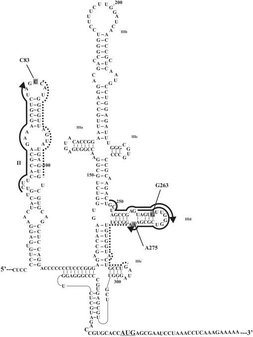
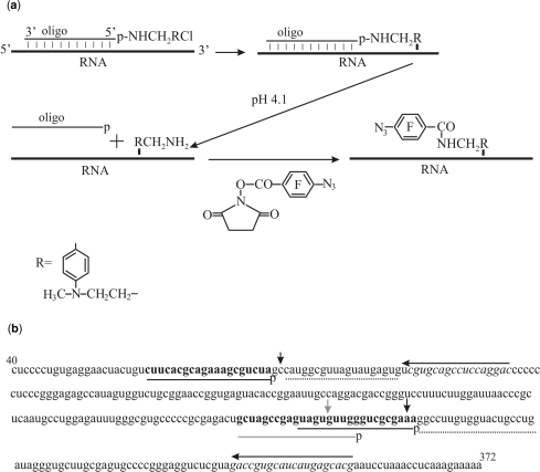
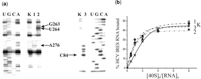
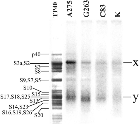
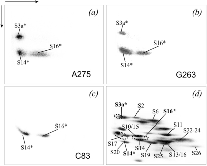
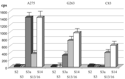
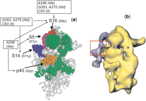
The 5'-untranslated region of the hepatitis C virus (HCV) RNA contains a highly structured motif called IRES (Internal Ribosome Entry Site) responsible for the cap-independent initiation of the viral RNA translation. At first, the IRES binds to the 40S subunit without any initiation factors so that the initiation AUG codon falls into the P site. Here using an original site-directed cross-linking strategy, we identified 40S subunit components neighboring subdomain IIId, which is critical for HCV IRES binding to the subunit, and apical loop of domain II, which was suggested to contact the 40S subunit from data on cryo-electron microscopy of ribosomal complexes containing the HCV IRES. HCV IRES derivatives that bear a photoactivatable group at nucleotide A275 or at G263 in subdomain IIId cross-link to ribosomal proteins S3a, S14 and S16, and HCV IRES derivatized at the C83 in the apex of domain II cross-link to proteins S14 and S16.


Figures

Figure 1.
Figure 1.
Structural organization of the HCV IRES (7), the initiation AUG codon is underlined. RNA sequences complementary to deoxy-oligomers used for site-specific modification of the HCV IRES are marked with thick lines with arrows indicating the 5′-phosphates derivatized with alkylating groups. RNA sequences complementary to helper oligomers used together with the alkylating derivatives are marked with dotted lines. Nucleotides of the HCV IRES cross-linked to the oligonucleotide derivatives are shaded.
Figure 2.
Figure 2.
Scheme of site-specific introduction of a photoactivatable group into specific RNA sites based on the site-specific alkylation of the RNA with [4-(N-2-chloroethyl-N-methylamino)benzyl]-phosphoramides of oligodeoxyribonucleotides. (a) Order of chemical reactions. (b) Nucleotide sequence of HCV IRES. Target sequences for the deoxy-oligomer derivatives are given in bold, the respective complementary oligodeoxyribonucleotides are shown with either gray or black lines under the sequence, and the letters ‘p’ indicate the terminal 5′-phosphates derivatized with alkylating groups. Helper oligomers are shown by dotted lines. Lines with arrows above the sequence indicate primers used for reverse transcription (arrows show the direction of primer extension). RNA sequences complementary to the primers are given in italics. Either gray or black vertical arrows show cross-linked nucleotides.
Figure 3.
Figure 3.
Identification of sites of cross-linking of oligonucleotide derivatives to the HCV IRES by reverse transcription and binding properties of the HCV IRES derivatives. (a) Extension of [5′-32P]-labeled primers complementary to the HCV IRES sequences 331–350 (left panel) and 103–120 (right panel). Lanes 1, 2 and 3, primer extension on HCV IRES alkylated with derivatives of deoxy-oligomers complementary to the sequences 259–276, 248–267 and 62–81, respectively. Lanes K, primer extension with control HCV IRES incubated under conditions of alkylation but without oligomer derivatives. Lanes U, G, C, A, sequencing of HCV IRES. Arrows indicate positions of the reverse transcription stops caused by the cross-links. (b) Isotherms of binding of control unmodified HCV IRES (K) and its derivatives containing a perfluorophenyl azide group at A275, G263 or C83 to 40S subunits (1, 2 and 3, respectively). The initial concentration of the HCV IRES or its derivatives was 1.0 × 10⁻⁷ M. Relative error was about 10%.
Figure 4.
Figure 4.
Autoradiogram of the 40S ribosomal proteins cross-linked to the 32P-labeled derivatives of HCV IRES by 1D PAGE in the presence of SDS. Nucleotides bearing the cross-linker are indicated. Lane K, proteins isolated from an irradiated control complex of unmodified HCV IRES with 40S subunits. Lane TP40, silver stained gel; positions of the 40S ribosomal proteins are indicated (43,44).
Figure 5.
Figure 5.
Analysis by 2D PAGE of proteins cross-linked to 32P-labeled HCV IRES derivatives in the binary complexes with the 40S subunits. (a–c), The autoradiograms correspond to the experiments with HCV IRES derivatives containing the cross-linker at nucleotide A275, G263 or C83 (marked in the respective panels). (d), Coomassie stained gel corresponding to (b) (as example); the positions of the proteins are indicated (44, 45). The locations of the radioactive spots corresponding to the cross-linked proteins are indicated on the stained gel by dotted lines. The cross-linked proteins are highlighted by an asterisk (*), and are also in bold in (d).
Figure 6.
Figure 6.
Analysis of 40S proteins cross-linked to 32P-labeled HCV IRES derivatives (nucleotides bearing the cross-linker are indicated at the top) by immunoprecipitation using antibodies against mammalian 40S ribosomal proteins (indicated at the bottom).
Figure 7.
Figure 7.
Structural model of the mammalian 40S ribosomal subunit (a) and of the complex of the 40S subunit with HCV IRES (b) obtained from cryo-EM data. View from the solvent side. (a) Model of the 40S subunit of the elongating ribosome [adapted from (39), PDB accession number 2ZKQ]. Locations of ribosomal proteins cross-linked to HCV IRES nucleotides belonging to domain II and subdomains IIId (this study) and IIIe (17) are shown (p40 in orange, S5 in purple, S14 in dark blue and S16 in red), prokaryotic homologues of these proteins are indicated in brackets. All other proteins that have prokaryotic counterparts are shown in green and proteins specific for eukaryotes are not shown; gray balls represent the 18S rRNA. (b) Surface representation of the 40S ribosomal subunit in complex with the HCV IRES [adapted from (21)]. The cryo-EM map is in yellow. The difference map corresponding to the HCV IRES is superimposed and presented in purple. The suggested location of hairpins IIId/e is boxed. Asterics show sites of conformational changes in the 40S subunit caused by HCV IRES binding.

References

    1. Brocard M, Paulous S, Komarova AV, Deveaux V, Kean KM. Evidence that PTB does not stimulate HCV IRES-driven translation. Virus Genes. 2007;35:5–15. - PubMed
    1. Wasley A, Alter MJ. Epidemiology of hepatitis C: geographic differences and temporal trends. Semin. Liver Dis. 2000;20:1–16. - PubMed
    1. Pawlotsky JM. Pathophysiology of hepatitis C virus infection and related liver disease. Trends Microbiol. 2004;12:96–102. - PubMed
    1. Rosenberg S. Recent advances in the molecular biology of hepatitis C virus. J. Mol. Biol. 2001;313:451–464. - PubMed
    1. Tsukiyama-Kohara K, Iizuka N, Kohara M, Nomoto A. Internal ribosome entry site within hepatitis C virus RNA. J. Virol. 1992;66:1476–1483. - PMC - PubMed

Publication types

MeSH terms