. 2009 Mar;20(3):535-44.
doi: 10.1681/ASN.2008040377. Epub 2009 Jan 7.

Divergent role of donor dendritic cells in rejection versus tolerance of allografts

Affiliations

Divergent role of donor dendritic cells in rejection versus tolerance of allografts

Takuya Ueno et al. J Am Soc Nephrol. 2009 Mar.

Abstract

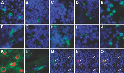
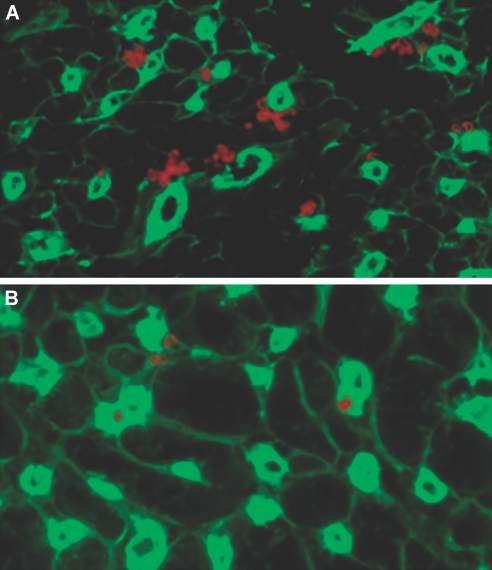
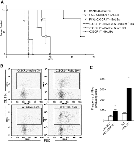
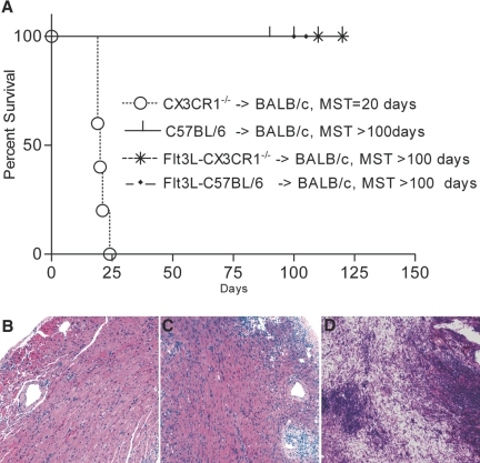
Little is known about heart tissue/donor dendritic cells, which play a key role in mounting alloimmune responses. In this report, we focus on three primary features of donor dendritic cells: their generation, their trafficking after transplantation, and their role in regulating tolerance versus rejection. Using transgenic mice as donors of heart allografts enabled us to monitor trafficking of donor dendritic cells after transplantation. Donor dendritic cells rapidly migrated into secondary lymphoid tissues within 3 h of transplantation. We found that the chemokine receptor CX3CR1 regulates the generation of heart tissue dendritic cells constitutively. Compared with wild-type hearts, CX3CR1(-/-) hearts contained fewer dendritic cells, and heart allografts from CX3CR1(-/-) donors survived significantly longer without immunosuppression. Unexpectedly, though, co-stimulatory blockade with anti-CD154 or CTLA4-Ig induced long-term survival for wild-type heart allografts but not for CX3CR1(-/-) heart allografts. Increasing the dendritic cell frequency in CX3CR1(-/-) hearts by treatment with Flt3L restored the anti-CD154-induced prolongation of CX3CR1(-/-) heart allograft survival. Compared with wild-type donors, depleting transgenic donors of dendritic cells before heart transplantation also markedly worsened chronic rejection under anti-CD154 treatment. These data indicate the importance of the CX3CR1 pathway in the generation of heart tissue dendritic cells and the divergent role of tissue/dendritic cells in rejection versus tolerance.


Figures

Figure 1.
Figure 1.
dDC Trafficking. LN (A through E) and spleens (F through J) of recipients of DTR-GFP-DC hearts were recovered at 3 h and at days 1, 3, 5, and 7, respectively, and were examined for the presence of GFP+ cells by anti-GFP staining (green); DAPI (blue) was used to counterstain nuclei. (K) Naive DTR-GFP-DC hearts stained for capillaries by anti-CD31 (Cy3, red) showed that DC cluster proximal to the vasculature; the endogenous GFP+ cells (green) are shown within the vicinity of the capillaries. (L) Heart samples from GFP+ donors were also stained with anti-GFP primary antibody (green) and DAPI to reveal cell nuclei (blue), and double-stained cells were found in these hearts, thereby demonstrating their presence in donor organs before transplantation. Sections of LN from day 5 after transplantation reveal that dDC, stained with anti-GFP (M, green), express donor MHC class I (N, using anti-H2b class I, red) and that cells coexpress GFP and class I (O, orange-yellow). DAPI was used to reveal cell nuclei (blue), ruling out the possibility that dDC were phagocytosed by recipient macrophages or DC. Magnifications: ×40 in A through L; ×20 in M through O.
Figure 2.
Figure 2.
The CX3CR1 pathway and formation of heart tissue DC. (A and B) Compared with CX3CR1−/− hearts (B), naive WT hearts (A) contain a much greater percentage and number of DC. DC were counted per 20 sections of naive heart tissue and were 75 ± 5 and 43 ± 4 for WT and CX3CR1−/−, respectively (n = 3 mice/group; P < 0.02). Sections of naive hearts were immunostained for CD11c. CD11c+ cells (red) co-localized with endothelial cells (stained with anti-CD31 FITC, green) were identified in WT and CX3CR1−/− hearts.
Figure 3.
Figure 3.
CX3CR1−/− donors and allograft survival. (A) Prolongation of heart allograft survival using CX3CR1−/− donors compared with WT donors (MST 17 and 8 d, respectively; n = 6 mice/group; P < 0.01). Transplanting hearts from Flt3L-treated CX3CR1−/− donors results in a similar tempo of rejection as compared with using Flt3L-treated WT donors but an accelerated rejection compared with when untreated CX3CR1−/− donors are used, highlighting the importance of reduced dDC numbers and the consequent prolongation of CX3CR1−/− heart allograft survival (n = 3 to 4 mice/group; P < 0.01). (B) Flt3L enhances heart DC numbers in both CX3CR1−/− and WT mice (representing two separate experiments; n = 4/group; P < 0.03). (C) Results of our ELISpot assay show that IFN-γ production is markedly higher in recipients of WT allografts (versus CX3CR1−/−) after in vitro stimulation with C57BL/6 antigen. In both groups, treatment with Flt3L significantly increased IFN-γ production (n = 3 to 4 mice/group; *P < 0.03).
Figure 4.
Figure 4.
dDC and their importance in the induction of tolerance. (A) Whereas recipients of CX3CR1−/− hearts exhibited refractoriness to the effect of MR1, hearts from WT donors exhibited long-term allograft survival (MST of 20 and >100 d, respectively; n = 5 mice/group; P < 0.01). Hearts from CX3CR1−/− implanted with the Flt3L hybridoma exhibited restoration of prolongation of heart allograft survival by MR1 (MST >100; n = 5 mice; P < 0.1). Compared with WT donors treated with MR1, treatment of WT donors with Flt3L had no effect on the course of MR1 treatment (MST >100 d in both groups). To provide a suggestive mechanism for the necessity of tissue DC, we also co-transplanted hearts from WT and CX3CR1−/− mice into BALB/c recipients treated with MR1. (B and C) Both WT (B) and CX3CR1−/− (C) heart allografts showed preserved muscles with mild cellular infiltrates, although CX3CR1−/− hearts seem to have more dense infiltration (C). (D) Of note, single CX3CR1−/− heart allografts under MR1 showed massive infiltration with muscle necrosis, whereas WT allografts showed little inflammation and intact myocytes (data not shown).
Figure 5.
Figure 5.
dDC and Treg in allografts. (A and B) Compared with WT donor hearts (A) (recovered from allograft recipients under MR1 at day 14 after transplantation), CX3CR1−/− donor hearts (B) showed a much lower number of Foxp3-stained cells. Foxp3+ cells were counted per 20 sections of heart tissue, which were 98 ± 5 and 12 ± 5, for WT and CX3CR1−/− allografts, respectively (n = 4 mice/group; P < 0.01).
Figure 6.
Figure 6.
dDC depletion with DT in DTR-GFP-DC mice. An advantage of using the DTR-GFP-DC mouse was that DC in this model could efficiently be depleted by DT. Because of the low affinity of DT for the rodent heparin-binding EGF-like growth factor, murine cells, unlike primate cells, are resistant to killing by DT. Transfer of a primate DTR into mice via transgenesis confers DT sensitivity to murine cells. Naive DTR-GFP-DC mice were administered an injection of 250 ng of DT intraperitoneally. Cells extracted from hearts and spleens of treated mice were subjected to our FACS analysis, and cells were gated on CD11c and GFP. Compared with untreated mice, DT efficiently depleted GFP+ cells in both heart and spleen.
Figure 7.
Figure 7.
Lack of dDC and worsening of chronic rejection. WT mice that received DT and MR1 were used as controls. Mice from both groups were sacrificed at day 100 after transplantation, and heart allografts were examined for cellular infiltrates; CD4+, CD8+, and Foxp3+ cells; and the severity of chronic rejection. Samples shown were recovered from DTR-GFP-DC donors treated with DT and MR1 (right) as well as from WT donors treated with DT and MR1 (left). (A, B, E, and F) The percentage of CD4+ and CD8+ infiltrating lymphocytes in the DTR-GFP-DC donors treated with DT (A and E, respectively) was significantly higher than those of WT donors (B and F). (A through D) Examination of the localization of Foxp3 expression revealed that whereas most infiltrating CD4+ cells overlap with Foxp3+ cells (B and D) in the WT donors, infiltrating CD4+ cells in DTR-GFP-DC donors treated with DT are predominantly non-Foxp3+ cells (A and C). (G and H) Interestingly, compared with WT donors (H), DTR-GFP-DC donors treated with DT showed substantially more severe chronic rejection, as is shown by increased cell numbers and a greater degree of smooth muscle necrosis/inflammation, and intimal thickening (G). Quantitative analysis of graft arteriolosclerosis demonstrated that luminal stenosis of coronary arteries in the DT-treated group (G) was significantly higher as compared with that of the WT group (H). The percentage of luminal stenosis for DT-treated and WT grafts was 85.0 ± 4.0 and 18.0 ± 3.5, respectively (n = 3 to 4 mice/group, P < 0.002).

References

    1. Larsen CP, Morris PJ, Austyn JM: Migration of dendritic leukocytes from cardiac allografts into host spleens: A novel pathway for initiation of rejection. J Exp Med 171: 307–314, 1990 - PMC - PubMed
    1. Barker CF, Billingham RE: The role of afferent lymphatics in the rejection of skin homografts. J Exp Med 128: 197–221, 1968 - PMC - PubMed
    1. Pedersen NC, Morris B: The role of the lymphatic system in the rejection of homografts: A study of lymph from renal transplants. J Exp Med 131: 936–969, 1970 - PMC - PubMed
    1. Imhof BA, Aurrand-Lions M: Adhesion mechanisms regulating the migration of monocytes. Nat Rev Immunol 4: 432–444, 2004 - PubMed
    1. Geissmann F, Jung S, Littman DR: Blood monocytes consist of two principal subsets with distinct migratory properties. Immunity 19: 71–82, 2003 - PubMed

Publication types

MeSH terms