Cone contacts, mosaics, and territories of bipolar cells in the mouse retina
- PMID: 19129389
- PMCID: PMC6664901
- DOI: 10.1523/JNEUROSCI.4442-08.2009
Cone contacts, mosaics, and territories of bipolar cells in the mouse retina
Abstract
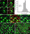
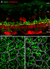
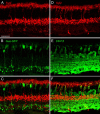
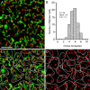
We report a quantitative analysis of the different bipolar cell types of the mouse retina. They were identified in wild-type mice by specific antibodies or in transgenic mouse lines by specific expression of green fluorescent protein or Clomeleon. The bipolar cell densities, their cone contacts, their dendritic coverage, and their axonal tiling were measured in retinal whole mounts. The results show that each and all cones are contacted by at least one member of any given type of bipolar cell (not considering genuine blue cones). Consequently, each cone feeds its light signals into a minimum of 10 different bipolar cells. Parallel processing of an image projected onto the retina, therefore, starts at the first synapse of the retina, the cone pedicle. The quantitative analysis suggests that our proposed catalog of 11 cone bipolar cells and one rod bipolar cell is complete, and all major bipolar cell types of the mouse retina appear to have been discovered.
Figures


References
-
- Badea TC, Nathans J. Quantitative analysis of neuronal morphologies in the mouse retina visualized by using a genetically directed reporter. J Comp Neurol. 2004;480:331–351. - PubMed
-
- Boycott B, Wässle H. Parallel processing in the mammalian retina. The Proctor Lecture. Invest Ophthalmol Vis Sci. 1999;40:1313–1327. - PubMed
-
- Boycott BB, Wässle H. Morphological classification of bipolar cells in the macaque monkey retina. Eur J Neurosci. 1991;3:1069–1088. - PubMed
Publication types
MeSH terms
Substances
LinkOut - more resources
Full Text Sources
Other Literature Sources
Molecular Biology Databases
