. 2009 Jan 7;29(1):234-49.
doi: 10.1523/JNEUROSCI.5273-08.2009.

Lipocalin-2 is an autocrine mediator of reactive astrocytosis

Affiliations

Lipocalin-2 is an autocrine mediator of reactive astrocytosis

Shinrye Lee et al. J Neurosci. .

Abstract

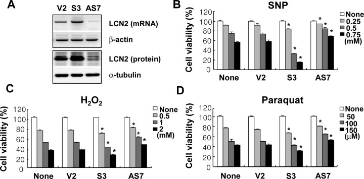
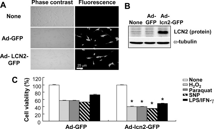
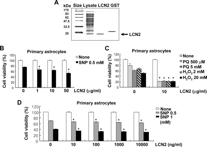
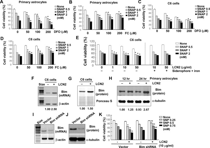
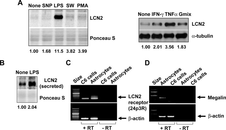
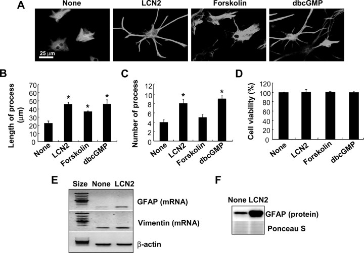
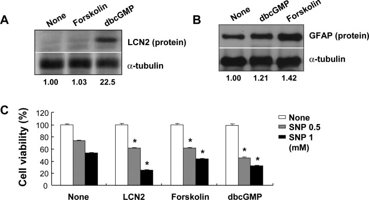
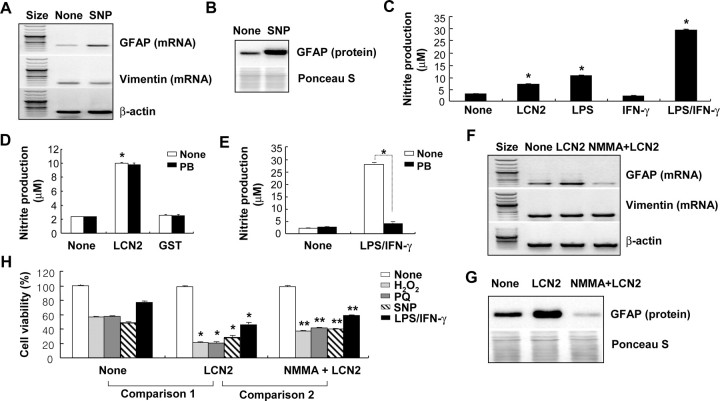
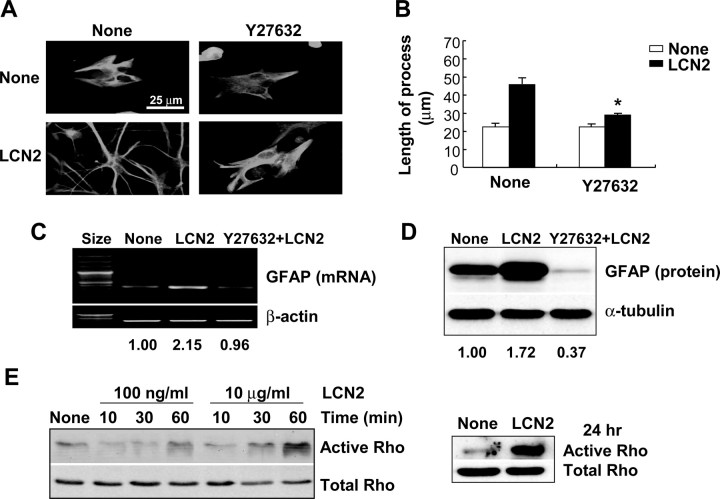
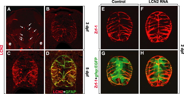
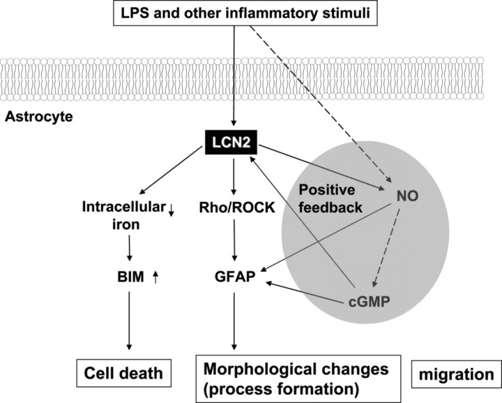
Astrocytes, the most abundant glial cell type in the brain, provide metabolic and trophic support to neurons and modulate synaptic activity. In response to a brain injury, astrocytes proliferate and become hypertrophic with an increased expression of intermediate filament proteins. This process is collectively referred to as reactive astrocytosis. Lipocalin 2 (lcn2) is a member of the lipocalin family that binds to small hydrophobic molecules. We propose that lcn2 is an autocrine mediator of reactive astrocytosis based on the multiple roles of lcn2 in the regulation of cell death, morphology, and migration of astrocytes. lcn2 expression and secretion increased after inflammatory stimulation in cultured astrocytes. Forced expression of lcn2 or treatment with LCN2 protein increased the sensitivity of astrocytes to cytotoxic stimuli. Iron and BIM (Bcl-2-interacting mediator of cell death) proteins were involved in the cytotoxic sensitization process. LCN2 protein induced upregulation of glial fibrillary acidic protein (GFAP), cell migration, and morphological changes similar to characteristic phenotypic changes termed reactive astrocytosis. The lcn2-induced phenotypic changes of astrocytes occurred through a Rho-ROCK (Rho kinase)-GFAP pathway, which was positively regulated by nitric oxide and cGMP. In zebrafishes, forced expression of rat lcn2 gene increased the number and thickness of cellular processes in GFAP-expressing radial glia cells, suggesting that lcn2 expression in glia cells plays an important role in vivo. Our results suggest that lcn2 acts in an autocrine manner to induce cell death sensitization and morphological changes in astrocytes under inflammatory conditions and that these phenotypic changes may be the basis of reactive astrocytosis in vivo.


Figures

Figure 1.
Figure 1.
Inverse correlation between the level of lcn2 expression and the sensitivity of astrocytes to cytotoxic agents. C6 glial cells with an increased or decreased lcn2 expression were established by stable transfection of lcn2 sense (S3) or antisense (AS7) cDNA. The increased or decreased lcn2 expression in the stable transfectants compared with cells transfected with an empty vector (V2) was confirmed by RT-PCR (top) and Western blot analysis (bottom) (A). β-Actin or α-tubulin was also detected to confirm the equal loading of the samples. Compared with the empty vector transfectant (V2), the lcn2 sense (S3) or antisense (AS7) stable transfectants showed an increased or decreased sensitivity, respectively, to cytotoxic agents such as SNP, H2O2, and paraquat (B–D). Cells were treated with the indicated concentrations of cytotoxic agents for 24 h, and cell viability was assessed by MTT assay. The asterisks indicate statistically significant differences from the empty vector transfectant (V2) treated with the same concentrations of cytotoxic agents (V2 vs S3 or V2 vs AS7) (p < 0.05). The results are mean ± SD (n = 3). The results in this and all similar experiments were repeated several times, and one representative done in triplicate is shown.
Figure 2.
Figure 2.
Augmented cell death in the astrocytes that were infected with adenoviral vector expressing lcn2. C6 glial cells were infected with adenoviral vectors expressing GFP (Ad-GFP) or lcn2 fused with GFP (Ad-lcn2-GFP) for 2 d, and then observed under fluorescence microscope (A). Magnification, ×200. Overexpression of lcn2-GFP fusion protein was confirmed by Western blot analysis using rabbit polyclonal anti-LCN2/NGAL antibody in the virus-infected cells (B). Virus-infected cells were treated with cytotoxic agents such as H2O2 (1 mm), paraquat (100 μm), SNP (0.5 mm), or a combination of LPS (100 ng/ml) and IFN-γ (50 U/ml) for 24 h, and then cell viability was assessed by MTT assay (C). The asterisks indicate statistically significant differences from the GFP-expressed cells (Ad-GFP) exposed to the same stimulus (p < 0.05; Ad-GFP vs Ad-lcn2-GFP). Values represent mean ± SD.
Figure 3.
Figure 3.
Recombinant LCN2 protein sensitized astrocytes to cell death. GST-fused LCN2 protein was expressed in BL21 cells, which was then cleaved by thrombin to release the pure LCN2 protein (A). The GST protein eluted was run on the same gel for comparison. Treatment with recombinant lcn2 protein for 24 h enhanced the sensitivity of astrocytes to NO toxicity and oxidative stress (B, C). Primary astrocyte cultures were exposed to the indicated concentration of recombinant LCN2 proteins with SNP, paraquat (PQ), or H2O2 for 24 h, and then cell viability was evaluated by MTT assay (B, C). The LCN2-induced sensitization of astrocytes to NO toxicity was dose dependent, as measured by MTT assay after 24 h (D). The results are mean ± SD (n = 3). The asterisks indicate statistically significant differences from the control treated with the same cytotoxic agent in the absence of LCN2 (p < 0.05).
Figure 4.
Figure 4.
Role of iron and Bim in the LCN2 effects. Primary astrocytes or C6 glial cells were treated with DFO (A, C) or FC (B, D) in the presence of indicated concentrations of the NO donor SNAP for 24 h, and then cell viability was assessed by MTT assay. The iron chelation (DFO) and donation (FC) increased and decreased NO-induced cell death, respectively (A–D). The LCN2-induced cell death sensitization was abolished by the concurrent addition of siderophore–iron complex (E). *p < 0.05 compared with the same concentration of SNAP treatment alone. The expression of proapoptotic Bcl-2 family protein BIM was significantly increased by the treatment with recombinant LCN2 protein (10 μg/ml) for 12 or 24 h. This was assessed by RT-PCR (F) and Western blot analysis (G, H) in C6 glia cells (F, G) and primary astrocyte cultures (H). The results were subjected to densitometric analysis, and normalized relative intensity of the bands is shown below (F–H). Stable knockdown of Bim expression by transfection of C6 glia cells with Bim-specific shRNA construct decreased the apoptotic sensitivity compared with the empty vector transfectant. Knockdown of Bim expression was confirmed by RT-PCR (I) and Western blot analysis (J) of the transfectants. Bim shRNA-transfected C6 glia cells were treated with LCN2 protein (10 μg/ml) in the presence of SNP (0.25, 0.5, or 0.75 mm) for 24 h, and then cell viability was assessed by MTT assay (K). β-Actin/α-tubulin detection or Ponceau S staining was done to confirm the equal loading of the samples. Error bars indicate SD. *p < 0.05 compared with the same concentration of SNP treatment alone in empty vector transfectants. **p < 0.05 compared with the same concentration of SNP treatment along with LCN2 in empty vector transfectants.
Figure 5.
Figure 5.
Expression of lcn2 and lcn2 receptor (24p3R) in C6 glia cells and primary astrocytes. The expression of lcn2 was strongly increased by LPS (100 ng/ml) or TNF-α (100 ng/ml) in primary astrocytes after 24 h treatment as determined by Western blot analysis. Serum withdrawal for 8 h (SW), PMA (100 μg/ml), IFN-γ (50 U/ml), ganglioside mixture (Gmix) (50 μg/ml), but not SNP (0.5 mm), treatment for 24 h slightly increased the protein level (A). Secreted LCN2 protein was also detected by Western blot analysis of culture media after similar treatment with LPS (100 ng/ml) for 24 h (B). Ponceau S staining or α-tubulin detection was conducted to confirm the equal loading of the samples. Normalized intensity of the bands is shown below (A, B). RT-PCR analysis revealed that C6 glial cells and primary astrocyte cultures expressed lcn2 receptor (24p3R), which has been shown to mediate the apoptotic effect of lcn2 (C). Megalin expression was detected in primary astrocytes, but not C6 cells (D). The lcn2 receptor (24p3R) or megalin expression was not detected in the reaction without reverse transcriptase (−RT). β-Actin was used as an internal control.
Figure 6.
Figure 6.
The effect of recombinant LCN2 protein on the morphology and GFAP expression of astrocytes. Addition of recombinant LCN2 protein (10 μg/ml) induced morphological changes in primary astrocyte cultures after 24 h (A), reminiscent of those in reactive astrocytosis in vivo. Treatment with forskolin (100 nm) or dbcGMP (1 mm) for 24 h also induced a similar morphological change. Cells were double-stained for GFAP (green) and Hoechst 33342 (nuclei; blue) (magnification, ×400) (A). Results are one representative of more than three independent experiments. The length (B) or number (C) of astrocyte processes was measured by examining several randomly chosen fields under fluorescence microscope (B, C). No significant cytotoxicity of the stimuli used was confirmed by MTT assay (D). Error bars indicate SD. After primary astrocytes were treated with recombinant LCN2 protein (10 μg/ml) for 24 h, GFAP or vimentin mRNA levels were determined by RT-PCR (E), or GFAP protein levels were assessed by Western blot analysis (F). Ponceau S staining or β-actin detection was done to confirm the equal loading of the samples. *p < 0.05 compared with untreated control.
Figure 7.
Figure 7.
Role of cAMP and cGMP in the expression of LCN2/GFAP and cell death of astrocytes. After primary astrocytes were treated with forskolin (100 nm) or dbcGMP (1 mm) for 24 h, LCN2 or GFAP protein was detected by Western blot analysis (A, B). Normalized intensity of the bands is shown below (A, B). Primary astrocytes were exposed to LCN2 protein (10 μg/ml) or other stimuli (forskolin, 100 nm; dbcGMP, 1 mm) in the presence of SNP (0.5 or 1 mm) for 24 h, and then cell viability was assessed by MTT assay (C). Error bars indicate SD. *p < 0.05 compared with the same concentration of SNP treatment alone.
Figure 8.
Figure 8.
Involvement of NO in the LCN2 effects in astrocytes. After primary astrocytes were treated with SNP (0.5 mm) for 24 h, GFAP/vimentin mRNA or GFAP protein was detected by RT-PCR (A) or Western blot analysis (B), respectively. The expression of GFAP was significantly increased by SNP (0.5 mm) in primary astrocytes. Primary astrocytes were stimulated with recombinant LCN2 protein (10 μg/ml), LPS (100 ng/ml), IFN-γ (50 U/ml), or a combination of LPS (100 ng/ml) and IFN-γ (50 U/ml) for 24 h, and then nitrite production was assessed by Griess reagent (C). *p < 0.05 compared with no treatment. Primary astrocytes were also pretreated with polymyxin B (PB) (10 μg/ml) for 30 min before the treatment with LCN2 protein (10 μg/ml), GST protein (10 μg/ml) (D), or a combination of LPS (100 ng/ml) and IFN-γ (50 U/ml) (E) for 24 h, and then nitrite production was assessed by Griess reagent (D, E). *p < 0.05 compared with untreated control (C, D) or as indicated (E). Alternatively, primary astrocytes were treated with LCN2 protein (10 μg/ml) for 24 h or pretreated with NMMA (500 μm) for 30 min before the treatment with recombinant LCN2 protein (10 μg/ml) for 24 h. The expression of GFAP or vimentin was then detected by RT-PCR (F) or Western blot analysis (G) as indicated. After a similar treatment of primary astrocytes with LCN2 and NMMA in the presence of various cytotoxic agents [H2O2 (1 mm), paraquat (PQ) (100 μm), SNP (0.5 mm), and LPS (100 ng/ml) plus IFN-γ (50 U/ml) for 24 h], cell viability was assessed by MTT assay (H). NMMA (500 μm) alone did not exert any cytotoxicity (data not shown). Error bars indicate SD. The single asterisks indicate statistically significant differences between treatments with cytotoxic agents in the absence and presence of LCN2 (*p < 0.05; None vs LCN2; comparison 1). The double asterisks indicate statistically significant differences between treatments with cytotoxic agents in the presence of LCN2 and NMMA plus LCN2 (**p < 0.05; LCN2 vs NMMA plus LCN2; comparison 2).
Figure 9.
Figure 9.
Role of Rho/ROCK pathway in the LCN2-induced morphological changes of astrocytes. Primary astrocytes were pretreated with Y27632 (ROCK inhibitor; 100 μm) in the absence or presence of recombinant LCN2 protein (10 μg/ml) for 24 h. Cells were double-stained for GFAP (green) and Hoechst 33342 (cell nuclei; blue) (magnification, ×400) (A). Measurement of the length of the longest astrocyte processes for each treatment was done (B). Error bars indicate SD. *p < 0.05 compared with LCN2 treatment alone. GFAP mRNA (C) or protein levels (D) were also evaluated by RT-PCR or Western blot analysis, respectively, after stimulation with LCN2 in the absence or presence of Rho kinase inhibitor Y27632 (100 μm) for 24 h. β-Actin or α-tubulin was detected to confirm the equal loading of the samples. Results of densitometric analysis are shown below (C, D). After primary astrocytes were treated with LCN2 protein (100 ng/ml or 10 μg/ml) for the indicated time periods or 24 h, the activation state of Rho was assessed by a pulldown assay (E). The results are representative of three or more independent experiments.
Figure 10.
Figure 10.
Stimulation of astrocyte migration by LCN2. Primary astrocytes were treated with recombinant LCN2 protein (10 μg/ml) for the indicated time periods, and then either wound healing assay (A) or Boyden chamber assay (B) was performed to evaluate cell migration (magnification, ×100). Quantification of astrocyte migration was conducted after wound healing assay (C) or Boyden chamber assay (D). Error bars indicate SD. *p < 0.05 compared with untreated control at the same day.
Figure 11.
Figure 11.
Expression of lcn2 in spinal cord of zebrafish embryos and its functional role in determination of radial glia morphology. A–D, Anti-LCN2 antibody labeled radial glia in the spinal cord of zebrafish embryos. All images are transverse sections of zebrafish CNS, labeled by anti-LCN2 antibody, dorsal to top. A, B, LCN2 antibody labeled microglia-like cells in forebrain (A, arrows) and radial glia in spinal cord at 3 dpf (B). The “e” indicates an eye. C, D, Double labeling of spinal cord with anti-LCN2 (red) and -GFAP antibodies (green) at 5 dpf (C, D, same section). LCN2 was expressed in the GFAP+ radial glia. E–H, Overexpression of rat lcn2 increased the number and thickness of radial glial processes. All images are transverse sections of spinal cord of Tg(gfap:egfp) embryos, dorsal upward. Anti-Zrf-1 antibody labeling (E, F) and combined anti-Zrf-1 (red) and gfap:EGFP (green) images of the same sections (G, H) of control embryo which did not receive any RNA injection (E, G) and lcn2 RNA-injected embryo (F, H).
Figure 12.
Figure 12.
Schematic diagram showing the bifunctional role of LCN2 in cell death and morphological changes of astrocytes. Reactive astrocytes under inflammatory condition secrete lcn2, which may feed back on astrocytes to induce morphological as well as functional changes. lcn2 induces morphological changes of reactive astrocytes and promotes their migration through the Rho–ROCK pathway. lcn2 also renders astrocytes more sensitive to cell death signals by regulating iron metabolism and Bim pathway, which may provide a basis for the self-regulatory elimination of reactive astrocytes in vivo. NO and cGMP appear to form a positive feedback loop that amplifies the lcn2–Rho–ROCK–GFAP pathway. The morphological changes of astrocytes appear to be closely related with the phenotypic changes into cell death-prone astrocytes.

References

    1. Abe K, Misawa M. Astrocyte stellation induced by Rho kinase inhibitors in culture. Brain Res Dev Brain Res. 2003;143:99–104. - PubMed
    1. Anzenberger U, Bit-Avragim N, Rohr S, Rudolph F, Dehmel B, Willnow TE, Abdelilah-Seyfried S. Elucidation of megalin/LRP2-dependent endocytic transport processes in the larval zebrafish pronephros. J Cell Sci. 2006;119:2127–2137. - PubMed
    1. Aschner M. Astrocytes as mediators of immune and inflammatory responses in the CNS. Neurotoxicology. 1998;19:269–281. - PubMed
    1. Avalos AM, Arthur WT, Schneider P, Quest AF, Burridge K, Leyton L. Aggregation of integrins and RhoA activation are required for Thy-1-induced morphological changes in astrocytes. J Biol Chem. 2004;279:39139–39145. - PubMed
    1. Barres BA, Barde Y. Neuronal and glial cell biology. Curr Opin Neurobiol. 2000;10:642–648. - PubMed

Publication types

MeSH terms

LinkOut - more resources