. 2009 Mar;20(5):1374-87.
doi: 10.1091/mbc.e08-05-0474. Epub 2009 Jan 7.

Essential role of hIST1 in cytokinesis

Affiliations

Essential role of hIST1 in cytokinesis

Monica Agromayor et al. Mol Biol Cell. 2009 Mar.

Abstract

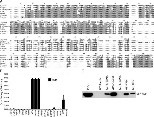
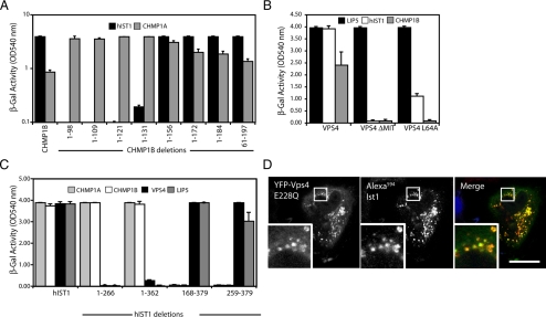
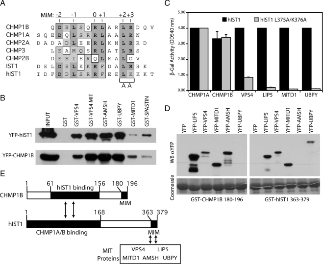
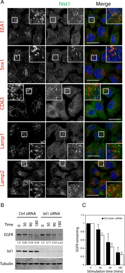
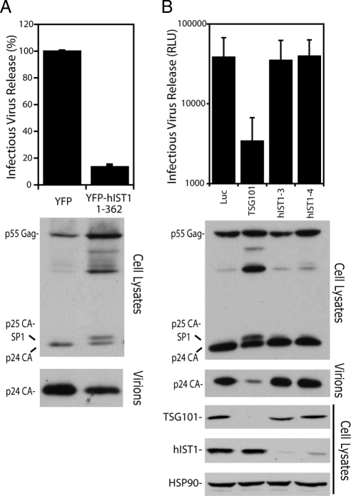
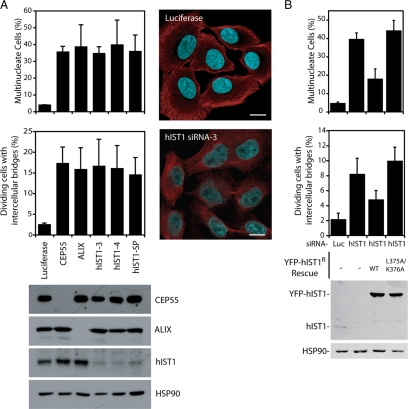
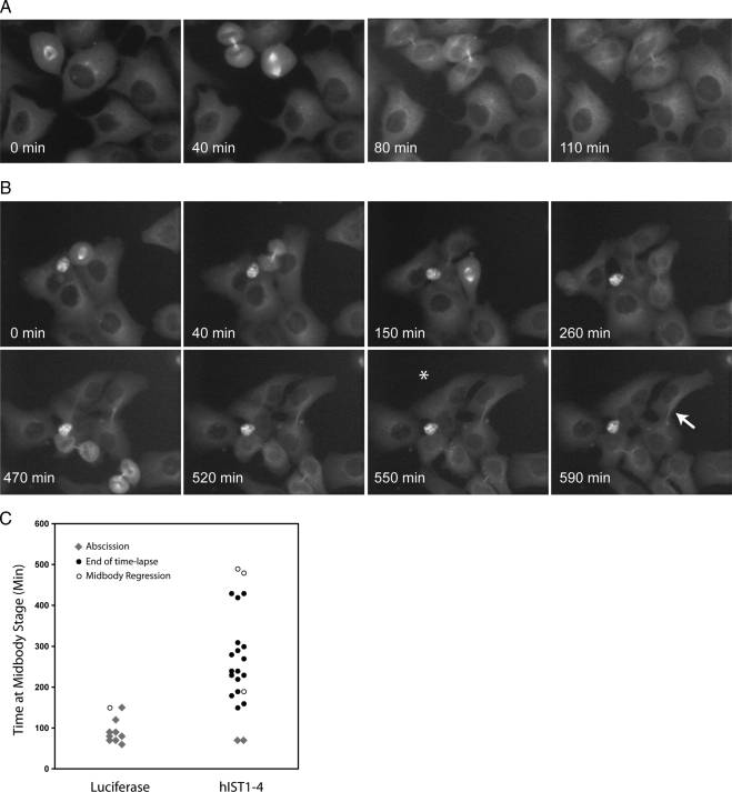
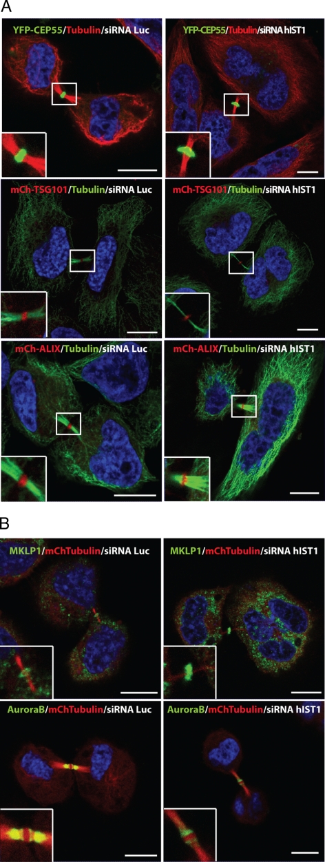
The last steps of multivesicular body (MVB) formation, human immunodeficiency virus (HIV)-1 budding and cytokinesis require a functional endosomal sorting complex required for transport (ESCRT) machinery to facilitate topologically equivalent membrane fission events. Increased sodium tolerance (IST) 1, a new positive modulator of the ESCRT pathway, has been described recently, but an essential function of this highly conserved protein has not been identified. Here, we describe the previously uncharacterized KIAA0174 as the human homologue of IST1 (hIST1), and we report its conserved interaction with VPS4, CHMP1A/B, and LIP5. We also identify a microtubule interacting and transport (MIT) domain interacting motif (MIM) in hIST1 that is necessary for its interaction with VPS4, LIP5 and other MIT domain-containing proteins, namely, MITD1, AMSH, UBPY, and Spastin. Importantly, hIST1 is essential for cytokinesis in mammalian cells but not for HIV-1 budding, thus providing a novel mechanism of functional diversification of the ESCRT machinery. Last, we show that the hIST1 MIM activity is essential for cytokinesis, suggesting possible mechanisms to explain the role of hIST1 in the last step of mammalian cell division.


Figures

Figure 1.
Figure 1.
hIST1 binds to CHMP1A, CHMP1B, VPS4, and LIP5. (A) Sequence alignment showing the high degree of similarity between IST1 genes from different organisms. The shading on the alignment indicates conserved amino acids. (B) hIST1 fused to the VP16 activation domain was tested for interactions with the human components of ESCRT-I, -II, -III, and ESCRT-III–associated proteins by yeast two-hybrid assays. Error bars indicate the SD from the mean of triplicate measurements. (C) Coprecipitation assays showing binding of hIST1 to CHMP1A, CHMP1B, CHMP2A, VPS4, and LIP5. One percent of the starting cell lysate (INPUT) and 10% of the volume eluted from the beads were analyzed by Western blot with α-GFP antibody.
Figure 2.
Figure 2.
(A) Mapping by yeast two-hybrid analysis of the regions needed in CHMP1B for interaction with IST1. Binding to CHMP1A was used as a positive control; the regions of VPS4 required for interaction with IST1. LIP5 was used as a positive control (B); and the regions of IST1 required for interaction with CHMP1A, CHMP1B, VPS4, or LIP5 (C). Error bars indicate the SD from the mean of triplicate measurements. (D) Micrographs showing that endogenous hIST1 is recruited to aberrant endosomes in the presence of YFP-VPS4 E228Q. Left, images acquired with YFP (pseudocolored in green); middle, images acquired with Alexa594 (pseudocolored in red); and right, overlaid images. A higher magnification of the boxed areas is shown in all panels and DNA is shown in blue. Bar, 10 μm.
Figure 3.
Figure 3.
IST1 contains a functional MIM. (A) Sequence alignment of the MIMs from the indicated human ESCRT-III proteins and the human and yeast IST1 genes. Positions of the six MIM residues contacting Vps4 MIT domain are indicated on top of the alignment and conserved amino acids are shaded in gray. (B) Coprecipitation assays showing binding of the indicated MIT containing proteins to YFP-CHMP1B or YFP-hIST1. One percent of the starting cell lysate (INPUT) and 10% of the volume eluted from the beads were analyzed by Western blot with α-GFP antibody. (C) hIST1 residues in MIM's position +2 and +3 were mutated (hIST1 L375A/K376A), and interactions with ESCRT-III and MIT-containing proteins were monitored using yeast two-hybrid assays. Error bars indicate the SD from the mean of triplicate measurements. (D) Cell lysates from bacteria expressing CHMP1B's MIM (GST-CHMP1B 180-196) or hIST1's MIM (GST-hIST1 363-379) were tested by coprecipitation assays for binding to the indicated MIT-containing proteins fused to YFP and binding was monitored with α-GFP mAb (WBαYFP). Bottom, GST fusion protein expression in cell lysates with Coomassie staining. (E) Schematic representation of CHMP1B and hIST1 proteins, showing the regions needed for their interaction (arrows), and the position of their MIMs.
Figure 4.
Figure 4.
Analysis of hIST1's function in endosomal sorting. (A) Endogenous hIST1 (pseudocolored in green, middle column) and the indicated endosomal markers (pseudocolored in red, left column) were studied by immunofluorescence. A higher magnification of the boxed areas is shown in all panels, and DNA is shown in blue. Bar, 10 μm. (B) HeLa cells treated with either nonspecific control or hIST1-specific siRNA were serum starved and stimulated for the indicated times with EGF. Lysates were analyzed with α-EGFR, α-hIST1, and α-tubulin. (C) Graphic representation of the quantification obtained via infrared imaging after immunoblotting with α-EGFR. Error bars indicate the SD from the mean of triplicate measurements.
Figure 5.
Figure 5.
hIST1 effect on HIV budding. (A) Overexpression of a hIST1 fragment lacking the C terminus of the protein fused to YFP inhibits HIV budding. (B) siRNA depletion of endogenous hIST1 has no effect on virus release. TSG101-specific and luciferase control siRNAs were used as positive and negative controls, respectively. Error bars indicate the SD from the mean of three independent experiments. The inhibition of viral particle release was analyzed in cell lysates and extracellular virions by Western blot with α-HIV Gag antibody. The positions on the blot of the precursor Pr55Gag and the mature CA protein are indicated. The bottom images in B show the siRNA-mediated silencing of the endogenous hIST1 and TSG101 and a protein loading control as determined by Western blot analysis with α-TSG101, α-hIST1, and α-HSP90.
Figure 6.
Figure 6.
hIST1 is required for cytokinesis. (A) HeLa cells were transfected with siRNA targeting CEP55, ALIX, hIST1 (hIST1-3, hIST1-4, and Dharmacon's siGENOME SMARTpool (hIST1-SP), or a luciferase control and scored for multinucleation. Representative micrographs are shown (B) in which tubulin is labeled in red. HeLa cells stably expressing YFP (−), siRNA-resistant YFP-hIST1 (WT), or siRNA-resistant YFP-hIST1 L375A/K376A were treated with the indicated siRNA and scored for multinucleation or dividing cells with intercellular bridges. Error bars indicate the SD from the mean of triplicate measurements. siRNA-mediated depletion of the indicated endogenous protein was examined by immunoblotting with α-CEP55, α-ALIX, or α-hIST1. Protein loading control was determined with α-HSP90.
Figure 7.
Figure 7.
Cytokinesis fails at the terminal stage in hIST1-knockdown cells. Selected frames from time-lapse microscopy of HeLa cells stably expressing mCherry-tubulin treated with Luciferase (A) or hIST1 (B) siRNA. The white star in panel B shows a cell that has divided into two cells that remain connected for 510 min before eventually regressing to form a binucleated cell. The white arrow highlights an example of two cells that remain connected until the end of the time-lapse, showing a delay in cytokinesis. Elapsed times are provided in each panel. Movies showing time-lapse images are provided in Supplemental Figures S6 and S7. Images were recorded every 10 min, starting 48 h after siRNA transfection. (C) Quantitative analysis of time-lapse microscopy. When possible, the amount of time from telophase to abscission was measured (gray diamond). When abscission does not occur due to a delay in cytokinesis and the end of filming (black circle) or due to midbody regression to form a multinucleated cell (open circle) we plotted the maximum time that the cells were observed connected by a midbody (time at midbody stage). Luciferase-treated cells n = 10; hIST1-depleted cells n = 22.
Figure 8.
Figure 8.
Cellular localization of endogenous hIST1 during mitosis in human HeLa cells. hIST1 accumulates near the spindle during telophase and is detected at the midbody during cytokinesis. HeLa cells were immunostained for hIST1 (green), α-tubulin (red) and DNA (blue). The far right column shows a higher magnification of the boxed areas in the merged column of cells in telophase and cytokinesis. Bar, 10 μm.
Figure 9.
Figure 9.
Characterization of the midbody composition in cells that lack hIST1. (A) Representative micrographs describing midbody localization of YFP-CEP55 (green), mCh-TSG101 (red), or mCh-ALIX (red) in cells stably expressing these fusion proteins and treated with siRNA targeting luciferase or hIST1. After siRNA treatment, cells were fixed and stained with α-tubulin and Alexa594 (red) or Alexa488 (green) conjugated secondary antibodies. (B) Similarly, HeLa cells stably expressing mCh-Tubulin (red) treated with siRNA targeting luciferase or hIST1 were immunostained for MKLP1 or AuroraB (green). In all cases, enlargements depict the midbody localization of the studied proteins. DNA is shown in blue. Bar, 10 μm.

References

    1. Agromayor M., Martin-Serrano J. Interaction of AMSH with ESCRT-III and deubiquitination of endosomal cargo. J. Biol. Chem. 2006;281:23083–23091. - PubMed
    1. Azmi I., Davies B., Dimaano C., Payne J., Eckert D., Babst M., Katzmann D. J. Recycling of ESCRTs by the AAA-ATPase Vps4 is regulated by a conserved VSL region in Vta1. J. Cell Biol. 2006;172:705–717. - PMC - PubMed
    1. Azmi I. F., Davies B. A., Xiao J., Babst M., Xu Z., Katzmann D. J. ESCRT-III family members stimulate Vps4 ATPase activity directly or via Vta1. Dev. Cell. 2008;14:50–61. - PubMed
    1. Babst M., Katzmann D. J., Estepa-Sabal E. J., Meerloo T., Emr S. D. Escrt-III: an endosome-associated heterooligomeric protein complex required for mvb sorting. Dev. Cell. 2002a;3:271–282. - PubMed
    1. Babst M., Katzmann D. J., Snyder W. B., Wendland B., Emr S. D. Endosome-associated complex, ESCRT-II, recruits transport machinery for protein sorting at the multivesicular body. Dev. Cell. 2002b;3:283–289. - PubMed

Publication types

MeSH terms