. 2009 Mar;20(5):1428-40.
doi: 10.1091/mbc.e08-01-0001. Epub 2009 Jan 7.

Centrosome/spindle pole-associated protein regulates cytokinesis via promoting the recruitment of MyoGEF to the central spindle

Affiliations

Centrosome/spindle pole-associated protein regulates cytokinesis via promoting the recruitment of MyoGEF to the central spindle

Michael Asiedu et al. Mol Biol Cell. 2009 Mar.

Abstract

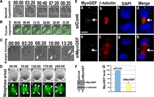
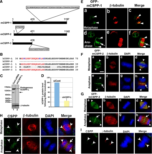
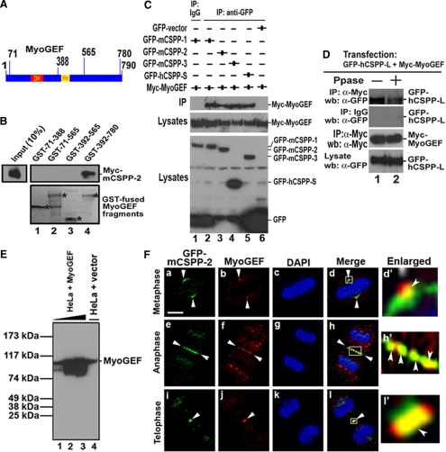
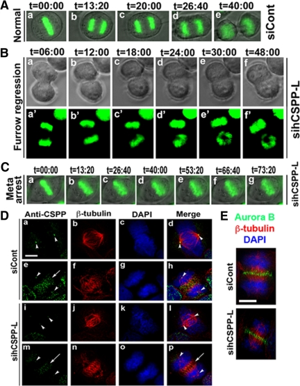
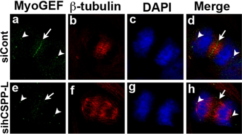
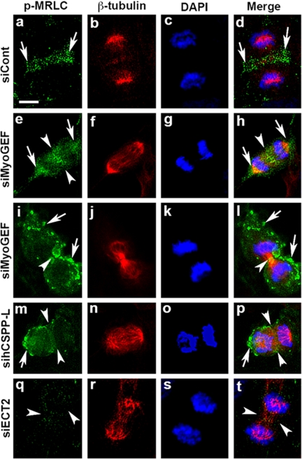
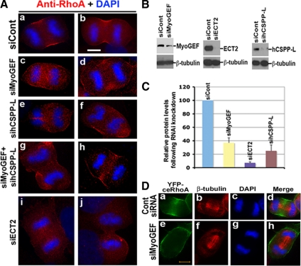
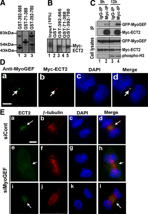
Cooperative communications between the central spindle and the contractile ring are critical for the spatial and temporal regulation of cytokinesis. Here we report that MyoGEF, a guanine nucleotide exchange factor that localizes to the central spindle and cleavage furrow, interacts with centrosome/spindle pole-associated protein (CSPP), which is concentrated at the spindle pole and central spindle during mitosis and cytokinesis. Both in vitro and in vivo pulldown assays show that MyoGEF interacts with CSPP. The C-terminus of MyoGEF and N-terminus of CSPP are required for their interaction. Immunofluorescence analysis indicates that MyoGEF and CSPP colocalize at the central spindle. Depletion of CSPP or MyoGEF by RNA-interference (RNAi) not only causes defects in mitosis and cytokinesis, such as metaphase arrest and furrow regression, but also mislocalization of nonmuscle myosin II with a phosphorylated myosin regulatory light chain (p-MRLC). Importantly, CSPP depletion by RNAi interferes with MyoGEF localization at the central spindle. Finally, MyoGEF interacts with ECT2, and RNAi-mediated depletion of MyoGEF leads to mislocalization of ECT2 and RhoA during cytokinesis. Therefore, we propose that CSPP interacts with and recruits MyoGEF to the central spindle, where MyoGEF contributes to the spatiotemporal regulation of cytokinesis.


Figures

Figure 1.
Figure 1.
Depletion of MyoGEF caused defects in furrow ingression. (A) Normal cytokinesis. Live cell images show normal cytokinesis in HeLa cells transfected with control siRNA. (B) Furrow regression. Time-lapse images show that furrow does not successfully ingress in HeLa cells transfected with MyoGEF siRNA and a plasmid encoding H2B-GFP (green). (C) Ectopic furrowing. Time-lapse images show ectopic furrow formation (arrow in e) in a HeLa cell transfected with MyoGEF siRNA. (D) Metaphase arrest. Time-lapse images show that a HeLa cell transfected with MyoGEF siRNA and a plasmid encoding GFP-α-tubulin (green) was arrested at metaphase. Arrowheads indicate the spindles. (E) Depletion of MyoGEF did not affect central spindle formation. HeLa cells treated with siCont (a–d) or siMyoGEF (e–h) were fixed with methanol/acetone and subjected to immunofluorescence with antibodies specific for MyoGEF (green) and β-tubulin (red). The nuclei were stained with DAPI (blue). Bar, 20 μm. (F) HeLa cells treated with control siRNA (siCont; lane 1) or MyoGEF siRNA (siMyoGEF; lane 2) were subjected to immunoblot analysis with antibodies specific for MyoGEF and β-tubulin. (G) The immunoblot images in F were quantified using the NIH Image program. The numbers in A–D indicate the elapsed time (min:sec).
Figure 2.
Figure 2.
Identification of mCSPP. (A) Schematic diagram of three isoforms of mCSPP proteins. The major differences among mCSPP-1, -2, and -3 are shown: a 51-amino acid insert in the middle region of mCSPP-1; an 8-amino acid insert in the N-terminal region of mCSPP-2; 19 amino acids that are present in mCSPP-3 at the C-terminus, but not in mCSPP-1 and mCSPP-2. The number indicates the amino acids. (B) Alignments of the N-terminal amino acid sequences of human CSPP and mouse CSPP isoforms. The amino acids in red were used as antigen to raise an antibody specific for CSPP. (C) The polyclonal antibody specific for CSPP recognizes endogenous hCSPP-L. Treatment with hCSPP-L siRNA decreases the expression of hCSPP-L (cf. lane 1 with lane 2). (D) The immunoblot images in C were quantified using the NIH Image program. (E–G) GFP-mCSPP-1 localizes to the spindle pole and central spindle (E), whereas GFP-mCSPP-2 (F) and -3 (G) localize to the spindle microtubules and central spindle. The nuclei were stained with DAPI (blue). Bar, 20 μm. (H and I) Localization of endogenous CSPP during cytokinesis. HeLa cells were fixed and subjected to immunofluorescence with antibodies specific for CSPP (green) and β-tubulin (red) or γ-tubulin (red). Endogenous CSPP localizes to the spindle pole and central spindle (H) Endogenous CSPP also colocalizes with γ-tubulin at the spindle pole (I). The nuclei were stained with DAPI (blue). Bar, 20 μm. Note that cells in E–G were fixed with 4% paraformaldehyde, whereas cells in H and I were fixed with methanol/acetone (1:1).
Figure 3.
Figure 3.
Interaction between MyoGEF and CSPP. (A) Schematic diagram of MyoGEF. The numbers indicate amino acids. DH and PH, Dbl-homology domain and pleckstrin homology domain, respectively. (B) An in vitro GST pulldown assay using GST-MyoGEF fragments and in vitro–translated mCSPP-2. GST-MyoGEF fragments are indicated by asterisks. (C) Myc-MyoGEF interacted with GFP-CSPP in vivo. HeLa cells were transfected with plasmids encoding Myc-MyoGEF and GFP-mCSPP-1, GFP-mCSPP-2, GFP-mCSPP-3, GFP-hCSPP-S, or GFP alone. The transfected cells were subjected to coimmunoprecipitation with anti-GFP antibody (α-GFP) followed by immunoblot analysis with anti-Myc antibody. (D) Myc-MyoGEF interacted with GFP-hCSPP-L in vivo. HeLa cells transfected with plasmids encoding GFP-hCSPP-L and Myc-MyoGEF were subjected to immunoprecipitation with anti-Myc antibody, followed by immunoblot with anti-GFP antibody. Note that treatment with λ-phosphatase (Ppase) decreases the interaction between Myc-MyoGEF and GFP-hCSPP-L (cf. lane 1 with lane 2 in top panel). (E) Characterization of MyoGEF antibody. HeLa cells were transfected with an empty vector (lane 4) or a MyoGEF plasmid (without any tags; lanes 1-3). Increasing amount of HeLa cell lysates from cells exogenously expressing MyoGEF was used (lanes 1–3). Same amount of cell lysates was used in lanes 3 and 4. (F) Colocalization of GFP-mCSPP-2 and endogenous MyoGEF to the central spindle and midbody. HeLa cells expressing GFP-mCSPP-2 were fixed with methanol/acetone and subjected to immunofluorescence with anti-MyoGEF antibody (red). The nuclei were stained with DAPI (blue). (d′, h′, and l′) Enlarged images from panels d, h, and l, respectively. Bar, 20 μm.
Figure 4.
Figure 4.
Depletion of CSPP led to defects in mitosis and cytokinesis. (A) Normal cytokinesis in HeLa cells transfected with control siRNA and a plasmid encoding H2B-GFP (green). (B) Furrow regression in HeLa cells transfected with hCSPP-L siRNA and a plasmid encoding H2B-GFP (green; a′–f′). (C) Metaphase arrest in HeLa cells transfected with hCSPP-L siRNA and a plasmid encoding H2B-GFP (green). (D) Depletion of hCSPP-L did not affect central spindle formation. HeLa cells were transfected with control siRNA (siCont; a–h) or hCSPP-L siRNA (sihCSPP-L; i–p). The transfected HeLa cells were fixed with methanol/acetone and subjected to immunofluorescence with antibodies specific for CSPP (green) and β-tubulin (red). The nuclei were stained with DAPI (blue). (E) Cells treated with siCont or sihCSPP-L were subjected to immunofluorescence with antibodies specific for aurora B (green) and β-tubulin (red). Bar, (D) 20 μm; (E) 10 μm. Meta, metaphase. The numbers in A–C indicate the elapsed time (min:sec).
Figure 5.
Figure 5.
Depletion of CSPP affected MyoGEF localization. HeLa cells were transfected with control siRNA (siCont; a–d) or hCSPP-L siRNA (sihCSPP-L; e–l). The transfected HeLa cells were fixed with methanol/acetone and subjected to immunofluorescence with antibodies specific for MyoGEF (green) and β-tubulin (red). The nuclei were stained with DAPI (blue). Bar, 20 μm.
Figure 6.
Figure 6.
Depletion of MyoGEF or CSPP resulted in p-MRLC mislocalization during cytokinesis. HeLa cells were transfected with control siRNA (a–d), MyoGEF siRNA (e–l), hCSPP-L siRNA (m–p), or ECT2 siRNA (q–t). Seventy-two hours after transfection, the transfected cells were fixed with 4% paraformaldehyde and stained with antibodies specific for β-tubulin (red) and p-MRLC (green). The chromosomes were stained with DAPI (blue). Bar, 20 μm.
Figure 7.
Figure 7.
Depletion of MyoGEF or CSPP affected the distribution of active RhoA during cytokinesis. (A) U2OS cells treated with siRNAs as indicated were fixed with TCA and subjected to immunofluorescence with an antibody specific for RhoA (red). The nuclei were stained with DAPI (blue). Bar, 20 μm. (B) U2OS cells treated with siRNAs as indicated were subjected to immunoblot analysis with antibodies as indicated. (C) The immunoblot images in B were quantified using the NIH Image program. (D) Depletion of MyoGEF interfered with YFP-ceRhoA distribution during cytokinesis. HeLa cells were transfected with a YFP-ceRhoA–expressing plasmid and control siRNA (a–d) or siMyoGEF (e–h). Forty-eight hours after transfection, the transfected cells were fixed with 4% paraformaldehyde and stained with an antibody specific for β-tubulin (red). The nuclei were stained with DAPI (blue). Bar, 20 μm.
Figure 8.
Figure 8.
Depletion of MyoGEF affected ECT2 localization at the central spindle. (A) GST-tagged MyoGEF fragments (indicated by asterisks). (B) The GST pulldown assay using GST-tagged MyoGEF fragments and in vitro–translated Myc-ECT2. (C) HeLa cells were transfected with plasmids encoding GFP-MyoGEF and Myc-ECT2. At 0 or 12 h after release from thymidine block, the transfected cells were subjected to coimmunoprecipitation with anti-Myc antibody, followed by immunoblot analysis with antibodies as indicated. (D) Colocalization of endogenous MyoGEF and Myc-ECT2 to the midbody. (E) HeLa cells were treated with control siRNA (siCont; a–d) or MyoGEF siRNA (siMyoGEF; e–l). Seventy-two hours after transfection, the transfected cells were processed for immunofluorescence with antibodies specific for β-tubulin (red) and ECT2 (green). The chromosomes were stained with DAPI (blue). Note that cells in D were fixed with methanol/acetone (1:1), whereas cells in E were fixed with 4% paraformaldehyde. Bar, 20 μm.

References

    1. Asiedu M., Wu D., Matsumura F., Wei Q. Phosphorylation of myogef on thr-574 by polo-like kinase 1 (PLK1) promotes myogef localization to the central spindle. J. Biol. Chem. 2008;283:28392–28400. - PMC - PubMed
    1. Bement W. M., Benink H. A., von Dassow G. A microtubule-dependent zone of active RhoA during cleavage plane specification. J. Cell Biol. 2005;170:91–101. - PMC - PubMed
    1. Birkenfeld J., Nalbant P., Bohl B. P., Pertz O., Hahn K. M., Bokoch G. M. GEF-H1 modulates localized RhoA activation during cytokinesis under the control of mitotic kinases. Dev. Cell. 2007;12:699–712. - PMC - PubMed
    1. Burgess D. R., Chang F. Site selection for the cleavage furrow at cytokinesis. Trends Cell Biol. 2005;15:156–162. - PubMed
    1. Burkard M. E., Randall C. L., Larochelle S., Zhang C., Shokat K. M., Fisher R. P., Jallepalli P. V. Chemical genetics reveals the requirement for Polo-like kinase 1 activity in positioning RhoA and triggering cytokinesis in human cells. Proc. Natl. Acad. Sci. USA. 2007;104:4383–4388. - PMC - PubMed

Publication types

MeSH terms