. 2009 Jan 13;106(2):450-5.
doi: 10.1073/pnas.0808092106. Epub 2009 Jan 7.

Artificial ligand binding within the HIF2alpha PAS-B domain of the HIF2 transcription factor

Affiliations

Artificial ligand binding within the HIF2alpha PAS-B domain of the HIF2 transcription factor

Thomas H Scheuermann et al. Proc Natl Acad Sci U S A. .

Abstract

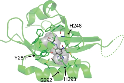
The hypoxia-inducible factor (HIF) basic helix-loop-helix Per-aryl hydrocarbon receptor nuclear translocator (ARNT)-Sim (bHLH-PAS) transcription factors are master regulators of the conserved molecular mechanism by which metazoans sense and respond to reductions in local oxygen concentrations. In humans, HIF is critically important for the sustained growth and metastasis of solid tumors. Here, we describe crystal structures of the heterodimer formed by the C-terminal PAS domains from the HIF2alpha and ARNT subunits of the HIF2 transcription factor, both in the absence and presence of an artificial ligand. Unexpectedly, the HIF2alpha PAS-B domain contains a large internal cavity that accommodates ligands identified from a small-molecule screen. Binding one of these ligands to HIF2alpha PAS-B modulates the affinity of the HIF2alpha:ARNT PAS-B heterodimer in vitro. Given the essential role of PAS domains in forming active HIF heterodimers, these results suggest a presently uncharacterized ligand-mediated mechanism for regulating HIF2 activity in endogenous and clinical settings.


Conflict of interest statement

The authors declare no conflict of interest.

Figures

Fig. 1.
Fig. 1.
Structural features of the HIF2 PAS-B* heterodimer. (A) Crystal structure of the high-affinity HIF2α PAS-B* (green) and ARNT PAS-B* (blue) heterodimer, including side chains that stabilize the heterodimer of PAS-B* proteins (sticks) and internally bound H2O molecules (red spheres). (B) The location of 8 ordered, internally bound water molecules (red mesh, FoFc omit map contoured at 4.0 σ), within the HIF2α PAS-B* cavity (gray transparent surface). The red asterisk (*) denotes the location of the protease-accessible R330 residue.
Fig. 2.
Fig. 2.
Characterization of a HIF2α PAS-B–ligand complex. (A) THS-044 chemical structure and the 15N/1H HSQC spectra of its complex with HIF2α PAS-B, demonstrating NMR slow-exchange kinetics. Overlaid spectra include 200 μM 15N-HIF2α PAS-B in the absence (black) and in the presence of 50 μM (blue) and 200 μM (red) THS-044. (B) Isothermal titration calorimetry of the wild-type HIF2α PAS-B domain and THS-044. (C) Limited trypsin proteolysis of THS-044-bound (Upper) and unliganded (Lower) wild-type HIF2α PAS-B. Full-length, uncut protein is indicated by open triangles and the large fragment produced by proteolysis at R330 by filled triangles. Protein bands marked with an asterisk (*) correspond to a 14.4 kDa molecular weight marker. (D) 2H exchange protection factors observed in THS-044-bound (Upper) and unliganded (Lower) HIF2α PAS-B. Bars indicate sites of β-strand (blue) and α-helix (red) secondary structure. Green asterisks denote sites for which data were omitted on account of spectral peak overlap and black asterisks indicate sites where the given protection factor is a lower limit, because we did not observe significant exchange on the 20-h timescale of measurement.
Fig. 3.
Fig. 3.
Structure of THS-044 in complex with HIF2α PAS-B*. THS-044 (gray sticks) occupies the apo-HIF2α PAS-B* internal cavity (transparent gray surface). The HIF2α PAS-B* HI loop in this structure was not modeled (dashed lines). For clarity, ARNT PAS-B* is not displayed.
Fig. 4.
Fig. 4.
Partial in vitro disruption of the wild-type HIF2 PAS-B heterodimer by THS-044. (A) PAS-B heterodimer NMR samples were prepared with a final concentration of 100 μM 15N-ARNT PAS-B and 300 μM unlabeled HIF2α PAS-B. 15N/1H HSQC spectra were collected in the presence of 0–500 μM THS-044 (purple to red gradient) and compared with a reference spectrum of 100 μM monomeric 15N-ARNT PAS-B (black). Arrows indicate 15N-ARNT PAS-B chemical shift changes induced by THS-044 binding to HIF2α PAS-B. (B) Wild-type PAS-B heterodimer disruption as assessed by monitoring the peak intensities of 200 μM 15N-ARNT PAS-B in the presence of 0–800 μM HIF2α PAS-B and in the absence (open circles) and presence of a 100 μM excess of THS-044 (filled circles). Error bars report the standard deviation observed among 23 sites examined in the 15N/1H HSQC spectra.

References

    1. Zagzag D, et al. Expression of hypoxia-inducible factor 1alpha in brain tumors: Association with angiogenesis, invasion, and progression. Cancer. 2000;88:2606–2618. - PubMed
    1. Bos R, et al. Levels of hypoxia-inducible factor-1alpha independently predict prognosis in patients with lymph node negative breast carcinoma. Cancer. 2003;97:1573–1581. - PubMed
    1. Wang GL, Jiang BH, Rue EA, Semenza GL. Hypoxia-inducible factor 1 is a basic-helix-loop-helix-PAS heterodimer regulated by cellular O2 tension. Proc Natl Acad Sci USA. 1995;92:5510–5514. - PMC - PubMed
    1. Jiang BH, Rue E, Wang GL, Roe R, Semenza GL. Dimerization, DNA binding, and transactivation properties of hypoxia-inducible factor 1. J Biol Chem. 1996;271:17771–17778. - PubMed
    1. Zhulin IB, Taylor BL, Dixon R. PAS domain S-boxes in Archaea, Bacteria and sensors for oxygen and redox. Trends Biochem Sci. 1997;22:331–333. - PubMed

Publication types

MeSH terms

Substances

LinkOut - more resources